Signalment:
Gross Description:
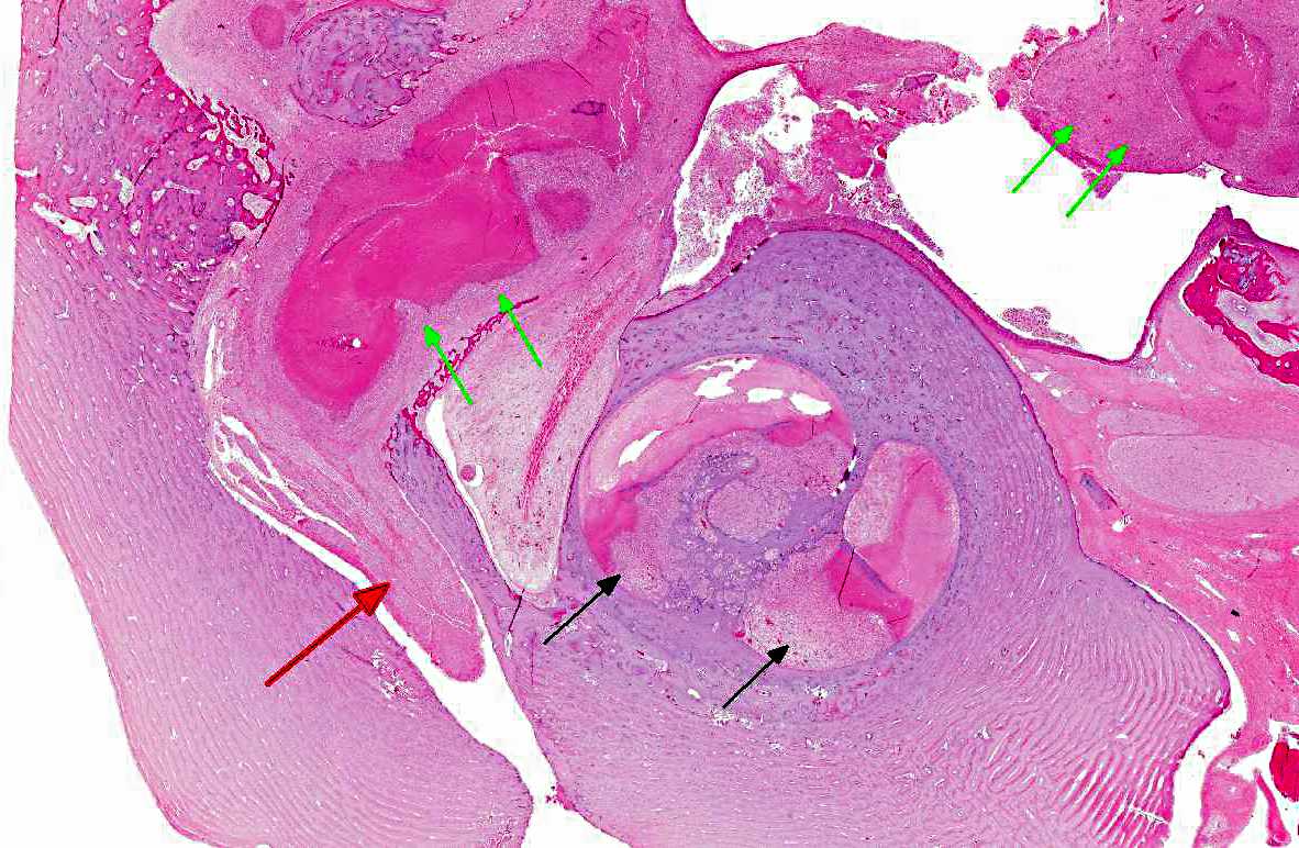
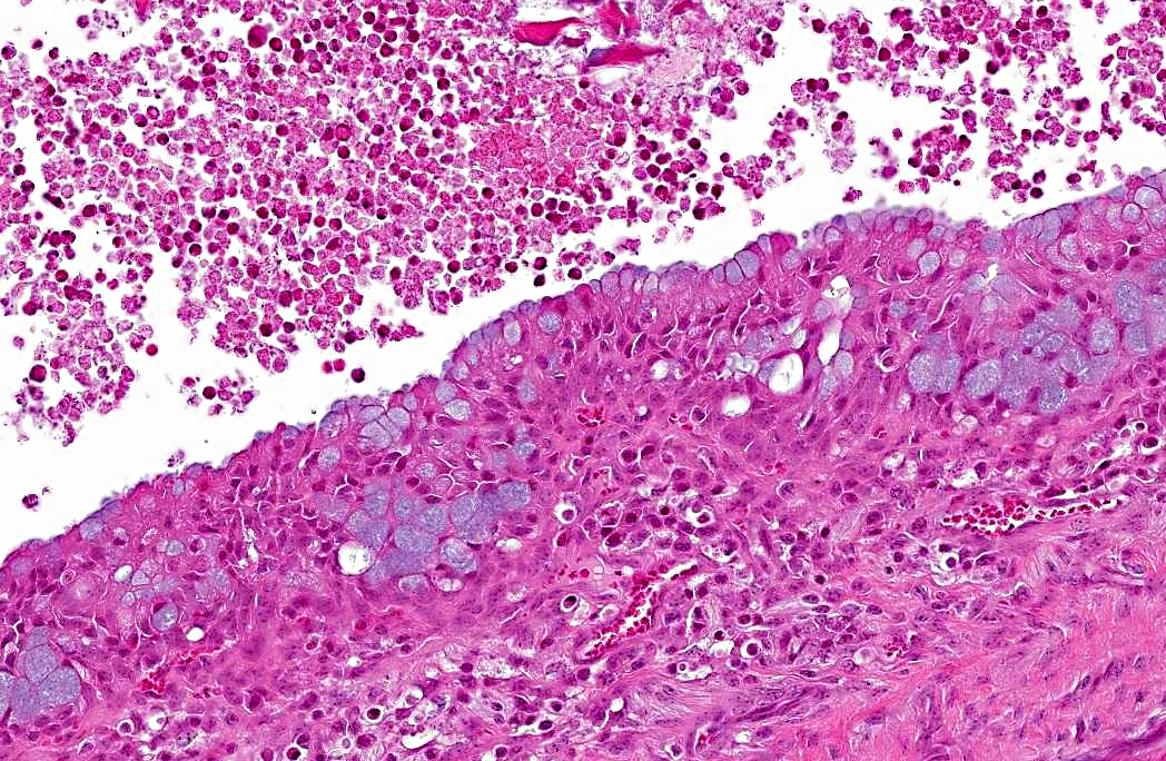
Histopathologic Description:
Morphologic Diagnosis:
Condition:
Contributor Comment:
Mycoplasma are extracellular bacteria that lack a cell wall and may be transmitted via infected secretions of the respiratory tract, genital tract, or mammary gland from infected cows or other calves.(1) Mycoplasma evades the host immune system by suppressing neutrophil and lymphocyte activation, as well as inducing lymphocyte apoptosis.(1) Mycoplasma otitis typically affects male dairy calves less than 2 months of age.(3) The pathogenesis of otitis interna and media due to Mycoplasma in calves is not completely understood but is thought to involve one or more of the following mechanisms: (1) ingestion of infected milk causing nasopharyngeal infection, with extension through the Eustachian tube to the middle ear, (2) immunosuppression or a primary viral infection (BVDV, IBRV) allowing overgrowth of resident flora, (3) direct extension from the external ear canal through the tympanic membrane to the middle and inner ear, or (4) hematogenous spread, which is more commonly seen in rodents, lambs, and swine.(3,5) Infection may then ascend via the vestibulocochlear and facial nerves to the brainstem to cause meningitis. Spleen, liver, kidney, and thymus from this case were tested for bovine viral diarrhea virus via a fluorescent antibody test. Testing for herpesvirus (IBRV) was not performed.
Mycoplasma bovis infections are of economic importance, causing widespread systemic disease, including pneumonia, arthritis, and mastitis.(1,3) Antigenic variation of the surface lipoproteins is thought to contribute to a poor response to antibiotic therapy and evasion of the host immune response.(1,2) Therefore, the cellular and humoral immune responses offer no protection, despite the development of antibody titers.(1) Ingestion of infected milk is thought to be the most likely route of infection, therefore, control measures include pasteurization of colostrum and milk being fed to calves, preventing direct contact between calves, and culling infected cows.(2)
JPC Diagnosis:
Conference Comment:
Recently some species have been reclassified from the rickettsial group into the genus Mycoplasma; these bacteria are referred to as hemoplasms, as they have a tropism for red blood cells rather than epithelium.(4) In addition to the diseases associated with Mycoplasma bovis that were adeptly described by the contributor, conference participants also discussed a variety of other Mycoplasma species of importance in veterinary medicine(4):
| Mycoplasma species | Hosts | Disease |
| M. mycoides subsp. mycoides (small colony type) | Bovine | Contagious bovine pleuropneumonia |
| M. bovis | Bovine | Mastitis, pneumonia, arthritis, otitis |
| M. agalactiae | Ovine, Caprine | Contagious agalactia (mastitis) |
| M. capricolum subsp. capripneumoniae | Caprine | Contagious caprine pleuropneumonia |
| M. capricolum subsp. capricolum | Ovine, Caprine | Septicemia, mastitis, polyarthritis, pneumonia |
| M. mycoides subsp. capri (includes strains previously classified as M. mycoides mycoides large colony type) | Ovine, Caprine | Septicemia, pleuropneumonia, mastitis,arthritis |
| M. ovioneumoniae | Ovine, Caprine | Pneumonia |
| M. pulmonis | Rodents--rat and mouse | Colonize nasopharynx and middle ear; affect respiratory and reproductive tracts and joints |
| M. hyopneumoniae | Swine | Enzootic pneumonia |
| M. hyosynoviae | Swine (10-30 weeks of age) | Polyarthritis |
| M. hyorhinis | Swine (3-10 weeks of age) | Polyserositis |
| M. suis | Swine | Mild anemia, poor growth rates |
| M. ovipneumoniae | mild pneumonia | |
| M. haemofelis | Feline | Feline infectious anemia |
| M. cynos | Canine | Implicated in kennel cough complex |
| M. haemocanis | Canine | Mild or subclinical anema; more severe signs in splenectomized animals |
| M. gallisepticum | Turkeys and Chickens | Chronic respiratory disease; infectious sinusitis |
| M. synoviae | Turkeys and Chickens | Infectious synovitis |
| M. meleagridis | Feline, Equine | Conjunctivitis in cats, pleuritis in horses |
| M. equigenitalium | Equine | Abortion |
Conference participants noted the presence of an inflamed mucosal lining, interpreted as a polyp, that did not appear to be attached to the rest of the tissue. The relationship between the polyp and the other structures was difficult to ascertain in the sections examined. Also, participants noted there is osteolysis of the petrous portion of the temporal bone.
References:

