Wednesday Slide Conference, Conference 8, Case 2
Signalment:
5 month old, intact female, Shar Pei, Canis familiaris, Dog.
History:
Limited history available. Alopecia and dermatitis, has been on cephalexin. epresentative samples submitted from face and legs.
Gross Pathology:
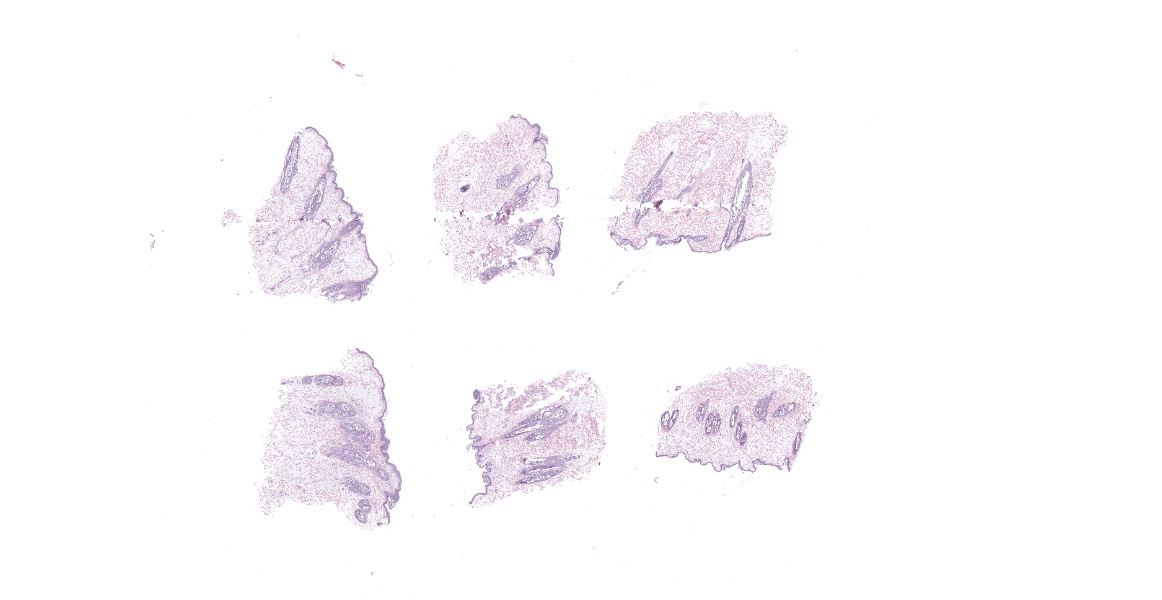
3 haired skin punch biopsy specimens, 5-6mm diameter, ranging from 3-4mm deep
Microscopic Description:
Haired skin: All 3 punch biopsy samples are similar.
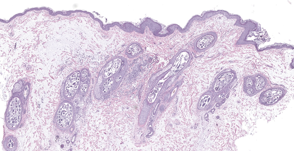
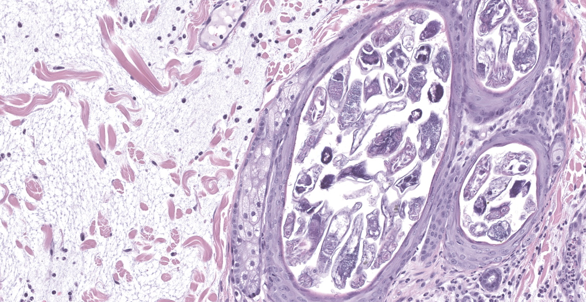
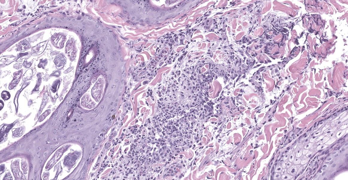
Diffusely, hair follicle lumina are moderately distended and filled with multiple longitudinal, transverse, and tangential sections of arthropods. These arthropods are elongate, up to 40µm diameter and 200µm in length; have a thin, eosinophilic, chitinous exoskeleton; short, jointed appendages; a hemocoel; striated musculature; digestive tract; and male or female reproductive tracts. There is minimal perifollicular as well as perivascular inflammation consisting of low numbers of lymphocytes, plasma cells, and eosinophils.
Dermal collagen fibers are diffusely fragmented and widely separated by abundant clear space and wispy, fibrillar, beaded, basophilic to amphophilic mucin. There are low numbers of histiocytes and lymphocytes scattered within the mucinous matrix.
There is clumping of melanin pigment within the follicular bulb matrix epithelium and to a lesser extent within the non-matrical follicular epithelium and follicular lumina.
Contributor’s Morphologic Diagnosis:
1. Haired skin: Follicular ectasia, moderate, diffuse, with mild lymphoplasmacytic and eosinophilic perifolliculitis and numerous follicular intraluminal Demodex canis mites
2. Haired skin: Dermal mucinosis, diffuse, marked
3. Haired skin, hair follicles: Matrical epithelial, follicular epithelial, and intraluminal melanin clumping
Contributor’s Comment:
This case represents three entities: juvenile-onset demodicosis, dermal mucinosis (a feature of “normal” skin in Shar Pei dogs), and color dilution.
Canine demodectic mange, also termed follicular mange or red mange, is one of the most common skin diseases of dogs.6-10 It is a noncontagious disease that occurs when commensal Demodex mites are allowed to proliferate resulting in overpopulation.1,5,6,9 Canine demodicosis is most commonly caused by D. canis, but D. injai can also cause disease.1,5,8,9 D. canis is approximately 300µm in length, while D. injai, the “long-bodied mite”, is 334-368µm in length.8 Predisposing factors to Demodex overpopulation in its host include multiple causes of host immunosuppression.1,6,8,9 Canine demodicosis can be either localized or generalized.1,5,8,9 Localized disease is mild and typically self-limiting with spontaneous resolution, whereas generalized disease may spontaneously resolve but can continue into adulthood if inadequately treated, and may be fatal.5,8 Canine demodicosis can also be either juvenile-onset or adult-onset.1,5,8 Disease is considered adult-onset if disease onset occurs at 4 years of age or older,8 although differentiation between these may be difficult.1 Generalized demodicosis is typically a juvenile-onset disease.4,8 Juvenile-onset demodicosis is thought to be due to a genetically mediated immunodeficiency resulting in decreased T-cell function.4,6,8,9 A recent study into the molecular pathogenesis of canine demodicosis found evidence that disease is associated with Demodex-induced host immune tolerance.6 Studies into this immune tolerance identified host cellular endoplasmic reticulum stress which in turn results in the accumulation of unfolded proteins (i.e., unfolded protein response) which regulates signaling pathways involved in Toll-like receptors (especially TLR2) and promotion of M2-phenotype immunosuppressive macrophages.7 Disease relapse, recurrence, or persistence is uncommon.1
Excessive accumulation of dermal mucin is termed cutaneous mucinosis.2,5,8,10 This condition is abnormal and rare in most dogs.5,8 In shar pei dogs, however, cutaneous mucinosis is due to a genetic mutation and is considered a normal feature which leads to their distinctive thick, wrinkled skin.5,8,9 The mucinous material in the shar pei dermis has been identified as hyaluronan (or hyaluronic acid [HA]).2,10 A study found that HA is produced in shar pei dermal fibroblasts in greater quantities than in control cells.10 Shar pei cutaneous mucinosis has further been linked to increased mRNA expression of the HAS2 isoform of hyaluronan synthase (HAS), resulting in increased transcription of HAS2 by dermal fibroblasts.2,9,10 The authors of that study suggest using the term “hereditary cutaneous hyaluronosis (HCH)” for the shar pei specific version of cutaneous mucinosis.2 Diffuse canine cutaneous mucinosis has also been associated with hypothyroidism; this is rarely reported but clinically and histologically striking.5,8,9 Cutaneous mucinosis is termed “myxedema” when associated with hypothyroidism.5,8,9 Increased focal areas of dermal mucin have been reported in association with numerous inflammatory and neoplastic processes, such as mast cell tumor, severe pyoderma, and eosinophilic diseases.5,8,9 A specific example of a cause of focal mucinosis in dogs is infection with the trombidioid mite Stralensia cynotis which causes characteristic mucinosis of the perifollicular dermis and pseudoepitheliomatous hyperplasia of the follicular epithelium (see WSC Conference 2, Case 4, 2018-2019).8,9
Color dilution has been reported in many species as well as in many breeds of dogs.3,8,9 The condition in dogs is inherited as an autosomal recessive trait and is due to abnormalities in melanin transfer and storage.3,5,8,9 In affected dogs, the coat color is pale, manifesting as blue, fawn, etc. This dilute color appearance is due to clumping of large melanin granules within hair follicles and characteristically within the epidermis.5,8,9 There is preliminary evidence that the genetic cause of color dilution in dogs is an autosomal recessive mutation in the melanophilin gene (MLPH).3,9 In dogs, the condition of color dilution may be associated with alopecia (i.e., color dilution alopecia), but is not always associated with alopecia.5,8,9 Color dilution alopecia should only be diagnosed if histologic findings of both color dilution (clumped melanin) and follicular dysplasia/distortion are present.5,9
Contributing Institution:
Tri-Service Research Laboratory
4141 Petroleum Dr
San Antonio, TX 78234
JPC Diagnosis:
1. Haired skin: Follicular ectasia, moderate, diffuse, with mild lymphoplasmacytic and eosinophilic perifolliculitis and numerous follicular intraluminal adult Demodex mites.
2. Haired skin, dermis: Mucinosis, diffuse, moderate.
JPC Comment:
Case 2 has several entities for participants to consider. Demodex mites and dermal mucinosis are abundantly present in these sections. These findings together are interesting and should prompt consideration of immunosuppression such as Cushing’s disease or severe hypothyroidism (myxedema) though the breed of this dog (Shar Pei) is an important detail as the contributor notes. Though these look similar histologically, the clinical presentation sorts these two camps out quickly. Other considerations for mite presence include long-term immunomodulatory drugs for control of allergic skin disease. In section, there are multiple examples of jointed appendages (figure #) and skeletal muscle that help to distinguish that hair follicle lumina are distended with many Demodex that the contributor nicely describes. Although not needed to distinguish the myxomatous dermis in this case, an Alcian blue pH 2.5 does highlight mucin nicely. One feature not present in this case is epidermal hyperplasia which is a secondary change due to the animal scratching (owing to mural folliculitis, rupture, and periadnexal mite fragments). Conference participants compared Demodex species across dogs and cats with D. cati being similarly follicularly-focused like D. canis while D. gatoi is found in the stratum corneum and is directly transferable between cats. The profound sebaceous hyperplasia induced by D. injai residing within sebaceous and ducts while lacking other epidermal changes explains the markedly greasy phenotype noted clinically.
The color dilution noted by the contributor prompted an interesting discussion among the group. Notably, many of the approved Shar Pei breed standard coats include dilute variants. As such, this could be a normal observation. We did not observe melanin clumping within epidermal melanocytes or the hair shaft in large quantity, though the hair bulb clumping is evident and worth describing. Though not in these section, distortion and fracture of the hair shaft is a helpful corroborating feature for color dilution alopecia.
Finally, Dr. Bradley touched on the role of inflammation in demodicosis. Although Demodex may be an incidental finding during routine skin biopsy, this case is clearly in excess of a single mite within a single follicle. Nonetheless, conference participants noted the overall muted inflammatory response in this case. Demodex is thought to play a role in the development of human rosacea in a two-fold manner. Foremost, proliferation of the mite activates TLR2 on inflammatory cells which upregulates production of cathelicidin (LL-37 peptide) which has both antimicrobial and proangiogenic effects.4 Activation of endothelial cells by LL-37 increases in conjunction with UVB damage from sunlight, which then promotes increased production of VEGF which has both proangiogenic and immunosuppressive effects on immune cells.4 In excess, VEGF binding causes loss of lymphocyte function and essential T cell exhaustion.4 Secondly, Demodex also express a surface glycan Thomsen-Nouveau Antigen (Tn Ag) which is recognized by the dendritic cell galactose-type lectin receptor. Once bound to this receptor, Tn Ag induces production of IL-10 and recruitment of regulatory T-cells that further tamp down inflammation and allow Demodex to evade a committed host response and proliferate further.4 These factors together hint at the interplay of VEGF, LL-37, and IL-10 in a complex feedback loop for which the flushed face, papules, and telangiectasia are only a hint of the sinister scheme occurring at the molecular level.
References:
1. Bowden DG, Outerbridge CA, Kissel MB, Baron JN, White SD. Canine demodicosis: a retrospective study of a veterinary hospital population in California, USA (2000-2016). Vet Dermatol. 2018;29(1):19-e10.
2. Docampo MJ, Zanna G, Fondevila D, et al. Increased HAS2-driven hyaluronic acid synthesis in shar-pei dogs with hereditary cutaneous hyaluronosis (mucinosis). Vet Dermatol. 2011;22(6):535-545.
3. Dr ögemüller C, Philipp U, Haase B, Günzel-Apel AR, Leeb T. A noncoding melanophilin gene (MLPH) SNP at the splice donor of exon 1 represents a candidate causal mutation for coat color dilution in dogs. J Hered. 2007;98(5):468-473.
4. Forton FMN. The Pathogenic Role of Demodex Mites in Rosacea: A Potential Therapeutic Target Already in Erythematotelangiectatic Rosacea?. Dermatol Ther (Heidelb). 2020;(10):1229–1253.
5. Gross TL, Ihrke PJ, Walder EJ, Affolter VK. Skin diseases of the dog and cat. 2nd ed. Oxford, UK:Blackwell Science; 2005:222-225, 380-383, 442-449, 482-483, 518-521, 526.
6. Kelly PA, Browne J, Peters S, et al. Gene expression analysis of Canine Demodicosis; A milieu promoting immune tolerance. Vet Parasitol. 2023;319:109954.
7. Kelly PA, McHugo GP, Scaife C, et al. Unveiling the Role of Endoplasmic Reticulum Stress Pathways in Canine Demodicosis. Parasite Immunol. 2024;46(4):e13033.
8. Mauldin EA, Peters-Kennedy J. Integumentary system. In: Maxie MG, ed. Jubb, Kennedy, and Palmer’s Pathology of Domestic Animals. Vol 1. 6th ed. Philadelphia, PA: Elsevier Saunders; 2016: 557, 678-682, 683.
9. Welle MM, Linder KE. The integument. In: Zachary JF, ed. Pathologic Basis of Veterinary Disease. 7th ed., St. Louis, MO; Elsevier; 2022:1120, 1134, 1135, 1142.e1, 1149.e3, 1179-1181, 1182.e1, 1233.
10. Zanna G, Docampo MJ, Fondevila D, Bardagí M, Bassols A, Ferrer L. Hereditary cutaneous mucinosis in shar pei dogs is associated with increased hyaluronan synthase-2 mRNA transcription by cultured dermal fibroblasts. Vet Dermatol. 2009;20(5-6):377-382.


