WSC Conference 4, Case 2
Signalment:
3-month-old, female Speckled Sussex, avian (Gallus gallus)
History:
Four birds from a backyard flock were losing weight and anorexic. This bird died suddenly.
Gross Pathology:
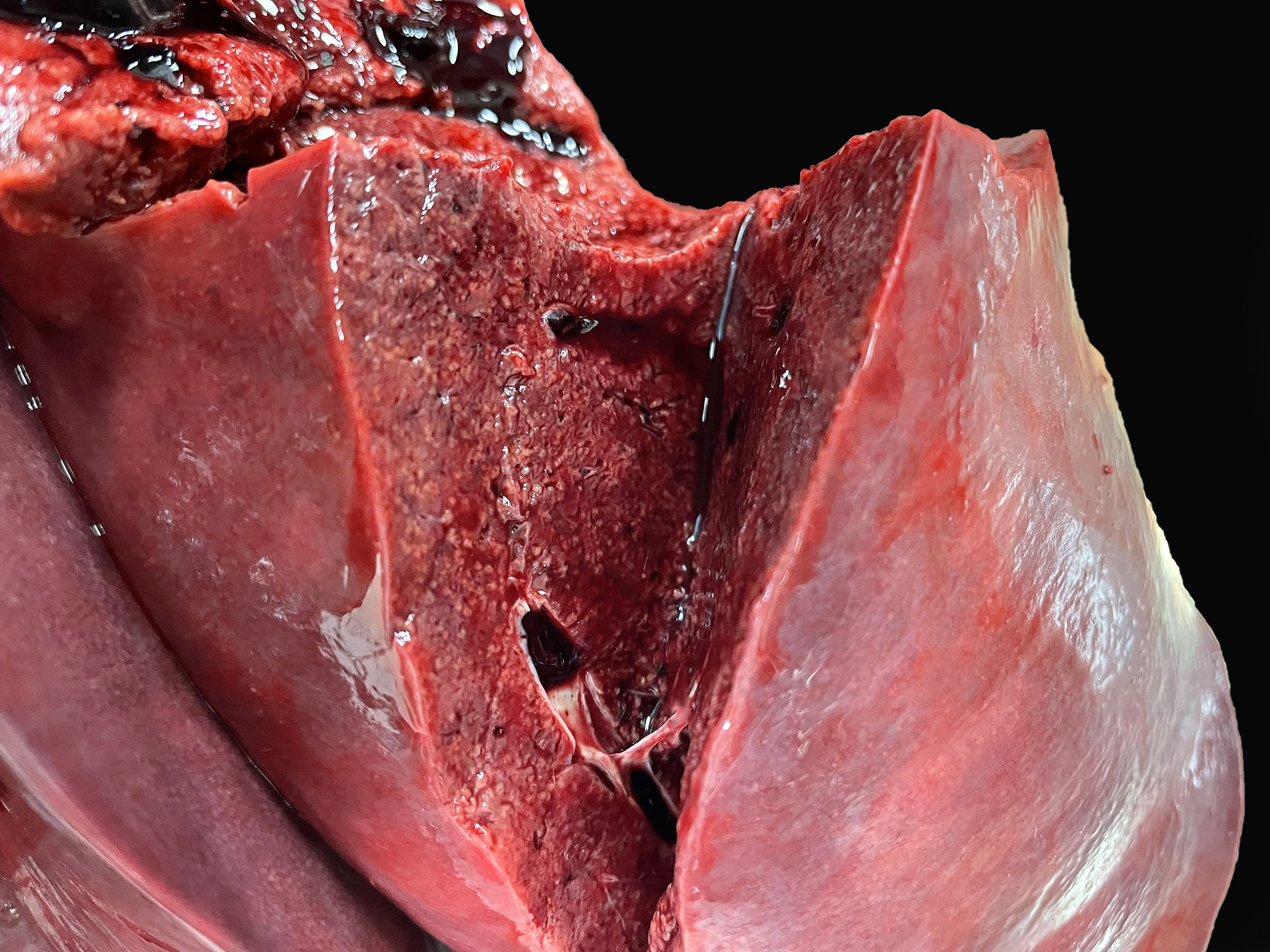
Necropsy was performed on a 3-month-old intact female chicken. It was dark brown with light speckling. The keel bone was prominent and pectoral muscles were atrophied. The eyes were sunken in the orbits. Loose white to green fecal material was present around the cloaca and ventral tail feathers. Diffusely the skin was bright red.
Laboratory Results:
Fecal examination revealed few eggs (0-3/10X) of Capillaria sp., Ascaridia sp. and Heterakis gallinarum.
Microscopic Description:
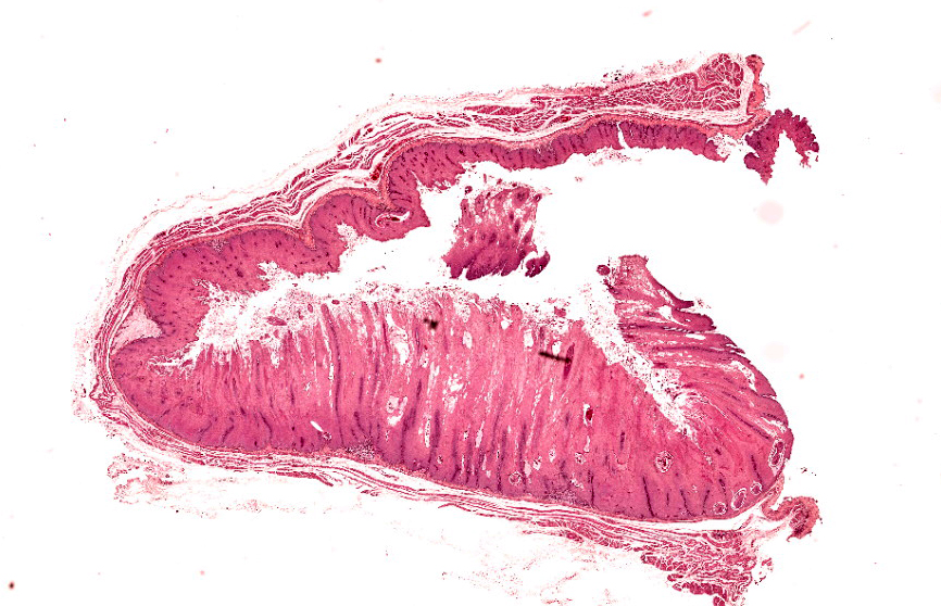
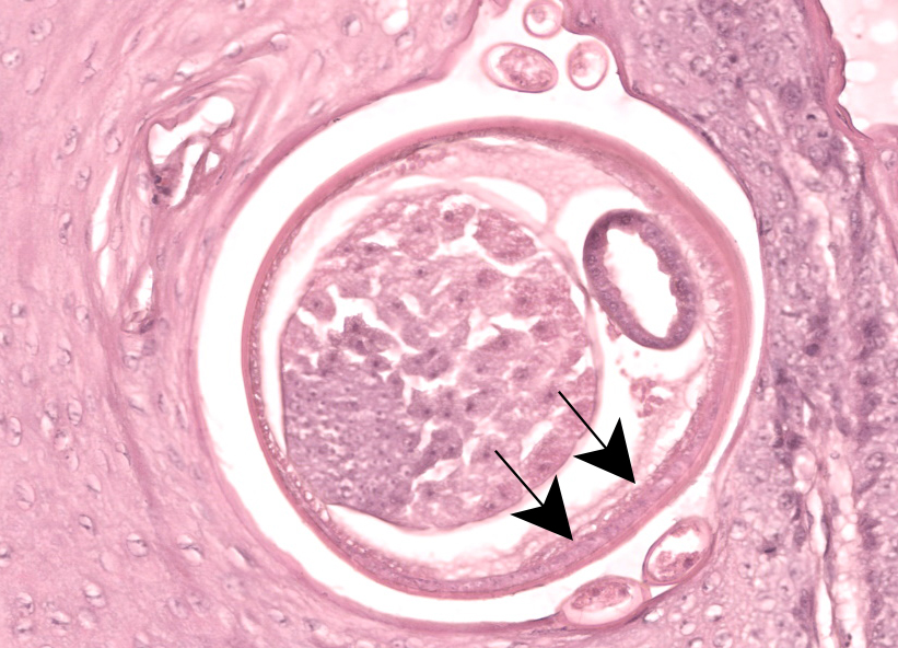
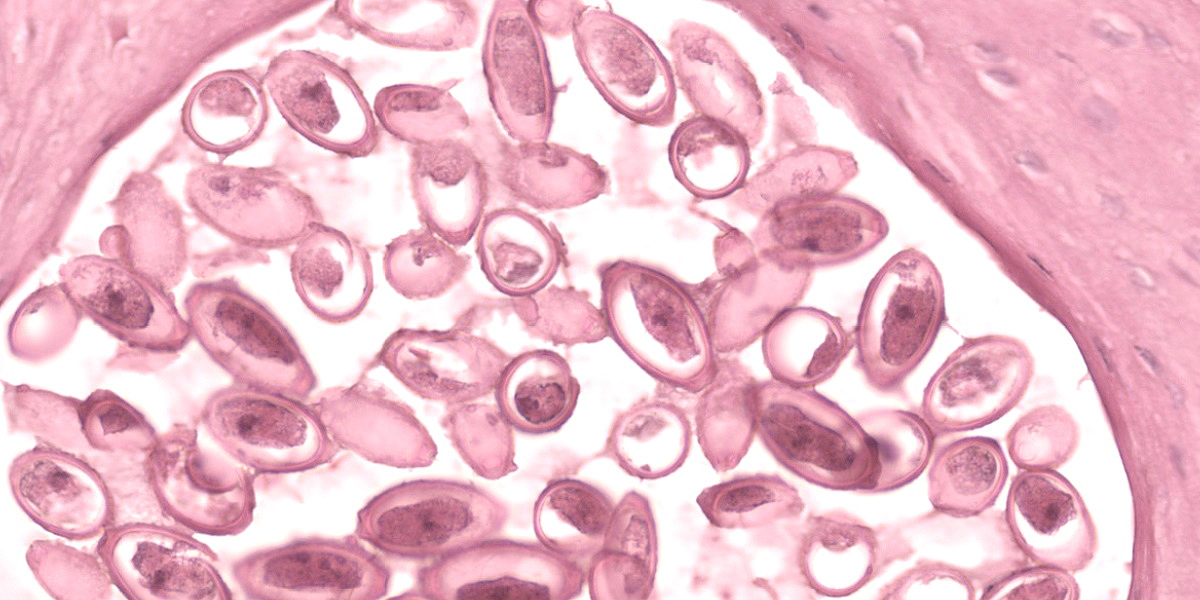
Crop: Numerous cross-sections of nematodes are embedded within the markedly thickened mucosa. The parasites are 160-350 micrometers in diameter and contain an approximately 6 micrometer smooth cuticle, coelmyarian musculature, hypodermal bacillary bands, a body cavity, a multinucleated intestine and ovaries. There are bioperculated, oval embryonated eggs (Capillaria sp.) within the space occupied by the nematode, or in spaces by themselves within the mucosal epithelium.
Contributor’s Morphologic Diagnosis:
Crop: Ingluvitis, proliferative, multifocal, moderate with intralesional nematode parasites, Capillaria sp.
Contributor’s Comment:
The crop and esophageal mucosa is markedly thickened and contain numerous cross sections of parasites and eggs. The cuticle, digestive tract and body cavity qualifies these as nematode parasites. The bioperculated, oval embryonated eggs present in the parasite, within and on the surface of the mucosa are consistent with Capillaria sp. Similar nematode parasites and eggs were also present in the small intestine and double operculated eggs (Capillaria sp.) and eggs consistent with Ascaridia and Heterakis gallinae infections are present on fecal flotation. Although chickens can harbor many species of Capillaria, only Capillaria annulatus and contortus are present in both the crop and small intestine. As nematode parasites were not recovered during necropsy, definitive identification was not possible.
This chicken had a severely thickened crop mucosa and was malnourished as evidenced by the prominent keel bone and pectoral muscle atrophy. The loose fecal material around the cloaca and ventral tail feathers are consistent with diarrhea, which is often associated with heavy intestinal Capillaria infections. Capillaria infections are usually of little consequence in most chickens; however, when there are heavy infections in the crop and esophagus by C. annulatus or contortus (the most pathogenic Capillaria species) the mucosa can become so thickened that swallowing can be difficult to impossible. This can result in high mortality as was present in this case. All 4 chickens in this backyard flock died and had lesions similar to those in this chicken. As both C. annulatus and contortus are transmitted by earthworms, backyard flocks in humid environments are extremely susceptible. Death of this chicken was subsequent to heavy Capillaria infections of the crop, esophagus and small intestine.
Contributing Institution:
Tuskegee University
Pathobiology Department
https://www.tuskegee.edu/programs-courses/colleges-schools/cvm
JPC Diagnosis:
Crop, mucosa: Hyperplasia, segmental, marked, with few adult female aphasmid nematodes and numerous eggs.
JPC Comment:
Capillariasis is a common disease of free-range chickens. Some species within the Capillaria genus infect the upper digestive system, particularly the crop and esophagus, while others cause intestinal or cecal disease.1 The adult worms are thin and filamentous, giving them the monikers “thread worms” or “hairworms,” and produce characteristic bi-operculate eggs. These worms burrow into their preferred mucosal lining, typically without causing clinical signs, but with increasing numbers, severe inflammation, diarrhea, wasting, and death may occur.
The life cycles vary among the Capillaria species; some have a direct life cycle, while other require an intermediate host, most commonly an invertebrate such as an earthworm.1 The most common Capillaria species that cause disease in chickens are C. contorta, C. annulata, C. anatis, C. bursata, C. caudinflata, and C. obsignata. As the contributor notes, of these, C. annulata and C. contorta are found in the crop and esophagus, C. anatis occurs in the ceca, and the balance inhabit the small intestine. Depending on the species and life cycle, chickens are infected by ingesting litter containing worm eggs or by ingesting an infected intermediate host.
As in this case, the typical histologic lesion of crop capillariosis is thickening and inflammation of the mucosa, while intestinal and cecal-focused Capillaria species may cause inflammation, hemorrhage, and erosion of the gastrointestinal linings.
Conference discussion centered around histologic differences between the avian crop and esophagus and the difficulty in differentiating between the two locations. While differences appear to be species-specific, in general the crop has no or few glands compared to the esophagus, where glands are typically more numerous. Conference participants felt it was quite difficult to determine if examined section was esophagus or crop, and many participants felt they would have had to wing it were it not for the contributor’s gross necropsy findings.
Conference participants also noted the very mild nature of the inflammatory infiltrate and postulated that this might be because larvae are typically restricted to the mucosa in crop capillariasis. Participants felt that the striking hyperplasia was the more important histologic finding and deserved pride of place in the JPC morphologic diagnosis.
References:
- Swayne DE, Glisson JR, McDougald LR, et.al. Diseases of Poultry. 13th ed. Blackwell Publishing Ltd;2008:1162-1164.


