CASE I:
Signalment:
4-year-old male neutered Labradoodle (Canis lupus familiaris)
History:
This patient presented to the emergency department for 2 days of vomiting and diarrhea with a historical seizure following weakness at home. Clinical examination revealed that the dog was obtunded, weak, in shock and hypoglycemic. He was treated for cerebral edema without response and required up to 10% dextrose to maintain normoglycemia. Bloodwork was suggestive of sepsis, but the abdominal ultrasound was not consistent with septic peritonitis and the fluid was too scant to obtain. The patient arrested before an echocardiogram could be performed and before blood cultures, urine cultures and ACTH stimulation test results returned. The tentative clinical diagnosis was sepsis of unknown source.
Gross Pathology:
The adrenal glands are reduced in size bilaterally and measure 1.5 x 0.4 x 0.5 cm on the right and 1.8 x 0.5 x 0.5 cm on the left. The adrenal cortices appear bilaterally attenuated (approximate cortex to medullary ratio of 1:5). The thyroid/parathyroid and pituitary glands are grossly within normal limits.
Laboratory Results:
Chemistry panel:
Glucose initially too low to read on in-house panel,
Sodium is 146 mmol/L (reference 142-152 mmol/L),
Potassium is 4.4 mmol/L (reference range 4.0-5.4 mmol/L),
Sodium: potassium ratio is 33 (reference range 28-37),
Cholesterol: 105 mg/dL (L) (reference range 131-345 mg/dL)
Albumin: 2.4 g/dL (L) (reference range 2.7-3.9 g/dL)
Cortisol levels are 0.6 µg/dL (L) (reference range 2.0-6.0 µg/dL)
CBC:
White blood cells are 2.5 K/µL (L) (reference range 4.9-17.6 K/µL),
Neutrophils are 0.1K/µL (L) (reference range 2.94-12.67 K/µL),
Lymphocytes are 2.15 K/µL (reference range 1.06-4.95 K/µL,
Monocytes are 0.05 K/µL (L) (reference range 0.13-1.15 K/µL)
Eosinophils are 0.2 K/µL (reference range 0.07-1.49 K/µL),
Microscopic Description:
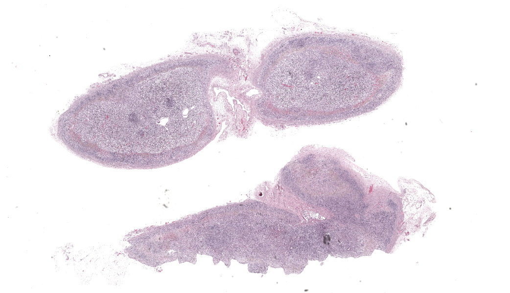
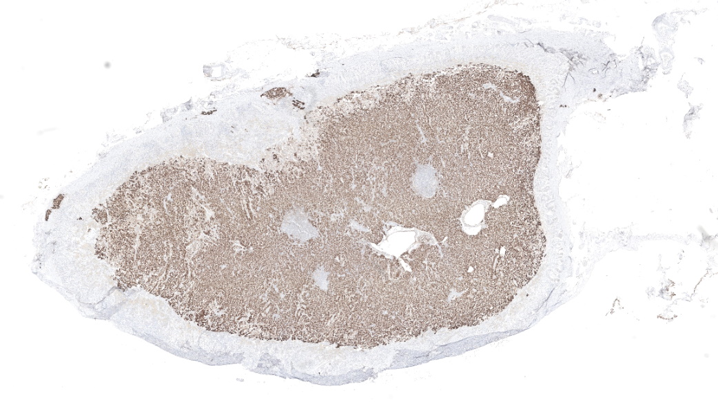
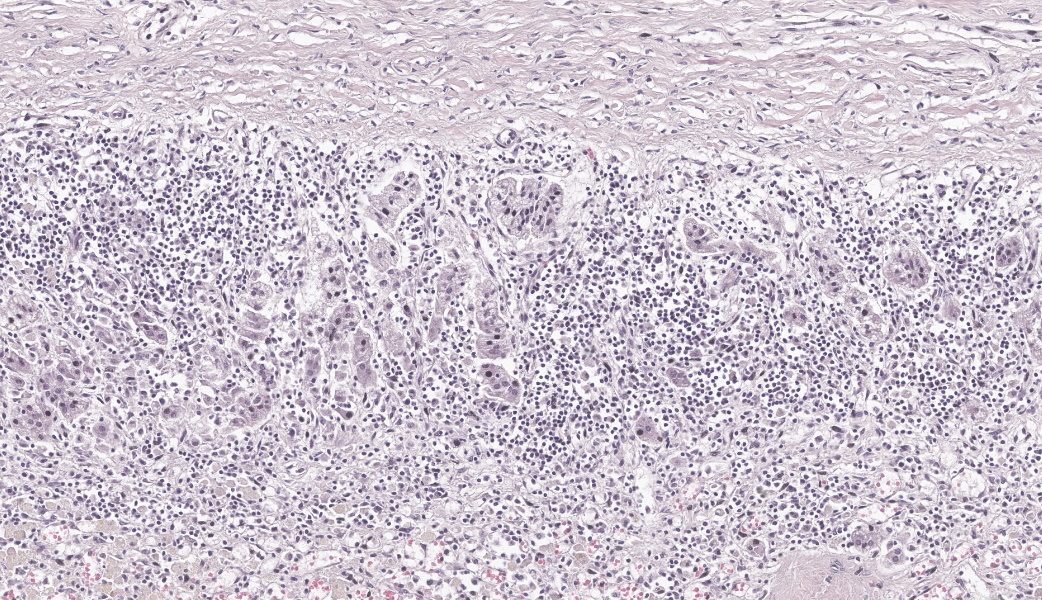
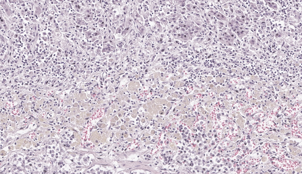
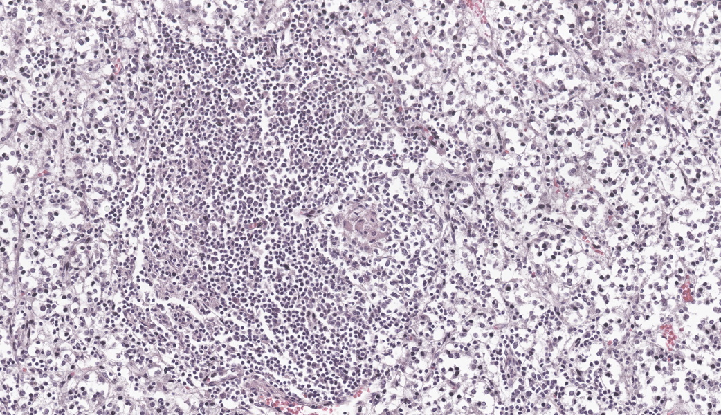
Two adrenal glands (one section of each) are examined. In each section, the cortex is severely attenuated, with a reduced cortex to medullary ratio (approximately 1:5). The zona glomerulosa can be visualized, however, the zonae fasciculata and reticularis are severely atrophied to absent with stromal collapse and rare remaining cortical cells. These remaining cells are interspersed by inflammation and exhibit cytoplasmic vacuolation. The deeper layers of the cortex are largely replaced by inflammatory cells, including macrophages, lymphocytes and plasma cells, intermixed with fibrous connective tissue (fibrosis). Fibrous tissue is highlighted with a Masson’s trichrome stain. Macrophages contain abundant, yellow to gold and brown granular pigment (presumed lipofuscin, hemosiderin). Small amounts of hemorrhage are intermixed with the inflammatory cells of the adrenal cortex. The medullary portion of the adrenal gland is intact, recognizable by cells of a neuroendocrine phenotype, containing a granular, amphophilic to basophilic, often vacuolated cytoplasm with separation into small packets by a fine fibrovascular stroma. Small amounts of scattered inflammation extend into the medulla, including lymphocytes, plasma cells and macrophages.
Contributor’s Morphologic Diagnosis:
Adrenal glands: Adrenalitis, histiocytic, lymphoplasmacytic, severe, with regional cortical atrophy, collapse and loss (zonae fasciculata and reticularis), lipofuscin and hemosiderin accumulation (consistent with idiopathic adrenocortical atrophy, primary atypical adrenal insufficiency, hypoadrenocorticism / Addison’s disease)
Contributor’s Comment:
Nonsuppurative adrenalitis with severe atrophy and loss of the adrenal cortex correlates with the gross findings of adrenal cortical attenuation. The zonae fasciculata and reticularis were primarily affected and were unable to be visualized within the section. The outer zona glomerulosa was also inflamed, however, remnant tissue was present, which may explain the atypical presentation of this patient (lack of electrolyte abnormalities on bloodwork). Approximately 85%-90% of the adrenal cortex must be nonfunctional before clinical signs can be observed.3,9 In this case, sodium and potassium levels were within normal limits on the chemistry panel. Concurrent hyponatremia and hyperkalemia with a sodium:potassium ratio of greater than 23:1 can be used for clinical identification of hypoadrenocorticism.2,3,9 Similar electrolyte changes can occur with gastrointestinal disease (including parasitism), renal and hepatic disease, congestive heart failure, pleural effusion and severe metabolic or respiratory acidosis.2
Primary adrenal insufficiency, (hypoadrenocorticism or Addison’s disease) is characterized by reduced mineralocorticoid and/or glucocorticoid production by the adrenal glands.2 Secondary and tertiary (or central) hypoadrenocorticism are associated with abnormalities in the pituitary gland which produces adrenocorticotropic hormone (ACTH) or the hypothalamus which produces corticotropic releasing hormone (CRH).2 Secondary hypoadrenocorticism can occur with pituitary destruction caused by neoplasia, inflammation or head trauma.3 Tertiary hypoadrenocorticism has not been definitively diagnosed in dogs.2 Hypoadrenocorticism in dogs has an estimated prevalence between 0.06 and 0.3%.2 Breed related predispositions have been reported, including a strong heritable component in standard poodles as well as Portuguese water dogs, Nova Scotia duck tolling retrievers, soft coated wheaten terriers and bearded collies.2,3,9 Females are at increased risk (1.9-2.6 times greater), however, this has not been identified in all studies, which may reflect different breed-genetic risk factors. The reported median age is 3-4 years, however, dogs with glucocorticoid deficiency alone (the “atypical” form) are commonly older (6-8 years old).2,4,8 Affected dogs usually have vague, non-specific clinical signs, and hypoadrenocorticism has been referred to as “the great pretender”.2,3 Clinical characteristics that can raise suspicion for hypoadrenocorticism include waxing and waning gastrointestinal signs that respond rapidly to fluid or glucocorticoid therapy and are exacerbated by stress.2 Patients with the atypical form are reported to have a longer duration of clinical signs, likely because this form is more challenging to identify.4,8
The baseline cortisol level of this patient was below the reference range and the ACTH stimulation test results were not available. This patient was not receiving any medication and other than a historical “sensitive stomach”, was reported to have been clinically normal prior to the onset of vomiting and seizures. “Typical” primary hypoadrenocorticism encompasses glucocorticoid deficiency accompanied by mineralocorticoid deficiency, whereas “atypical” is loosely described as glucocorticoid deficiency in the absence of mineralocorticoid deficiency.2 The atypical form represents a minority of cases (approximately 5-10%).3,9 One study found that dogs with the “atypical” form have decreased basal and ACTH stimulated aldosterone levels, suggesting mineralocorticoid deficiency despite the lack of electrolyte abnormalities.9 A subset of dogs without electrolyte abnormalities will subsequently develop them within a year of diagnosis.9 Standardized nomenclature for these terms has not been established. The Agreeing Language in Veterinary Endocrinology (ALIVE) project suggests referring to “typical” as hyponatremic and/or hyperkalemic primary hypoadrenocorticism and “atypical” as eunatremic, eukalemic primary hypoadrenocorticism.2 In order to avoid confusion, herin, “atypical” glucocorticoid deficient hypoadrenocorticism will be referred to as GDH and “typical” mineralocorticoid-glucocorticoid deficient hypoadrenocorticism as MGDH.8
Acute adrenocortical insufficiency (Addisonian crisis) can be a life-threatening condition that clinically presents as shock, dehydration and metabolic derangements (the latter with MGDH).2,4,6 Hypotension can be attributed to hypovolemia, poor vascular tone and lack of compensatory tachycardia. Mortality can occur with hyperkalemia, hyponatremia, dehydration and hypovolemic shock.2 Possible complications include gastrointestinal ulceration, DIC, sepsis caused by bacterial translocation from the gastrointestinal tract, and aspiration pneumonia (due to megaesophagus).2,6 Collapse in GDH has been reported with hypoglycemia, sepsis and GI hemorrhage.4 The mainstays of treatment for hypoadrenocorticism include fluid resuscitation, glucocorticoid administration, correcting hypoglycemia and electrolyte abnormalities, if present.4,6,9 A recent study found that intravenous CRI of hydrocortisone as a treatment for acute Addisonian crisis is well-tolerated and safe but had no clear benefit when compared with traditional glucocorticoid replacement.6 With appropriate treatment and recovery following a crisis and lifelong administration of medical therapy, the prognosis is excellent for normal life expectancy.6,9
Definitive diagnosis of hypoadrenocorticism requires histologic evaluation of the adrenal glands and correlation with clinical parameters. The adrenal cortex has three functional zones. The outer zona glomerulosa (25% of the cortex) produces mineralocorticoids (aldosterone) and supplies precursor cells for the inner 2 layers.2,3,9 The middle zona fasciculata (60% of the cortex) produces glucocorticoids, and the inner zona reticularis (15% of the cortex) produces androgens.2,9 Aldosterone regulates water, acid-base and electrolyte homeostasis via increasing sodium absorption and potassium secretion.2 Aldosterone mainly targets the kidney and is the most important hormone affecting renal potassium excretion.3 Immune mediated destruction is the most common cause of primary hypoadrenocorticism in dogs and is suspected in this case. Uncommon causes of adrenal gland destruction and hypoadrenocorticism include inflammatory or neoplastic diseases, amyloid deposition, infarcts and drug administration (e.g., mitotane, trilostane, sudden withdrawal of steroids). Any inflammatory, neoplastic, vascular, infectious, traumatic or developmental process affecting the hypophysis can result in secondary hypoadrenocorticism.1,2,9
Several immune response genes have been identified for determining susceptibility to Addison’s disease in humans. Supporting evidence of an autoimmune pathogenesis was demonstrated in dogs with hypoadrenocorticism, encompassing circulating autoantibodies against steroid synthesis enzyme 21 hydroxylase and p450 side chain-cleavage enzyme.1 Overlap of autoimmune diseases is common, and approximately 50% of humans with Addison’s disease have other autoimmune diseases.1 Dogs and humans with hypoadrenocorticism may have concurrent hypothyroidism, diabetes mellitus or hypoparathyroidism.1,9 A genetic component to canine hypoadrenocorticism is suspected, given the strong breed associations in epidemiologic studies.1,2 Molecular genetic studies have found that genes and signaling pathways similar to those implicated in humans may be associated with canine hypoadrenocorticism. Genetic variation in the genes encoding MHC class II proteins (which plays an important role in the adaptive immune system) have been associated with autoimmune diseases in multiple species.1 Immune response genes which may be important for increased susceptibility to hypoadrenocorticism in dogs include dog leukocyte antigen (DLA) and cytotoxic T-lymphocyte associated protein 4 (CTLA4).1,5 A genome wide microsatellite analysis in Portuguese water dogs showed a strong association with the DLA region of chromosome 12 and a marker close to CTLA4 on chromosome 37.1 Despite the clear association with genetic factors, canine hypoadrenocorticism is most likely a complex genetic disorder.1 In humans with primary adrenal insufficiency, many genetic causes have been identified and include disorders of steroidogenesis, defects in cholesterol biochemistry, peroxisomal defects, mitochondrial disorders, abnormal adrenal development, ACTH resistance and autoimmune polyglandular syndromes.7
Cortisol has many roles, including regulating circulating glucose concentrations, enhancing urinary calcium excretion and reducing absorption, maintaining the normal function of the intestinal mucosa, sustaining myocardial performance and increasing cardiac output, facilitating arteriolar constriction, helping to maintain normal blood pressure and volume, stimulating production of erythrocytes, release of platelets and neutrophils from the bone marrow and suppressing lymphocyte proliferation.2 Basal cortisol levels less than 55 nmol/L is highly sensitive for diagnosis of hypoadrenocorticism, thus is a useful screening test. The cortisol levels in this case were 0.6 ug/dL or 17 nmol/L, well under the cutoff of 55 nmol/L. The ACTH stimulation test is the test of choice for diagnosis of hypoadrenocorticism.2 Hypoglycemia was also present in the submitted case, which is a common abnormality with hypoadrenocorticism due to glucocorticoid insufficiency. In one review, hypoglycemia was found in 31% of GDH cases, presumably due to decreased hepatic gluconeogenesis and increased peripheral sensitivity to insulin.2 Clinical signs referable to hypoglycemia included profound weakness and seizures.2
In one study comparing GDH and MGDH, hypocholesterolemia and hypoalbuminemia were more commonly observed in the GDH group.8 Hypocholesterolemia may be attributable to reduced gastrointestinal lipid absorption, reduced mobilization of fatty acids due to low cortisol or increased utilization from high ACTH concentrations.8 Interestingly, hypocholesterolemia and hypoalbuminemia were also observed in this case. Hypercalcemia is observed in approximately 30% of dogs with hypoadrpenocorticism and was not observed in this case.9 A stress leukogram (neutrophilia, lymphopenia, monocytosis and eosinopenia) occurs with cortisol release. In hypoadrenocorticism, a stress leukogram may not be present in the face of systemic illness. In addition, a reverse stress leukogram can be observed in cases of hypoadrenocorticism, which comprises eosinophilia and lymphocytosis.2,8,9 Reduced cortisol increases bone marrow eosinophil production and decreases eosinophil migration into tissues, resulting in increased eosinophil levels in the blood. Cortisol can also reduce the numbers of circulating lymphocytes, thus cortisol deficiency results in increased numbers of lymphocytes.2 However, these changes are not present in a significant percentage of cases and have limited diagnostic value.2 In this case, the components of a reverse stress leukogram were not present, however, neutrophils and monocytes were below the reference ranges and sepsis was suspected, but could not be confirmed.
Contributing Institution:
Schwarzman Animal Medical Center, Department of Anatomic Pathology, www.amcny.org
JPC Diagnosis:
Adrenal gland: Adrenalitis, lymphoplasmacytic and histiocytic, chronic, diffuse, marked, with severe pancortical atrophy and loss.
JPC Comment:
This week’s moderator was LTC Daniel Bland, JPC Class of ’23, a previous WSC coordinator, POLA ’25 course director, and the current Director of Pathology at the Walter Reed Army Institute of Research.
The contributor has provided their typical outstanding review of their submitted case, this time of Addison’s disease in dogs (and many details of the disease in humans). As the reviews submitted by the staff of the Animal Medical Center are always thorough, it remains to the JPC to proceed in a totally different direction. So, let’s briefly review hypoadrenocorticism in the cat.
Hypoadrenocorticism is considered rare in cats, although the non-specific and often intermittent clinical signs likely have led to underdiagnosis. Predisposing factors appear to be both primary and secondary in this species. Primary diseases result in the destruction of the adrenal cortex and include both adrenal cortical tumors and metastatic neoplasia, such as lymphoma; immune-mediated targeted destruction of the adrenal cortex; and congenital disease has been suggested in cases seen in cats less than 12 months of age. Secondary causes include tumors of the pituitary gland resulting in diminished ACTH secretion, as well as the sudden withdrawal of corticosteroids following their long-term administration.10 Feline hyperadrenocorticism is associated with waxing and waning clinical signs, with non-specific signs such as lethargy, anorexia, weakness, and polyuria/polydipsia predominating. Laboratory findings in affected animals include hypochloremia, hyponatremia, and hypokalemia, with azotemia and hyperphosphatemia typically accompanying the condition in dehydrated animals. Interestingly, in this species, a study evaluating decreased sodium-potassium ratios in affected cats found that this ratio did not contribute to the diagnosis in any cases, and decreased Na: K ratios were seen in a range of other diseases and several types of effusions.11 As in the dog, baseline cortisol levels and ACTH stimulation are paramount in the diagnosis of feline hypoadrenocorticism, and plasma ACTH is useful in identifying pituitary-associated diseases, such as neoplasia.10
Hypoadrenocorticism is a rare condition in other domestic species and has been identified in horses and ferrets. Rapid withdrawal of long-term steroids has been documented as a cause in these species (as would be expected in any species). In foals, it has been associated with sepsis, and in adult horses, ischemic injury as seen in endotoxic shock, associated with the Waterhouse-Friedrichsen-like syndrome.13 In the ferret, it is typically iatrogenic, as long-term corticosteroid therapy is often employed in non-surgical patients with insulinoma to boost glucose levels, and occasionally when bilateral adrenalectomy is performed to treat adrenal neoplasia. In these cases, rapid withdrawal of glucocorticoids in hypoglycemic animals or mineralocorticoid replacement (given monthly in bilaterally adrenalectomized ferrets) may result in Addisonian crises. (BH Williams, personal communication).
Referemces:
- Boag AM, Catchpole B. A review of the genetics of hypoadrenocorticism. Top Companion Anim Med. 2014 Dec;29(4):96-101.
- Guzmán Ramos PJ, Bennaim M, Shiel RE et al. Diagnosis of canine spontaneous hypoadrenocorticism. Canine Med Genet. 2022 May 3;9(1):6.
- Klein SC, Peterson ME. Canine hypoadrenocorticism: part I. Can Vet J. 2010 Jan;51(1):63-9.
- Lathan P, Thompson AL. Management of hypoadrenocorticism (Addison's disease) in dogs. Vet Med (Auckl). 2018 Feb 9;(9):1-10.
- Massey J, Boag A, Short AD, et al. MHC class II association study in eight breeds of dog with hypoadrenocorticism. Immunogenetics. 2013 Apr;65(4):291-7.
- Mitropoulou A, Häuser MK, Lehmann H, et al. Comparison of Hydrocortisone Continuous Rate Infusion and Prednisolone or Dexamethasone Administration for Treatment of Acute Hypoadrenocortical (Addisonian) Crisis in Dogs. Front Vet Sci. 2022 Jan 25;(8):818515.
- Nisticò D, Bossini B, Benvenuto S, et al. Pediatric Adrenal Insufficiency: Challenges and Solutions. Ther Clin Risk Manag. 2022 Jan 11;(18):47-60.
- Thompson AL, Scott-Moncrieff JC, Anderson JD. Comparison of classic hypoadrenocorticism with glucocorticoid-deficient hypoadrenocorticism in dogs: 46 cases (1985-2005). J Am Vet Med Assoc. 2007 Apr 15;230(8):1190-4.
- Van Lanen K, Sande A. Canine hypoadrenocorticism: pathogenesis, diagnosis, and treatment. Top Companion Anim Med. 2014 Dec;29(4):88-95.
- Glebocka MJ, Boag A. Hypoadrenocorticism in cats: a 40-year update. J Feline Med Surg. 2024 Sep;26(9):1098612X241248381.
- Bell R, Mellor DJ, Ramsey I, Knottenbelt C. Decreased sodium: potassium ratios in cats: 49 cases. Vet Clin Pathol. 2005 Jun;34(2):110-4.
- Hart KA, Barton MH. Adrenocortical insufficiency in horses and foals. Vet Clin North Am Equine Pract. 2011 Apr;27(1):19-34.