Wednesday Slide Conference, Conference 6, Case 1
Signalment:
12.5 month old R672C heterozygous rat (Sprague-Dawley background)
History:
This R672C heterozygous rat was reported for bilateral hindlimb weakness. On exam, the rat was bright, alert, responsive, groomed, and had a BCS of 4/5. He was active and able to move around the cage using the fore limbs but dragged the hind limbs, which had severe proprioceptive deficits (right more severe than left). The withdrawal reflex was present but diminished in both hind limbs. The tail was also limp, and the rat was unable to freely move its tail (the tail dropped after picking it up and letting it go). There is no experimental history for this rat. The phenotype may include limb contracture, although the research group has not observed this phenotype.
Gross Pathology:
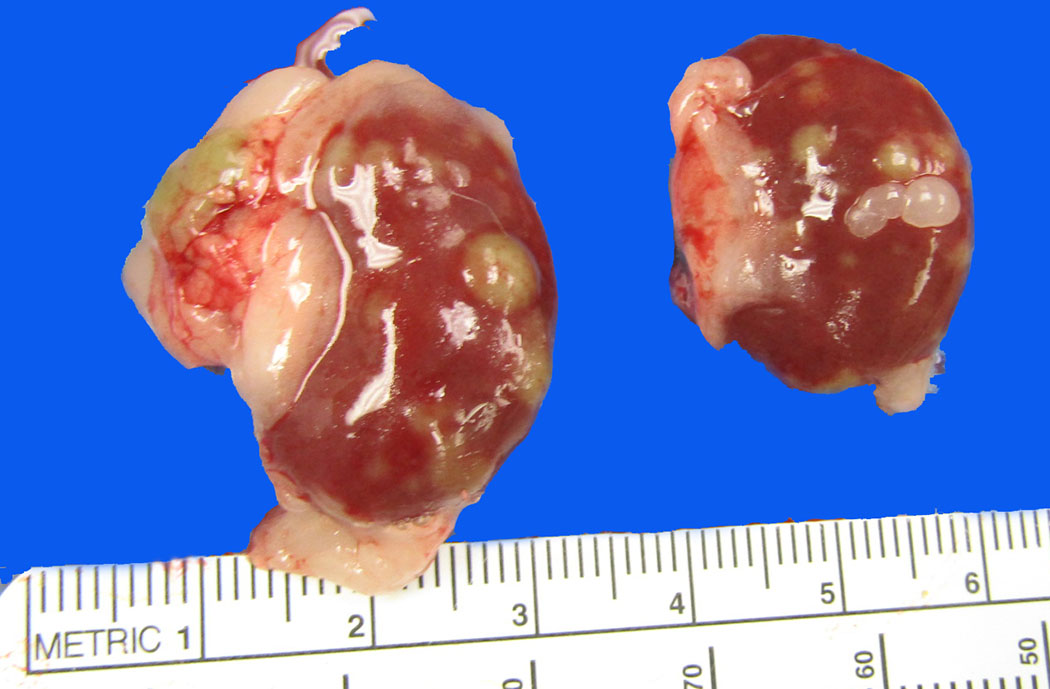
The rat is in fair postmortem condition and has a body condition score of 5/5 with substantial subcutaneous and visceral adipose stores. Within the abdomen, the spleen is markedly enlarged. There is a 5 mm x 1 mm green area on the serosal surface of the spleen that does not extend into the parenchyma. The kidneys have bilateral multifocal to coalescing green-tan foci, ranging in size from ~1mm to 5mm. Some of the foci are slightly raised; on cut section, they extend into the renal cortex and are semi-firm. The left and right renal cortex and medulla are moderately diffusely dark red in color with minimal corticomedullary distinction. The liver is mildly increased in size and is mottled red pink. There is a 1.5 cm x 1 cm green tubular soft tissue structure at the level of the ventral left lateral liver lobe. There is mild segmental reddening of the small intestine. Upon removal of the gastrointestinal tract, there are multiple 3-5 mm green soft tissue semi-firm structures within the visceral adipose tissue.
The vertebral column is removed from the level of the third lumbar vertebrae to the third sacral vertebrae. On cross section, within the vertebral canal, there is brown-green caseous material surrounding the spinal cord. Within the bone marrow of the femur, there is green caseous material that exudes on cut section.
The lungs are mottled light red to pink. The heart is mildly enlarged. Within the mediastinum, there are multiple, 5mm-7mm green soft tissue structures. Within the ventral neck, structures of the same appearance are observed, approximately at the level of the cervical lymph nodes. There are no other significant findings.
Laboratory Results:
Mediastinal mass culture: No growth on initial plate media. Leuconostoc pseudomesenteroides from enrichment broth only – presume contaminant.
Microscopic Description:
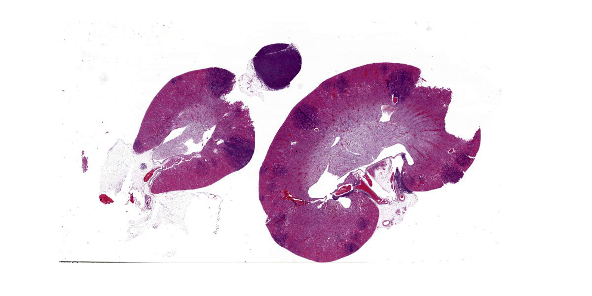
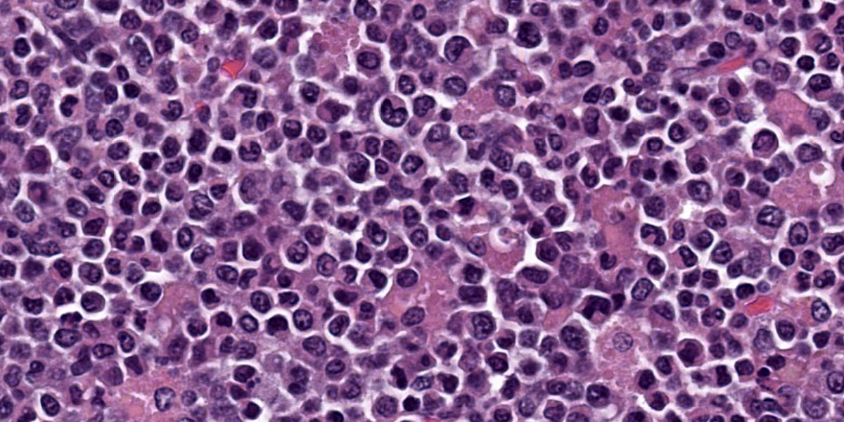
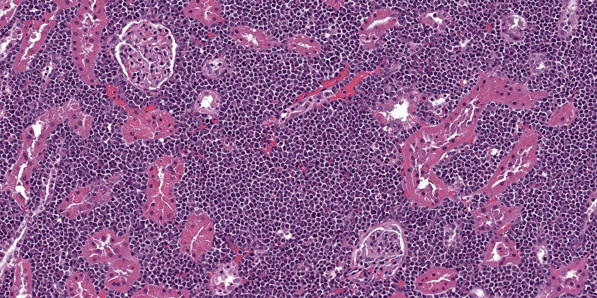
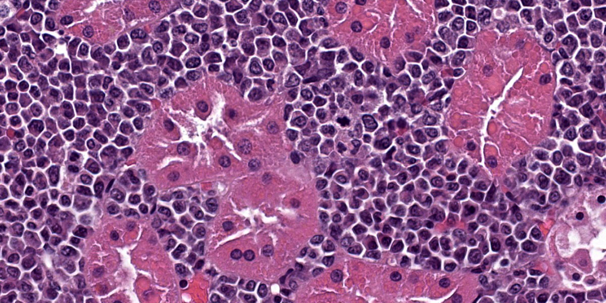
Kidney: Expanding the interstitium and separating the tubules, there is an unencapsulated and infiltrative neoplasm characterized by sheets of round cells with mild to moderate cytoplasm in a scant preexisting fibrovascular stroma. Nuclei are circular to ovoid and occasionally reniform, with a single distinct nucleolus. In some cells, there is an eccentric nucleus with eosinophilic cytoplasm. Cell borders are generally distinct. There is moderate anisocytosis and anisokaryosis. Mitoses are frequent. Many tubular epithelial cells contain intracytoplasmic eosinophilic droplets of varying size, up to 20 microns in diameter. The adrenal gland is effaced by similar neoplastic cells.
The round cell neoplasm also affects the following organs: bone marrow, head (Harderian gland, nasal cavity, salivary gland), meninges, lung, liver, kidney, lymph node, pancreas, spleen, submucosa of the bladder, fat, epaxial skeletal muscle, and nerve roots
Contributor’s Morphologic Diagnoses:
- Multiorgan hematopoietic neoplasm.
- Renal tubular epithelium: Intracytoplasmic hyaline droplets.
Contributor’s Comment:
The neoplasm in this rat is most consistent with a hematopoietic neoplasm. Hematopoietic neoplasms reported in rats include lymphoma, granulocytic leukemia (myeloid sarcoma), and histiocytic sarcoma.2 Hematopoietic neoplasms in general are much less common in the rat compared to the mouse.2 In one report, lymphoma had an approximately 1.5% incidence in the Sprague Dawley rat, of which large granular lymphocyte lymphoma (or mononuclear leukemia) was most common, and granulocytic leukemia had a 0.1-0.3% incidence.2 Granulocytic leukemia more often involves in the kidney in the rat in addition to other organs such as the liver, spleen and bone marrow, and cells with ring shaped nuclei or large, blastic cells may be seen. 2,3 Histiocytic sarcoma is the most common nonlymphoid hematopoietic neoplasm reported in the rat and is recognized as an age related neoplasm in the Sprague Dawley and other strains of rat.2, 5 This neoplasm has been reported to have an incidence of approximately 1% in Sprague Dawley rats, usually affects rats over 12 months of age, and most commonly affects the lung and liver.2,3 Other authors have described the neoplasm affecting hematopoietic organs including bone marrow, spleen and lymph nodes in the rat.5 Histiocytic sarcoma arises from histiocytes although the exact origin of the histiocytic cells remains uncertain.3 These cells arise from the mononuclear-phagocyte system lineage, and include pro-monocytes, monocytes, tissue histiocytes, and macrophages.3 Histologically, the neoplasm may have a granulomatous or sarcomatous appearance or present as sheets of round cells, and multinucleated giant cells are frequently observed.4 This neoplasm does not have a granulomatous or sarcomatous appearance, and multinucleated giant cells are not observed in the present case.
In the rodent, histiocytic sarcoma and occasionally other hematopoietic neoplasms may be associated with intracytoplasmic round eosinophilic droplets containing lysozyme in the tubular epithelial cells.3 In one study, 74 of 77 Sprague Dawley rats with
histiocytic sarcoma had renal hyaline droplet accumulation.3 The presence of this feature in rodents with histiocytic sarcoma appears to correlate with tumor burden and is likely associated with overproduction of endogenous protein by the neoplastic cells.3 Other differentials for eosinophilic droplets within the proximal tubular epithelium of the rat include α-2u globulin nephropathy.3 In the mouse and rat, chronic progressive nephropathy and other neoplasms may also be associated with hyaline droplet formation.1
Immunohistochemistry is helpful to confirm the diagnosis of hematopoietic neoplasms. Macrophages may require a panel of antibodies for diagnosis due to varying phenotypes in different tissue environments.6 Common antibodies used in the diagnosis of histiocytic sarcoma in the rat include CD68 (ED-1) and lysozyme, among others.5,6 Lysozyme is also reported in the diagnosis of granulocytic leukemia, in addition to myeloperoxidase (MPO).6 Antibodies useful in the diagnosis of lymphoma in the rat have also been reported.6 None of these antibodies, unfortunately, are optimized in our laboratory for the rat.
Contributing Institution:
University of Washington Veterinary Diagnostic Lab and Comparative Pathology Program
Department of Comparative Medicine
The Comparative Pathology Program (CPP) | Department of Comparative Medicine (washington.edu)
JPC Diagnoses:
- Kidney and lymph node: Hematopoietic sarcoma.
- Renal tubular epithelium: Intracytoplasmic hyaline droplets.
JPC Comment:
This week’s moderator was Dr. Michael Eckhaus from the National Institutes of Health who provided a lab-animal centric conference for participants.
In this first case, conference participants debated the exact origin of neoplastic cells, as the reniform nuclei may be seen in neoplasms of both histiocytic and granulocytic origin. To better characterize these neoplastic cells, we ran typical round cell immmunomarkers (IBA1, CD3, CD20, PAX5, MUM1, and lysozyme), all of which were negative. Unfortunately CD34 and myeloperoxidase, markers that should stain cells of myeloid origin were immunonegative as well. The gross description of the greenish nodules (figure 1-1) hints at a granulocytic (myeloid) leukemia origin, but we were unable to confirm their identity with more objective means. Based on the HE appearance, we ultimately agreed with the contributor on the morphologic diagnosis.
This case is also a nice example of hyaline droplets in the kidney of a rat (figure 1-5). While most participants recognized the hyaline droplets and associated them with a diagnosis of histiocytic sarcoma in this case, the droplets (and neoplastic cells) did not stain for lysozyme. Alpha-2u globulin nephropathy is another consideration for these droplets, though this animal did not have any history of chemical exposure. In Alpha-2u globulin nephropathy, hyaline droplets represent secondary lysosomes containing alpha-2u-globulin bound to a variety of chemicals and/or their metabolites with the complex being resistant to proteolytic degradation which promotes accumulation within the cytoplasm.7 Hyaline droplets are also a normal finding in male rats, albeit at a low background level far less than that seen in this case.
References:
- Decker JH, Dochterman LW, Niquette AL, et al. Association of Renal Tubular Hyaline Droplets with Lymphoma in CD-1 Mice. Toxicol Pathol. 2012; 40: 651-655.
- Frith CH. Morphologic Classification and Incidence of Hematopoietic Neoplasms in the Sprague-Dawley Rat. Toxicol Pathol. 1998; 16: 451-457.
- Frith CH, Ward JM, and Chandra M. The morphology, immunohistochemistry, and incidence of hematopoietic neoplasms in mice and rats. Toxicol Pathol. 1993; 21:206-218.
- Hard GC and Snowden RT. Hyaline Droplet Accumulation in Rodent Kidney Proximal Tubules: An Association with Histiocytic Sarcoma. Toxicol Pathol. 1991; 19: 88-97.
- Ogasawara H, Mitsumori K, Onodera, H et al. Spontaneous Histiocytic Sarcoma with Possible Origin from the Bone Marrow and Lymph Node in Donryu and F-344 Rats. Toxicol Pathol. 1993; 21: 63-70.
- Rehg JE, Bush D, and Ward JM. The Utility of Immunohistochemistry for the Identification of Hematopoietic and Lymphoid Cells in Normal Tissues and Interpretation of Proliferative and Inflammatory Lesions of Mice and Rats. Toxicol Pathol. 2012; 40: 345-374.
- Swenberg JA, Short B, Borghoff S, Strasser J, Charbonneau M. The comparative pathobiology of alpha 2u-globulin nephropathy. Toxicol Appl Pharmacol. 1989 Jan;97(1):35-46.
