CASE I:
Signalment
8-year-old, castrated, male, Boxer dog (Canis lupus familiaris)
History
This Boxer dog presented with 1.5-week history of mild ataxia and chronic weight loss and 1 day of seizures and drastic mental decline despite anticonvulsant therapy. He was dull, mentally inappropriate, and non-ambulatory tetraplegic, with persistent partial facial seizures. The owners elected euthanasia.
Gross Pathology
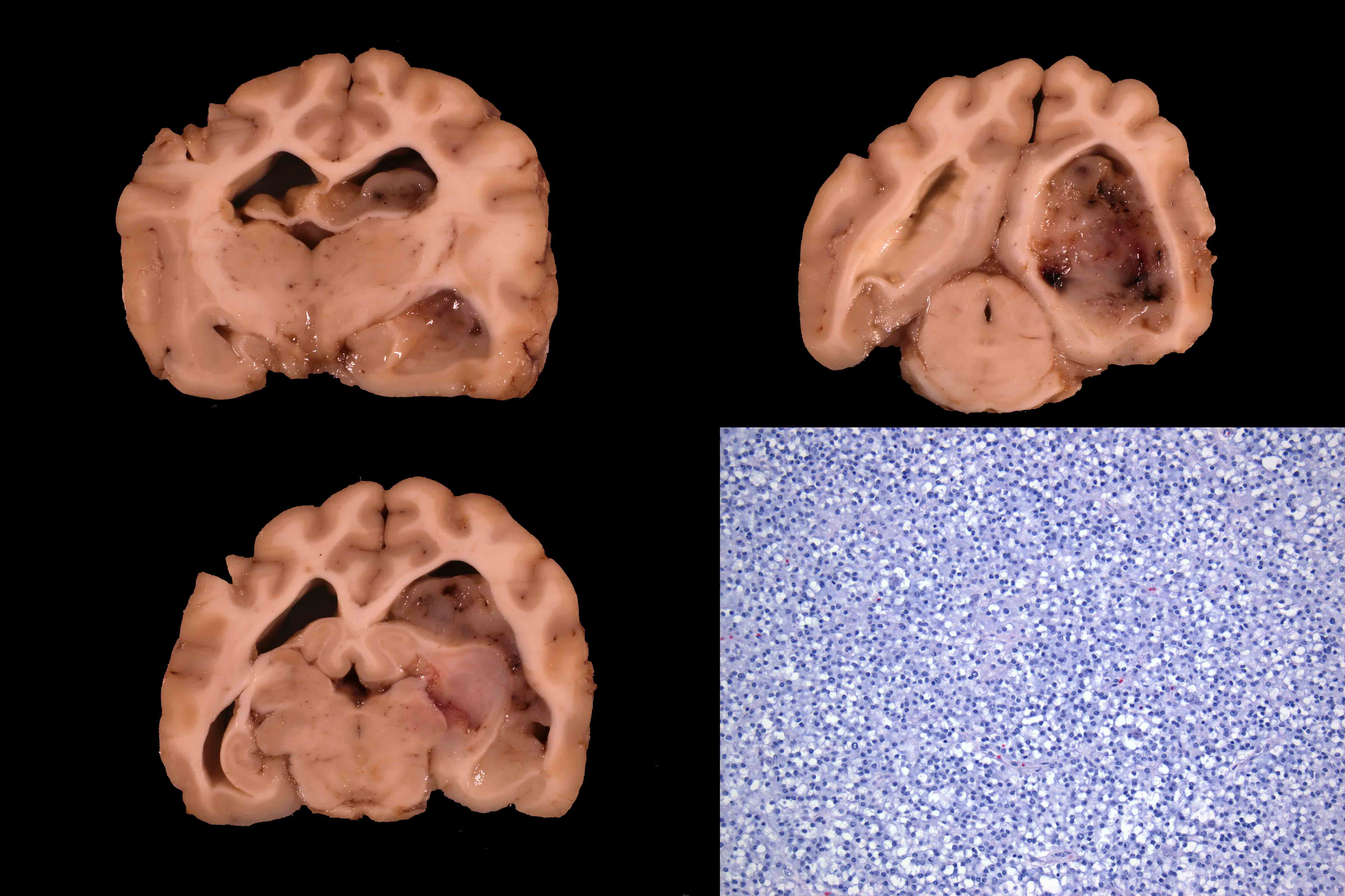
Filling and obstructing the left lateral ventricle was a gray, gelatinous, semi-firm mass, extending caudally from the level of the hippocampus to the occipital lobe. A similar, 2 mm, flattened, round, gray, gelatinous mass was also noted along the ventral aspect of the medulla oblongata. The cerebellum was caudally displaced with coning.
Laboratory Results
None available
Histopathic Description
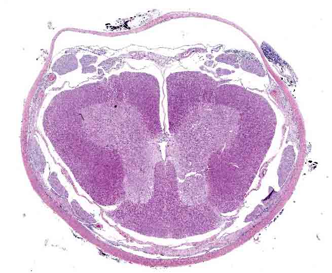
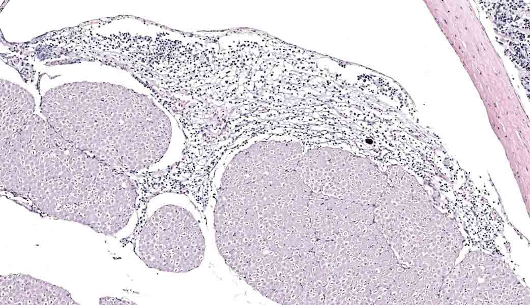
Spinal cord: Infiltrating and expanding the leptomeninges, surrounding spinal nerve roots, is an intradural, extramedullary, unencapsulated, poorly demarcated, densely cellular neoplasm composed of sheets of closely packed polygonal cells, forming a honey-comb pattern, on a scant fibrovascular stroma. Neoplastic cells have fairly distinct cell borders and moderate amounts of lightly staining cytoplasm or perinuclear halo. Nuclei are round, centrally located, and hyperchromatic with an indistinct nucleolus. Anisocytosis and anisokaryosis is moderate. There are 0 mitotic figures in 10 hpf. There is minimal change in the adjacent parenchyma.
Immunohistochemistry
Immunohistochemistry for Olig2 showed diffuse, strong, intranuclear immunoreactivity, indicating oligodendroglial differentiation. Immunohistochemistry for GFAP revealed strong cytoplasmic expression within parenchymal astrocytes, but no expression was seen in the neoplastic population (not shown).
Contributor's Morphologic Diagnoses:
Spinal cord: Oligodendroglioma
Contributor's Comment:
Oligodendrogliomas are common intracranial neoplasms in mature dogs with brachycephalic breeds overrepresented 2,4,6. These neoplasms typically present as a solitary, gelatinous, gray-tan mass within white matter of the frontal and temporal lobes of the cerebrum, occurring less frequently in the brainstem and spinal cord. Histologically, neoplastic oligodendroglial cells contain a lightly staining cytoplasm forming sheets of closely packed cells. Due to delayed post-fixation artifact, neoplastic cells may exhibit a non-staining appearance, or perinuclear halo, creating the classic fried egg or honeycomb pattern. Olig-2 is a reliably expressed immunohistochemical marker for non-neoplastic and neoplastic oligodendroglial cells, often used in combination with GFAP to rule out astrocytoma and E-cadherin/CD34 for meningiomas2,5. Other immunohistochemical markers have been utilized in veterinary literature with mixed results including CNPase, doublecortin and vimentin2,5,7. This neoplasm uncommonly breaks through the ependyma and disperses in the central nervous system (CNS) by migrating via cerebrospinal fluid and seeding structures along its path5,7-9. This "metastatic" capability of oligodendrogliomas into distant sites within the CNS is a well characterized, but rarely reported, manifestation in humans. This presentation of oligodendrogliomas in dogs is similarly rare, and its recognition and characterization has emerged only recently in veterinary literature.
Atypical presentations of canine oligodendrogliomas that have been described include the following: 1) multiple, concurrent oligodendrogliomas at distant sites within the central nervous system, 2) primary intraventricular oligodendroglioma, and 3) diffuse leptomeningeal oligodendrogliomatosis1,3,5,7. Brachycephalic breeds are overrepresented in all of these cases and their clinical signs reflect the volume and regions of the central nervous system that is affected. There have been three case reports of canine cerebral oligodendrogliomas arising within the ventricular system with minimal parenchymal involvement, and two reports of multifocal neoplastic infiltration with leptomeningeal extension. Oligodendrogliomatosis manifests as diffuse, intradural, extramedullary neoplastic infiltration with no primary parenchymal mass. Four cases of canine oligodendrogliomatosis have been reported.
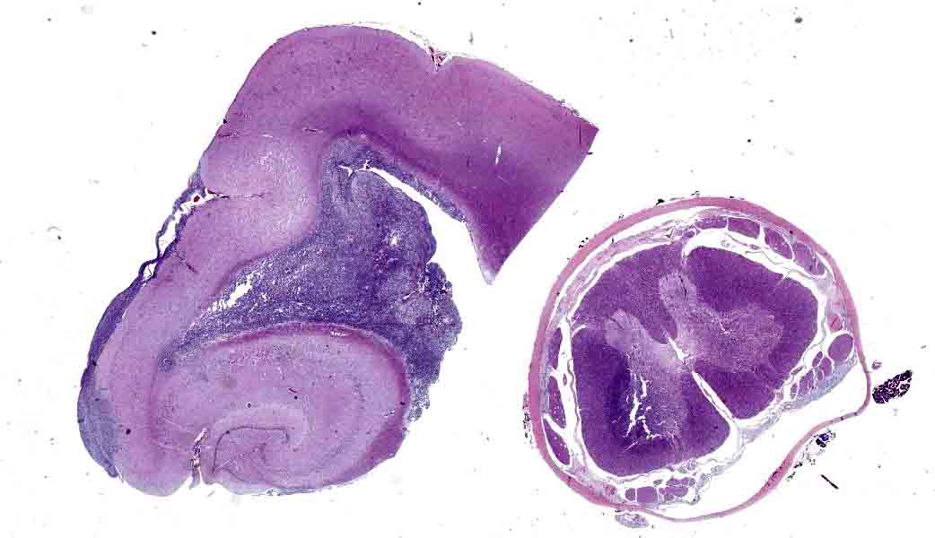
Conference participants received only a section of spinal cord with the neoplasm. Without knowledge of a primary neoplasm noted within the cerebrum, the intradural, extramedullary location of oligodendroglioma in this case may elicit a discussion and diagnosis of primary leptomeningeal oligodendrogliomatosis. However, oligodendroglioma in this dog was also noted within the brain, completely obstructing the left lateral ventricle, and histologically revealed continuous extension into the 4th ventricle as well as infiltrating the subarachnoid space along the ventral aspect of the medulla oblongata, and continuing as an intradural, extramedullary neoplasm throughout the entire spinal cord tract. Although there was minimal periventricular involvement in the brain by neoplastic cells, suggesting a predominantly intraventricular growth pattern, the tumor most likely metastasized from the large primary mass in the brain into the leptomeninges of the spinal cord. Recognition of these atypical manifestations of oligodendrogliomas suggests that examination of the spinal cord should routinely be performed to determine the full extent of canine oligodendrogliomal spread, as this may be an underrecognized disease manifestation.
Contributing Institution
Cummings School of Veterinary Medicine at Tufts University http://vet.tufts.edu/foster-hospital-small-animals/departments-and-services/pathology-service/
JPC Diagnoses
Lumbar spinal cord, subarachnoid space: Oligodendrogliomatosis.
JPC Comment
This week's moderator was LTC Curtis Cline, a highly experienced Army veterinary pathologist and graduate of the JPC's training program (and previous WSC coordinator as a second-year resident). Following completion of his residency, he served in the Training Office at the JPC, and followed that with a tour at the US Army Medical Research Institute of Infectious Diseases, where he worked in biocontainment as well as being assigned to the 1st Area Medical Laboratory. He is currently assigned as the Chief of Comparative Pathology at the US Army Medical Research Institute of Chemical Defense.
The contributor provides an excellent discussion of the different manifestations of how oligodendroglioma spreads in the dog. Since this case was submitted in 2016, the veterinary literature has added several excellent contributions to the classification and diagnosis of oligodendrogliomas in the dog.10-12
In 2018, the Comparative Brain Tumor Consortium published a revised classification scheme for the diagnosis of canine gliomas10, which not only serves to simplify the classification of astrocytomas and oligodendrogliomas in canine patients but also helps to bring their classification more in line with the human literature, facilitating future incorporation of clinical outcomes and genomic data. This new classification scheme reduces astrocytomas and oligodendrogliomas to low- versus high-grade tumors, and this article presents the criteria for reclassification of each. 10
Critical histologic criteria for the classification of oligodendrogliomas as high-grade versus low-grade are reduced to:
- necrosis (single-cell necrosis not considered),
- microvascular proliferation and pseudopalisading,
- the overt presence of features of malignancy (pleomorphism), and other evidence of significant cellular atypia
- mitotic count per 2.37 mm2 field.10
This neoplasm, both in the cerebral and spinal cord locations, has a very traditional "oligodendroglioma-like" appearance, with widely spaced cells, abundant clear cytoplasm, and monomorphic bland nuclei, giving it a "chicken wire" appearance. In the ventricular location, in spite of the low mitotic rate, the neoplasm demonstrates evidence of microvascular proliferation, a key criterion for the diagnosis for high-grade oligodendroglioma. According to this schema10, a high grade tumor is warranted in this case.
The contributor provides a concise review of immunohistochemical markers used to differentiate gliomas. A 2024 article by Rissi et al. provides an outstanding review of these markers and other markers used to differentiate primary and secondary nervous system neoplasms in the dog and cat. Oligodendrocytes typically stain strongly positive with the nuclear transcription factor OLIG2.11.
A confounding issue in using immunohistochemistry to diagnose primitive central nervous system tumors is the common origin of neurons, astrocytes, and glial cells from a similar primitive neuroglial precursor. OLIG2 is an excellent immunomarker to differentiate neoplastic glial cells from primitive neural elements. However, OLIG2 (despite a name that would lead one to assume it is specific for oligodendroglia) will simultaneously stain the nuclei of both astrocytes and oligodendrocytes due to their common ancestry. For this reason, it should always be paired with glial fibrillary acidic protein, which stains astrocyte cytoplasm and processes but not those of their oligodendroglial counterparts.10-12 (In most cases of glioma, even mixed gliomas), careful evaluation of the histology of these tumors on H&E sections, coupled with Olig-2 and GFAP, will provide a diagnosis and appropriate grade in most cases.
It should also be noted that time to fixation, as well as time in fixation, may affect these markers. OLIG2 appears more resistant to the antigen cross-linking associated with longer fixation periods, while other markers, including GFAP, CNPase, and Ki-67, should be interpreted cautiously. A lack of immunoreactivity should not be interpreted as a negative test for these markers without careful consideration.
Other immunomarkers that may be more useful in diagnosing oligodendrogliomas include CNPase, which stains the cytoplasm of oligodendrogliomas. MAP2 immunoreactivity may be tricky in some cases, as, like Olig-2, this marker may stain both astrocytic neoplastic cells within astrocytomas and oligodendrogliomas; in oligodendrogliomas, immunolabeling is predominantly perinuclear and cytoplasmic, whereas in astrocytomas, the marker predominantly stains the cytoplasmic processes.
Other less commonly used immunomarkers include nuclear transcription factors SOX2 and SOX10, platelet-derived growth factor alpha, and neural/glial antigen 2. However, more studies are required to establish more rigid interpretive guidelines and their utility in differentiating gliomas.11
The JPC diagnosis of "oligodendrogliomatosis" is based on the fact that the slide which was provided to the conference participants (and that described by the contributor above) contained only a section of lumbar spinal cord. The contributor did kindly provide a section of diencephalon with the tumor within the ventricle and meninges to answer questions about the origin of the tumor, following the expected discussion as to whether the spinal cord lesion represented a metastatic oligodendroglioma or a focus of leptomeningeal oligodendrogliomatosis. Based on the slide provided to participants, the origin of this lesion in the lumbar spinal cord cannot be definitively identified; hence we prefer a diagnosis of "oligodendrogliomatosis". Ultimately, with all sections and the gross images of this lesion considered, a diagnosis of metastatic oligodendroglioma becomes appropriate.
Finally, for neuropathologists and budding pathologists, the Davis Thompson Foundation published the sixth edition of its series on the Surgical Pathology of Tumors of Domestic Animals in 2026, covering tumors of the nervous system.12 Authored by members of the Comparative Pathology Tumor Board, this volume aligns with the newest literature.10-11 It provides an outstanding, easy-to-read overview of primary and secondary neoplasms of the central and peripheral nervous system, their diagnostic features, biologic behavior, and appropriate immunohistochemistry. It is well worth the investment.
References
- Canal S, Bernardini M, Pavone S, et al (2013). Primary diffuse leptomeningeal gliomatosis in 2 dogs. Can Vet J, 54: 1075-9.
- Johnson GC, Coates JR, and Wininger F. (2014) Diagnostic immunohistochemistry of canine and feline intracalvarial tumors in the age of brain biopsies. Vet Pathol, 51: 146-160.
- Koch MW, Sanchez MD, Long S, et al (2011). Multifocal oligodendroglioma in three dogs. JAAHA, 47: e77-85.
- Koestner A and Higgins RJ. "Tumors of the nervous system." Meuten DJ, Tumors in Domestic Animals, 4th Ed. 2002.
- Kovi RC, Wunschmann A, Armien AG, et al (2013). Spinal meningeal oligodendrogliomatosis in two Boxer dogs. Vet Pathol, 50(5): 761-4.
- Maxie MG and Youssef S. "Nervous system." Jubb, Kennedy, and Palmer's Pathology of Domestic Animals, 5th Ed. 2007.
- Rissi DR, Levine JM, Eden KB, et al (2015). Cerebral oligodendroglioma mimicking intraventricular neoplasia in three dogs. JVDI, 27(3): 396-400.
- Subramanian A, Harris A, Piggott K, et al (2002). Metastasis to and from the central nervous system - the "relatively protected site." Review. Lancet, 3: 498-507.
- Wesseling P, van den Bent M, and Perry A. (2015). Oligodendroglioma: pathology, molecular mechanisms and markers. Acta Neuropathol, 129: 809-827.
- Koehler JW et al. A Revised Diagnostic Classification of Canine Glioma: Towards Validation of the Canine Glioma Patient as a Naturally Occurring Preclinical Model for Human Glioma. J Neuropathol Exp Neurol. 2018 Nov 1;77(11):1039-1054.
- Rissi DR, Miller AD, Demeter EA, Church ME, Koehler JW. Diagnostic immunohistochemistry of primary and secondary central nervous system neoplasms of dogs and cats. J Vet Diagn Invest. 2024 Mar;36(2):153-168. doi: 10.1177/10406387231221858. Epub 2024 Jan 17. PMID: 38234003; PMCID: PMC10929637.
- Rissi D, editor. Surgical Pathology of Tumors of Domestic Animals, vol 6: Tumors of the Nervous System. Davis Thompson Foundation Publishing, 2026

Click the slide to view.
01-1. Brain, dog.
01-2. Brain and spinal cord, dog.
01-3. Spinal cord, dog.
01-4. Spinal cord, dog.
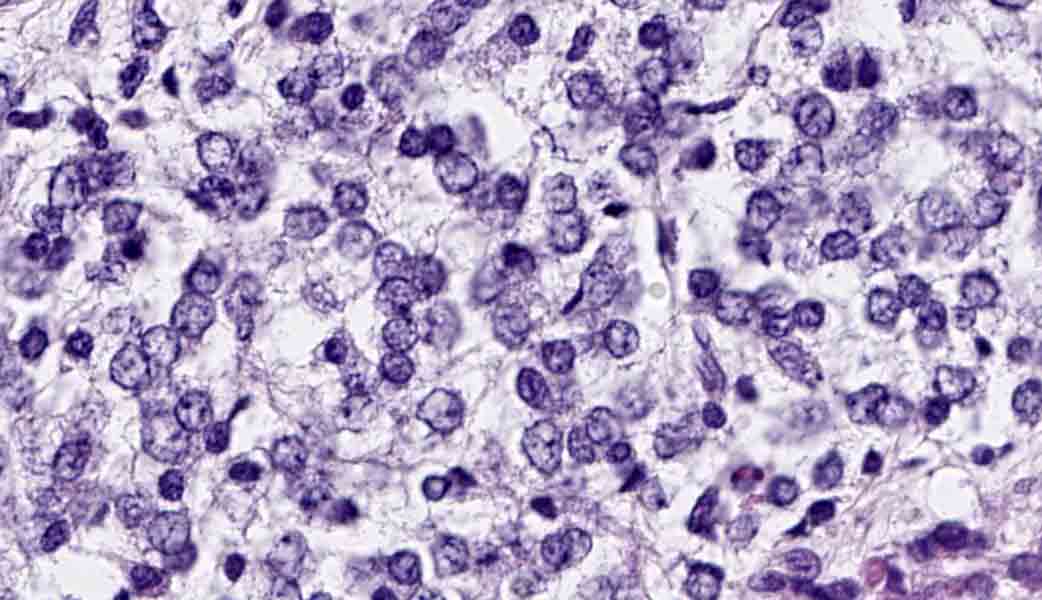
01-5. Ventricle, dog.
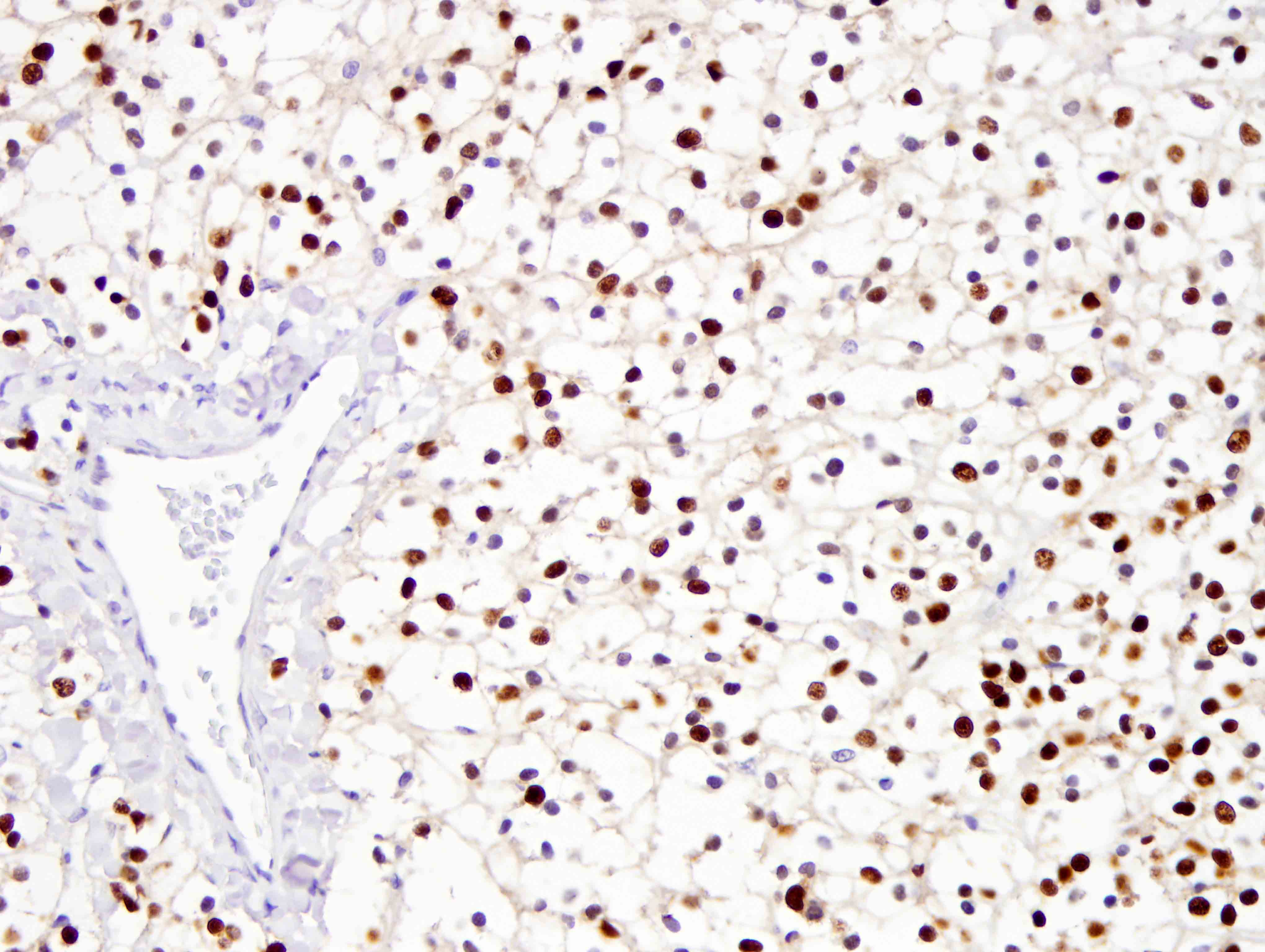
01-6. Ventricle, dog.
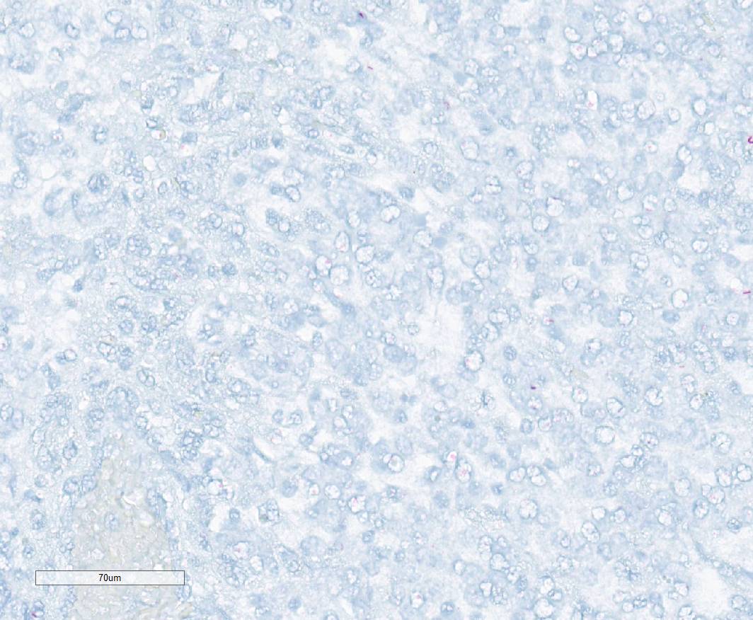
01-7. Ventricle, dog.
| Back | VP Home | Contact Us |