Signalment:  

Nine-year-old, female, Siamese cross, (Felis catus).The cat presented for chronic cystitis unresponsive to antibiotic therapy and dietary changes.  Pneumocystogram revealed a thickened uneven bladder wall with a small lumen.  The cat was referred to an internal medicine service with the primary differential diagnosis of neoplasia.  Ultrasound showed an irregular mass in the bladder wall at the trigone.  A “traumatic catheterization” of the bladder was performed and cytologic preparations were submitted for evaluation.


Gross Description:  

None.


Histopathologic Description:

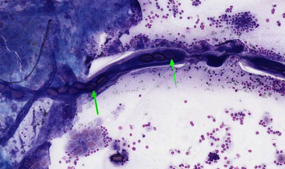
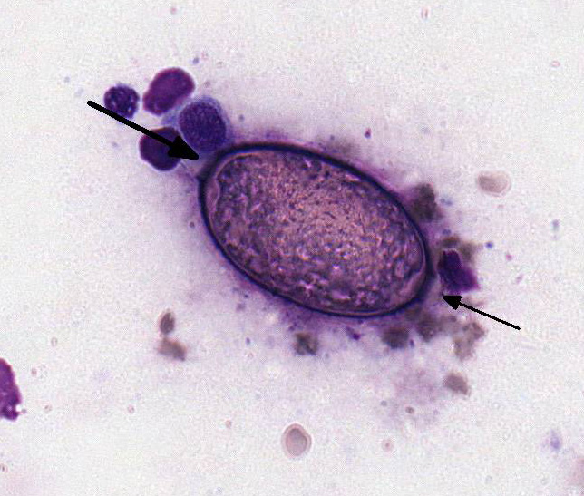
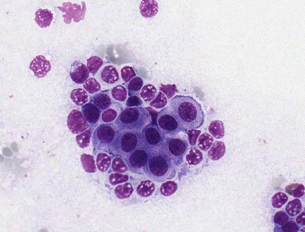
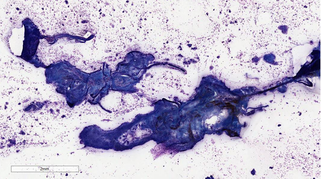
The preparation has large areas of dense cellularity.  There are numerous linear structures (nematode uterus) containing oval bioperculated ova are present within the dense areas.  Well-preserved and degenerate parasite ova are also scattered throughout the background.  The ova are approximately 60 microns in length with a granular interior and a thick refractile shell.  In the thinner areas of large cellular densities, the nucleated cells are present in organized sheets with well-defined cytoplasmic borders.  The cells have round nuclei with mild anisokaryosis and a scant to small amount of basophilic cytoplasm.  The background has a large number of lysed and degenerate cells with fewer small clusters of intact epithelial cells

showing the same morphology as those within the dense clusters.  There are areas of monolayer adjacent to the dense clusters with a moderate number of neutrophils and eosinophils.  Eosinophils, neutrophils, and small lymphocytes are found throughout the background.  Occasional small lymphocytes contain a few eosinophilic to azurophilic granules.


Table 1: Capillarid Species

Name

Location

Host

Eucoleus bouhmi

frontal sinus

fox, dog

Eucoleus aerophilus

bronchi

dog, cat, fox

Aonchotheca putorii

stomach, intestine

bear, hedgehog, raccoon, swine, bobcats, mustelids, cat

Aonchotheca spp

intestine

ruminants

Calodium hepaticum

liver

rodents, many occasional hosts including humans.

Pearsonema plica

urinary bladder

dog, fox, wolf

Pearsonema feliscati

urinary bladder

cat

embedded parasite.  The lifecycle for P. feliscati is not determined.2,6  It is assumed to be similar to P. plica which is thought to be through the earthworm as an intermediate host or a transport host such as a bird.  Adult worms are 2.5 to 5 cm in length.  The ova are elongate and approximately 60 microns in length and 27 microns in width.  They have bipolar plugs (bioperculate), a thick shell with “globular” ridges enclosing a single cell.10 The literature does not provide morphologic or biological characteristics other than the host to distinguish P. plica

and P. feliscati.  The cat is included in host species for both P. feliscati and P. plica in Georgis’ Parasitology for Veterinarians with no mention of differential characteristics.2  The capillarid in this case is given as P. feliscati based on host.


Morphologic Diagnosis:  

Transitionalcell hyperplasia with mild suppurative and eosinophilic inflammation and fragmented Pearsonema (Capillaria) feliscati nematode and numerous bio-perculated eggs.


Lab Results:  

The CBC at presentation to the specialty service was within reference interval.  The only abnormality in the chemistry profile was hyperglycemia.  The abnormalities in the urinalysis included glucosuria, mild pyuria and large numbers of variably sized trans-itional epithelial cells. showing the same morphology as those within the dense clusters. There are areas of monolayer adjacent to the dense clusters with a moderate number of neutrophils and eosinophils.  Eosinophils, neutrophils, and small lymphocytes are found throughout the background.  Occasional small lymphocytes contain a few eosinophilic to azurophilic granules.


Condition:  

Urinary capillariasis/Capillaria feliscati


Contributor Comment:  

Urinary nema-todiasis is caused by several genera and affects numerous species of domestic and wild animals (Table 1).2, 5,10  The nematode species found in the kidney, ureters or renal vasculature are associated with clinical disease.  The most spectacular of which is the giant kidney worm of dogs, Dio-ctophyme renale, that eventually destroys and replaces the renal parenchyma.  Nematodes of the genera Pearsonema (Capillaria), on the other hand, are found within the urinary bladder and are often an incidental finding with minimal clinical disease evident.2,10  The capillarid nematodes have been placed in several new genera based on location within the host, the most accepted of which are Eucoleus (nasal sinus and bronchi), Aonchotheca (intestine) and Pearsonema (urinary bladder) (Table 2)2Pearsonema plica and P. feliscati, infect the domestic dog, fox, wolf, and cat.  In most cases, the nematode is loosely attached to the urinary bladder mucosa and, less often, ureteral mucosa with mild inflammation and edema evident on histologic examination of the affected tissue.7,8  Clinical disease is uncommon but has been reported in the dog and fox and rarely in the cat.7,8  The prevalence of these parasites in the dog and cat is not known.  However, a prevalence of 76% and 59% has been reported in two dog breeding kennels in the United States.7 In this heavily parasitized population, hematuria, dysuria and pollakiuria were common findings in the dogs with confirmed infection.

In the cat, the disease is rarely reported in the United States;6  however, in Australia, an incidence of greater than 30% was found in one survey study.8  No evidence of clinical cystitis was seen in the infected cats.  On histological examination of the urinary bladder in the infected cats, the nematodes were superficially embedded in the mucosa with no breaching of the basal layer or basement membrane.  A moderate inflammatory infiltrate that included eosino-phils was seen in association with the

Table 2: Urinary Nematodes

Name

Host

Dioctophyme renale

dog, mink

Pearsonema plica, P. feliscati

dog, fox, wolf, cat

Stephanurus dentatus

swine

Trichosomoides crassicauda

rats

Crassicauda boopis

whales

embedded parasite.  The lifecycle for P. feliscati is not determined.2,6  It is assumed to be similar to P. plica which is thought to be through the earthworm as an intermediate host or a transport host such as a bird.  Adult worms are 2.5 to 5 cm in length.  The ova are elongate and approximately 60 microns in length and 27 microns in width.  They have bipolar plugs (bioperculate), a thick shell with “globular” ridges enclosing a single cell.10 The literature does not provide morphologic or biological characteristics other than the host to distinguish P. plica

and P. feliscati.  The cat is included in host species for both P. feliscati and P. plica in Georgis’ Parasitology for Veterinarians with no mention of differential characteristics.2  The capillarid in this case is given as P. feliscati based on host.


JPC Cytologic Interpretation: Presence of Peasonema spp nematode fragments and ova, mild transitional cell dysplasia, and low grade eosinophilic and neutrophilic inflammation, Siamese cross, Felis catus.


JPC Diagnosis:  

Presence of Peasonema spp nematode fragments and ova, mild transitional cell dysplasia, and low grade eosinophilic and neutrophilic inflammation, Siamese cross, Felis catus.


Conference Comment:  

The contributor provides a compelling cytologic specimen and outstanding review of capillarid nematodes affecting a wide variety of veterinary species. As mentioned by the contributor, the Capillaria genera have been reclassified based on the location of the adult parasite in the host; however, the genus name Capillaria is still widely used in the veterinary literature. Typically, infection with Pearsonema feliscati is of little pathologic significance to the cat and is usually considered an incidental finding. Most studies show no relationship between the presence of adult capillarids in the bladder and significant cystitis.8,9 Histologically, Pearsonema feliscati resides in the superficial epithelium of the urinary bladder and does not penetrate the basement membrane.5 However, if the ureters become occluded with adult nematodes, cats may display the clinical signs of post-renal obstruction. The highest reported number of adult worms present from a single urinary bladder was 25 and large numbers of eggs were found in all bladders containing five or more adults.9 In this case, the mild to moderate eosinophilic and neutrophilic inflammation may be secondary to a high parasite burden.

In both dogs and cats, Peasonema plica has been reported in the urinary bladder and ureter submucosa and is typically associated with mild subclinical inflammation and edema.1,7 There is a higher prevalence of the parasite in wild red foxes (Vulpes vulpes) in many European countries and it is associated with an increased pathogenicity. This can result in severe cystitis, pollakiuria, dysuria and hematuria in this species.1,3 In this case, both Pearsonema plica and Pearsonema feliscati have been implicated in urinary bladder infection in cats, conference participants could not distinguish between two cytologically. Conference participants readily identified numerous bioperculate eggs free within the sample and inside the reproductive tract of adult nematode fragments. Pearsonema spp are a subclassification of aphasmid nematodes of the family Trichuridae. Histologically, aphasmid nematodes are characterized by thin eosinophilic cuticle, reduced polymyarian-coelomyarian musculature, two hypodermal bacillary bands stichosome esophagus, a spiny sheath and oval bioperculate eggs.4 Cytologically, specific features of the adult nematode can be more difficult to appreciate than on histologic tissue section.  However, the presence of the highly characteristic ova confirms the presence of a urinary capillarid nematode.

Participants also noted vacuolation, binucleation, anisokaryosis, and prominent nucleoli within the reactive sloughed urothelium. Transitional epithelial cells are among the most pleomorphic cells in the body and can demonstrate marked reactive change in response to a variety of insults. For this reason, the conference moderator reminded participants that care must be taken prior to over-interpreting reactive urothelium as malignancy.


References:
1. Basso W, Spanhauer Z, Arnold D, Deplazes P. Capillaria plica (syn. Pearsonema plica) infection in a dog with chronic pollakiuria: Challenges in the diagnosis and treatment. Parasitol Int. 2014; 63:140142.
2. Bowen DD. Georgis’ Parasitology for Veterinarians. 2008. (Kindle Locations 9850-9856). Elsevier Health. Kindle Edition.
3. Fernandez-Aguilar X, et al. Pearsonema (syn Capillaria) plica associated cystitis in a Fennoscandian artic fox (Vulpes lagopus): A case report. Acta Vet Scand. 2010; 52:39.
4. Gardiner CH, Poynton SL. Aphasmids. In: Gardiner CH, Poynton SL, eds. An Atlas of Metazoan Parasites in Animal Tissues. Washington, DC: Armed Forces Institute of Pathology; 1999:40-43.
5. Lambertsen RH. Diseases of the common fin whale (Balaenoptera physalus): Crassicaudiosis of the urinary system. Journal of Mammology. 1986; 67(2):353-366.
6. Lautenslager JP. Internal helminthes of cats. Vet Clin North Amer. 1976; 6(3):353365
7. Senior DF, Solomon GB, Goldschmidt MH, et al. Capillaria plica infection in dogs. J Am Vet Med Assoc. 1980;176(9):901-905.
8. Waddell AH. Further observations of Capillaria feliscati in the cat. Aust Vet J. 1968;44(1):33-34.
9. Wilson-Hanson S, Prescott CW. Capillaria in the bladder of the domestic cat. Aust Vet J. 1982; 59:190-191.
10. Zajac, Anne M.; Conboy, Gary A. Veterinary Clinical Parasitology. 2011. (Kindle Locations 3752-3763). John Wiley and Sons. Kindle Edition.


Click the slide to view.


1-1. Urinary bladder, cat.


1-2. Urinary bladder, cat.


1-3. Urinary bladder, cat.


1-4. Urinary bladder, cat.


Back | VP Home | Contact Us |