Wednesday Slide Conference, Conference 5, Case 2
Signalment:
1-year-old, female Common Marmoset (Callithrix jacchus)
History:
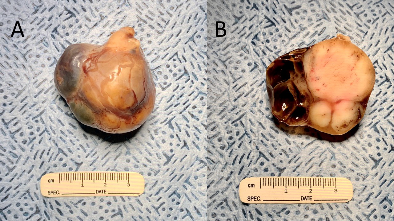
Animal was thought to be pregnant, but ultrasound revealed a large (~5cm diameter, cystic, abdominal mass in the area of the left kidney.
Gross Pathology:
The multilobular abdominal mass enveloped the entire left kidney. On cut section, portions were white-tan and first while others were cystic and filled with yellow-brown fluid.
Microscopic Description:
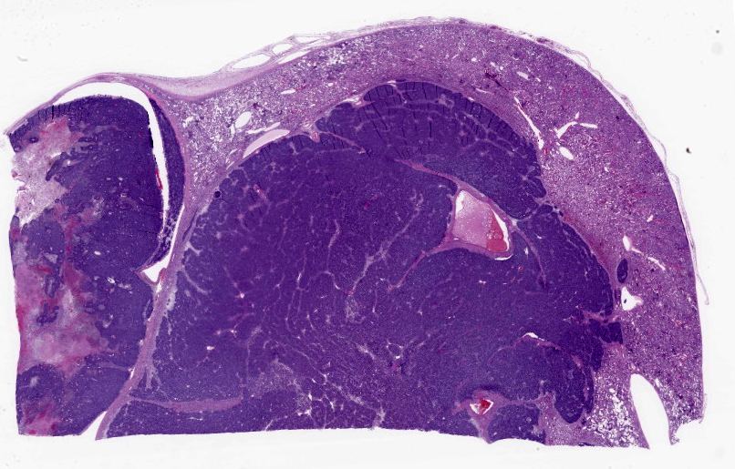
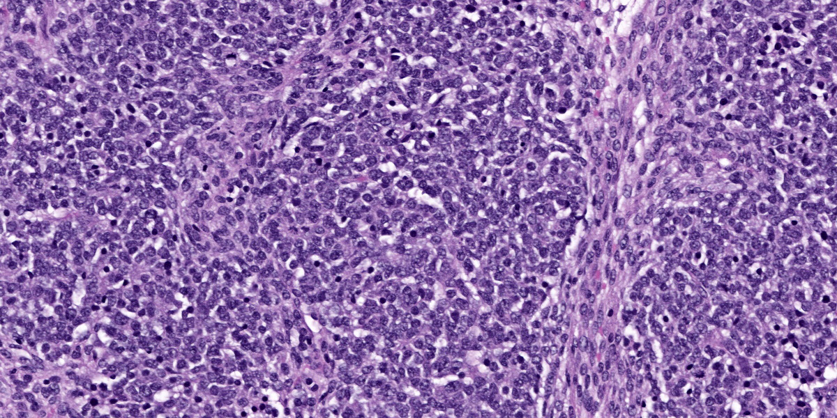
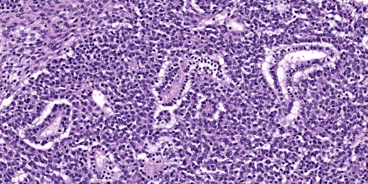
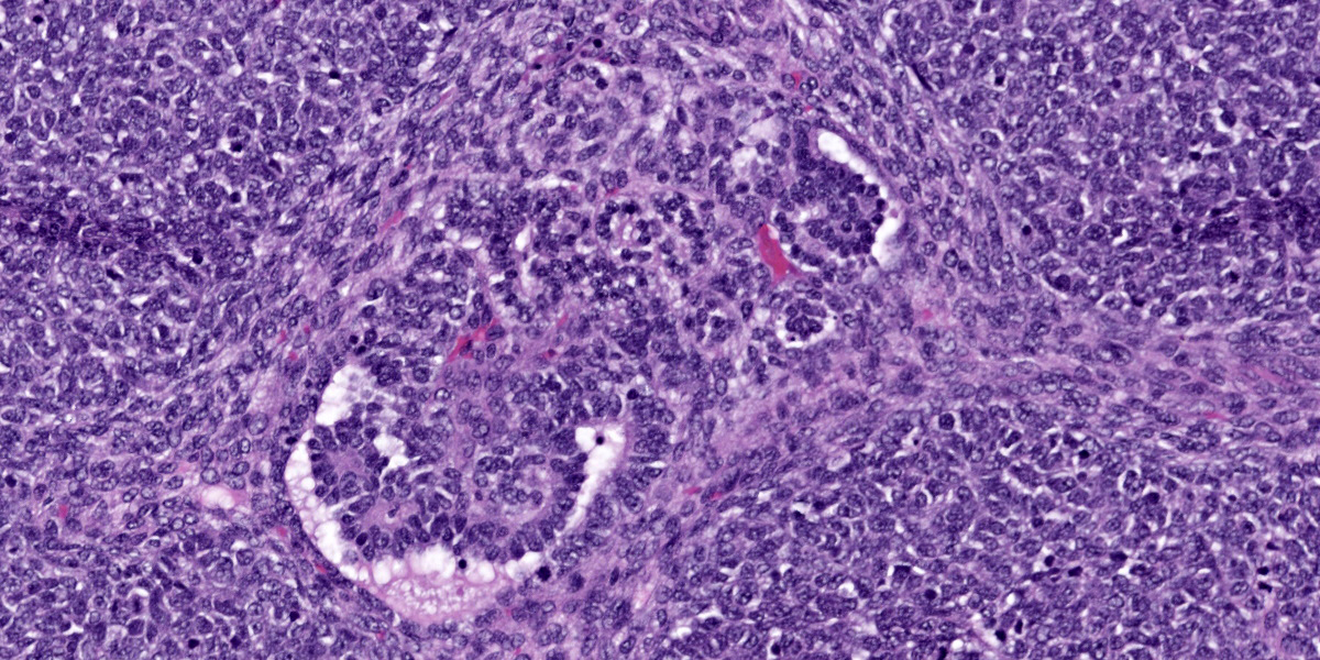
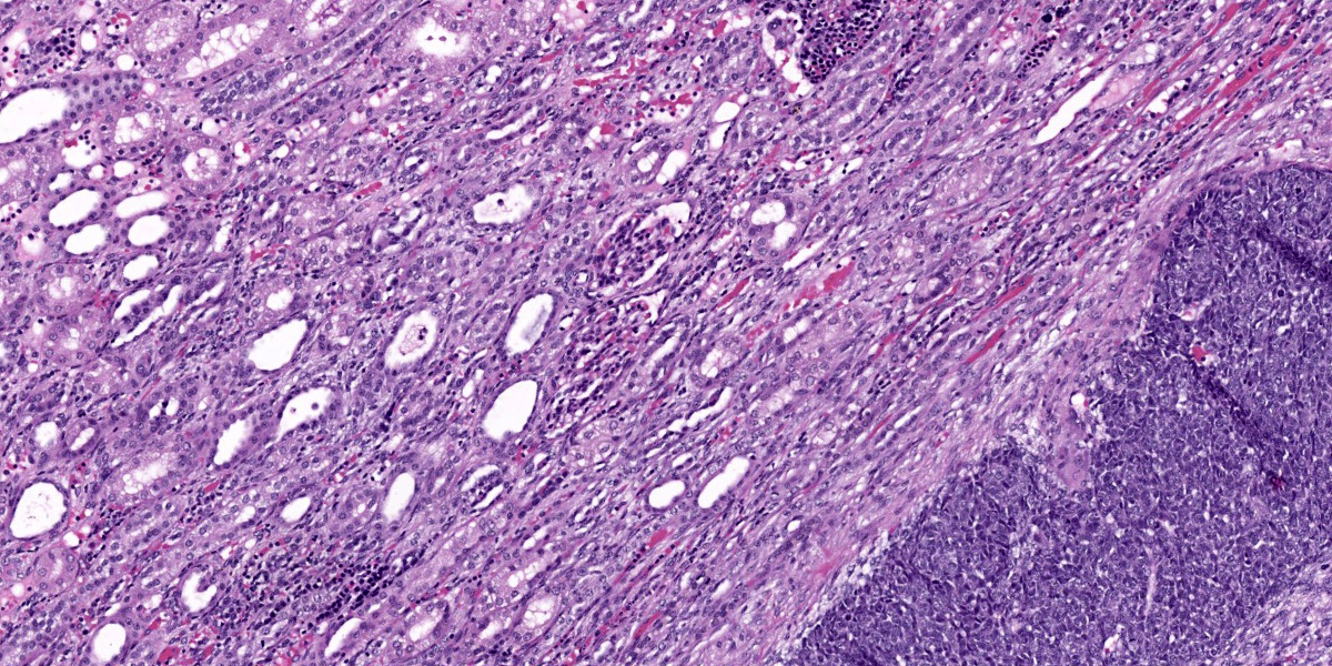
Kidney: Within the cortex, compressing and effacing adjacent renal parenchyma and extending to cut borders is a mostly encapsulated, well-demarcated, lobulated, and expansile neoplasm. The neoplasm is composed of a disorganized mixture of three distinct cell populations: epithelial, mesenchymal and blastemal. In all sections, the blastemal population is the primary component and consists of polygonal cells arranged in nests sometimes separated by connective tissue. These neoplastic cells have indistinct cell borders, a small amount of eosinophilic cytoplasm and a high nuclear to cytoplasmic ratio. Nuclei are round to oval with vacuolated chromatin and indistinct nucleoli. The mitotic rate is 1 per 40x field. In some sections, these cells cant be seen within blood vessels at the periphery of the neoplasm. The epithelial population is composed of cuboidal to columnar cells arranged in irregular and often infolded tubules. Occasionally, these tubules project tufts into lumina (primitive glomeruli). These neoplastic cells have variably distinct cell borders, a moderate amount of eosinophilic fibrillar cytoplasm, round to oval nuclei, loosely clumped chromatin, and rarely contain a distinct nucleolus. The mitotic rate is 0-1 per 40x field. The mesenchymal component often blends with the other two components, and generally consists of spindle cells present within a loose, myxoid, extracellular matrix (embryonal mesenchyme). Spindle cells are stellate to spindle with indistinct cell borders, a scant amount of eosinophilic fibrillar cytoplasm, oval to elongate nuclei with finely stippled chromatin and indistinct nucleoli. The mitotic rate is <1 per 40x field. In some sections, spindle cells differentiate along the lines of cardiomyocytes characterized by elongated cells with one to several round centrally located nucleus/nuclei with vesicular chromatin and eosinophilic, crossed-striated cytoplasm containing occasional intercalated discs. Cystic areas are present in some sections of the neoplasm and are lined by attenuated epithelial cells.
Necrosis and hemorrhage are present multifocally within the neoplasm and adjacent kidney. The kidney also contains tubular atrophy and foci of mixed inflammatory cells.
Contributor’s Morphologic Diagnoses:
Kidney: Nephroblastoma, Common Marmoset
Contributor’s Comment:
Often called Wilms’ tumor, nephroblastoma is the most common primary renal neoplasm in children, swine, chicken, and fish, the second most common primary renal tumor in cats, and the third most common in dogs.3,10 Nephroblastoma is commonly reported in rats, and can be experimentally induced by prenatal exposure to the carcinogen N-ethylnitrosourea (ENU).13
In nonhuman primates, primary neoplasms of the kidney have been reported infrequently as spontaneous cases and in association with exposure to radiation, chemical carcinogens, and parasites.7 The most commonly described renal neoplasms in nonhuman primates include carcinomas and adenomas.7 Only a few reports exist of nephroblastomas in Old World and New World monkeys including a report of nephroblastoma in a black-tufted marmoset4 and a single report describing a malignant nephroblastoma in a common marmoset.13 Interestingly, this case had similar features to that case report, including cystic areas and evidence of malignancy.
Nephroblastomas are true embryonal tumors that arise in primitive nephrogenic blastema and foci of renal dysplasia.3 The etiology and pathogenesis of nephroblastoma has still not been fully clarified. In children, the neoplasm is frequently associated with congenital abnormalities or syndromes, including cryptorchism, hemihypertrophy, hypospadias, and sporadic aniridia. Two loci on chromosome 11, locus 11p13 (WT1 gene) and locus 11p15 (WT2 gene), have been implicated in the genesis of Wilms’ tumors in children with developmental disorders. An abnormal WT1 gene is present in patients with WAGR syndrome (Wilms’ tumor, aniridia, genitourinary abnormalities, mental retardation) or Denys-Drash syndrome (Wilms’ tumor, progressive glomerulonephritis, male pseudohermaphroditism). A mutated WT2 gene can be observed in patients with Beckwith-Wiedemann syndrome or hemihypertrophy. However, the genetics of Wilms’ tumor appear to be multifactorial and probably include further chromosomal abnormalities. Familial Wilms’ tumor is rare and occurs in about 1% of cases and is not associated with mutations in chromosome 11.1,8,13
Contributing Institution:
Division of Laboratory Animal Resource
University of Pittsburgh
S1040 Thomas E. Starzl Biomedical Science Tower
200 Lothrop Street
Pittsburgh, PA 15261
http://www.dlar.pitt.edu/
JPC Diagnosis:
Kidney: Nephroblastoma.
Kidney: Nephritis, interstitial and lymphoplasmacytic, chronic, multifocal, mild with proteinosis.
JPC Comment:
Nephroblastoma remains a fan-favorite submission to the WSC and this case is a good example that clearly delineates the three components to describe (blastemal, mesenchymal, and epithelial) that are outlined in Figures 2-3, 2-4, and 2-5. In this case, the predominance of the blastemal cells with only rare tubular and glomerular differentiation was helpful, as trainees often have difficulty identifying blastemaConfirmatory IHCs for this case (WT1, CD56, pancytokeratin, vimentin, desmin) were not needed to arrive at the diagnosis.
Conference participants devoted a significant portion of the discussion to changes in the adjacent kidney. Participants noted occasional glomerular hypercellularity, synechiae, proteinosis, fibrosis, casts, and interstitial nephritis – we added a second morphologic diagnosis for the features we felt were best represented and not related to the neoplasm.
The constellation of glomerular changes and interstitial nephritis in this species suggests two potential concurrent pathologies. Spontaneous (chronic) progressive glomerulonephropathy (CPG) has been described previously in common marmosets2,6,12 as has “marmoset wasting syndrome” (MWS).9,11 Both CPG and MWS are common in this species (Callithrix jacchus) and can occur at any age. Additionally, both CPG and MWS cause chronic interstitial nephritis with predominantly lymphocytes and plasma cells and proteinosis as observed in this case. Low grade CPG can have subtle changes to the glomeruli, namely either hypercellularity or sclerosis of the mesangium. These changes are often absent in MWS however. In this case, we considered the possibility of low grade CPG given mildly hypercellular glomeruli with rare synechiae. In advanced cases of CPG, glomerular changes are marked and this condition can be more easily differentiated from MWS. Additionally, in cases of MWS, lymphoplasmacytic enteritis is invariably present. The presence or absence of enteric lesions was not mentioned in the clinical history, and MWS cannot be excluded as cause for the renal lesions in this case.
Finally, an important ruleout for this neoplasm is teratoma as the three cell types may be interpreted as primordial germ layers, particularly in cases where well-differentiated tissue (hair, cartilage, neural tissue, etc) is not included within the teratoma itself (see Conference 10, Case 1, 2023-2024).
References:
- Al-Hussain T, Ali A, Akhtar M. Wilms tumor: an update. Adv Anat Pathol. 2014 May;21(3):166-73.
- Brack M, Rothe H. Chronic Tubulointerstitial Nephritis and Wasting Disease in Marmosets (Callithrix jacchus). Veterinary Pathology. 1981;18(6_suppl):45-54.
- Cianciolo RE, Mohr FC. The urinary system. In: Maxie MG, ed. Jubb, Kennedy, and Palmer’s Pathology of Domestic Animals. Vol. 2, 6th ed. St. Louis, MO: Elsevier Limited; 2016:446-447.
- Ferreira Junior J.A., Rissi D.R., Elias M.A., Leonardo A.S., Nascimento K.A.,Macêdo J.T.S.A. & Pedroso P.M.O. Nephroblastoma in a black-tufted marmoset (Callithrix penicillata). Pesquisa Veterinária Brasileira. 2018. 38(11):2155-2158.
- Goens SD, Moore CM, Brasky KM, Frost PA, Leland MM, Hubbard GB. Nephroblastomatosis and nephroblastoma in nonhuman primates. J Med Primatol. 2005 Aug;34(4):165-70.
- Isobe K, Adachi K, Hayashi S, et al. Spontaneous Glomerular and Tubulointerstitial Lesions in Common Marmosets (Callithrix jacchus). Veterinary Pathology. 2012;49(5):839-845.
- Jones SR, Casey HW. Primary renal tumors in nonhuman primates. Vet Pathol. 1981 Apr;18(Suppl 6):89-104.
- Lowe LH, Isuani BH, Heller RM, Stein SM, Johnson JE, Navarro OM, Hernanz-Schulman M. Pediatric renal masses: Wilms tumor and beyond. Radiographics. 2000 Nov-Dec;20(6):1585-603.
- Ludlage E, Mansfield K. Clinical care and diseases of the common marmoset (Callithrix jacchus). Comp Med. 2003 Aug;53(4):369-82.
- Meuten DJ, Mansfield K. Clinical care and diseases of the common marmoset (Callithrix jacchus). Comp Med. 2003 Aug;53(4):369-82.
- Olstad KJ, Bleyer M. Other noninfectious conditions (inflammatory/degenerative/proliferative, immune-mediated/idiopathic/unknown in nonhuman primates. In: Atlas of Diagnostic Pathology in Nonhuman Primates. Kondova-Perseng I, Mansfield KG, Miller AD, editors. 211-228.
- Yamada N, Sato J, Kanno T, Wako Y, Tsuchitani M. Morphological Study of Progressive Glomerulonephropathy in Common Marmosets (Callithrix jacchus). Toxicologic Pathology. 2013;41(8):1106-1115.
- Zoller M, Matz-Rensing K, Fabrion A, Kaup F. Malignant nephroblastoma in a common marmoset (Callithrix jacchus). Vet Pathol. 2008:45:80-84.
