CASE I:
Signalment:
Two months old, male, Selle Français horse (Equus caballus)
History:
On 10.07.2024 the animal presented with acute colic, diarrhea and fever and was admitted to the clinic. Upon arrival, the foal was in a slightly reduced general condition but remained alert and continued nursing from the mare. Its body temperature had dropped to 38.4 °C, but the respiratory rate was increased to 40 breaths/minute. Further clinical examination showed icteric mucous membranes, increased pulmonary sounds bilaterally, mildly increased peristalsis, and profuse diarrhea. The foal did not urinate at all during its stay at the clinic, and no urine could be obtained even after catheterization. Thoracic ultrasound showed bilateral, severe comet trails, lung consolidation and mild pleural effusion. Abdominal ultrasound revealed amotile small intestines filled with gas and fluid, with thickened intestinal walls measuring up to 8 mm. The kidney appeared unremarkable, and the bladder could not be visualized.
Blood analysis revealed a severe azotemia (creatinine 1205 µmol/L) and electrolyte disturbances (Na 120 mmol/L, K 6 mmol/L, Cl 80 mmol/L), indicating severe renal damage. Pronounced hyperbilirubinemia (229.2 µmol/L) indicated hemolysis.
Leptospirosis was suspected and the foal was treated accordingly. The PCR test was positive for pathogenic Leptospira spp., confirming the suspected diagnosis. The condition of the foal worsened dramatically, and it died on 11.07.2024.
Gross Pathology:
The animal was in good body condition based on adequate skeletal muscle mass and adipose tissue stores. The mucous membranes and subcutaneous tissue, as well as the fibrous tissue in the various organs were diffusely discolored yellow. Bilaterally, the tarsocrural joints were markedly filled with yellow, clear fluid (synovia).
The trachea contained white foam up to the upper third portion. The lung showed multifocal to coalescing, up to 0.3 cm in diameter, dark red petechiae and ecchymosis in all lung lobes, 60 % of the parenchyma was affected. Upon opening the heart, the left ventricle revealed multifocal, up to 0.3 x 0.3 x 0.2 cm in size, subepicardial and subendocardial hemorrhages, in total 20 % of the ventricle was affected.
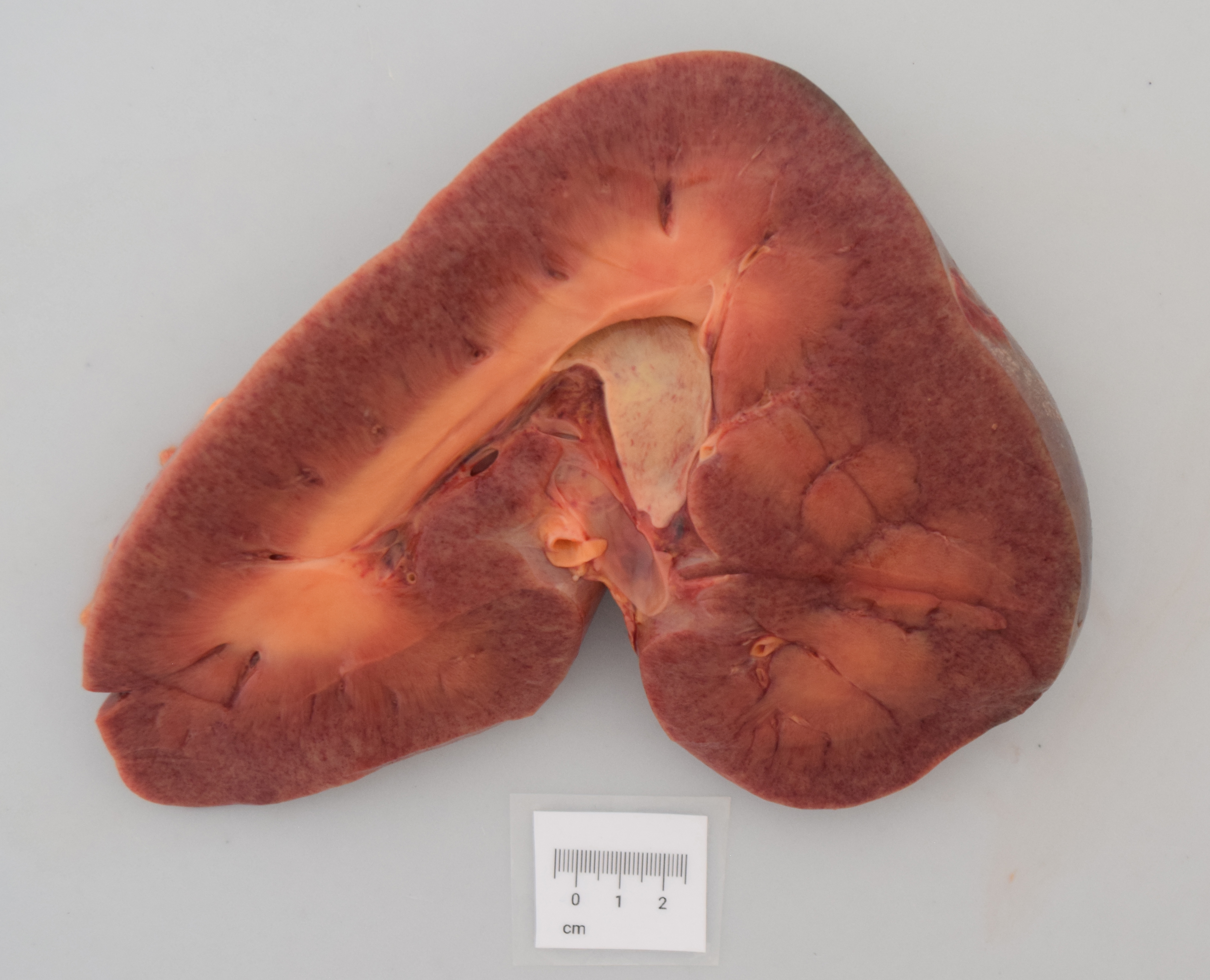
The abdomen contained 20 ml of yellow, clear fluid. The liver was diffusely pale. Both kidneys adhered to the capsule focally and multifocally the surface of both kidneys were patchy and pale (Figure 1). In the pars glandularis of the stomach, along the marco plicatus was a focal, 14.0 x 7.0 x 0.1 cm in size, acute ulceration. The other examined organs were macroscopically unremarkable.
Laboratory Results:
The bacteriological examination was done at the Institute of Veterinary Bacteriology University of Bern.
- RT-PCR for pathogenic leptospires performed on kidney tissue tested positive.
- General cultural examination of the kidney revealed no bacterial growth
Microscopic Description:
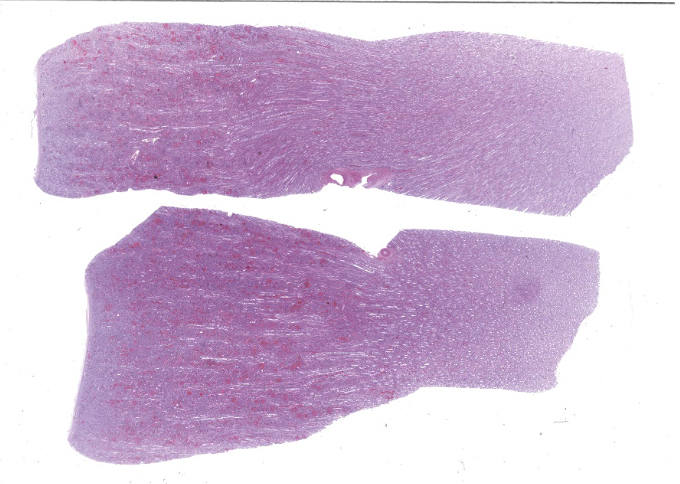
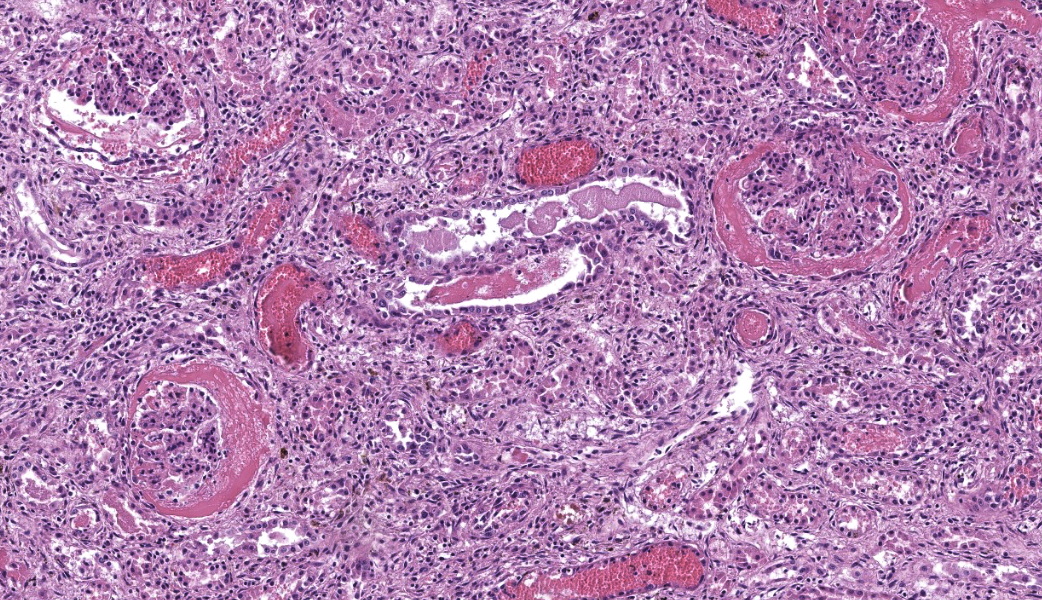
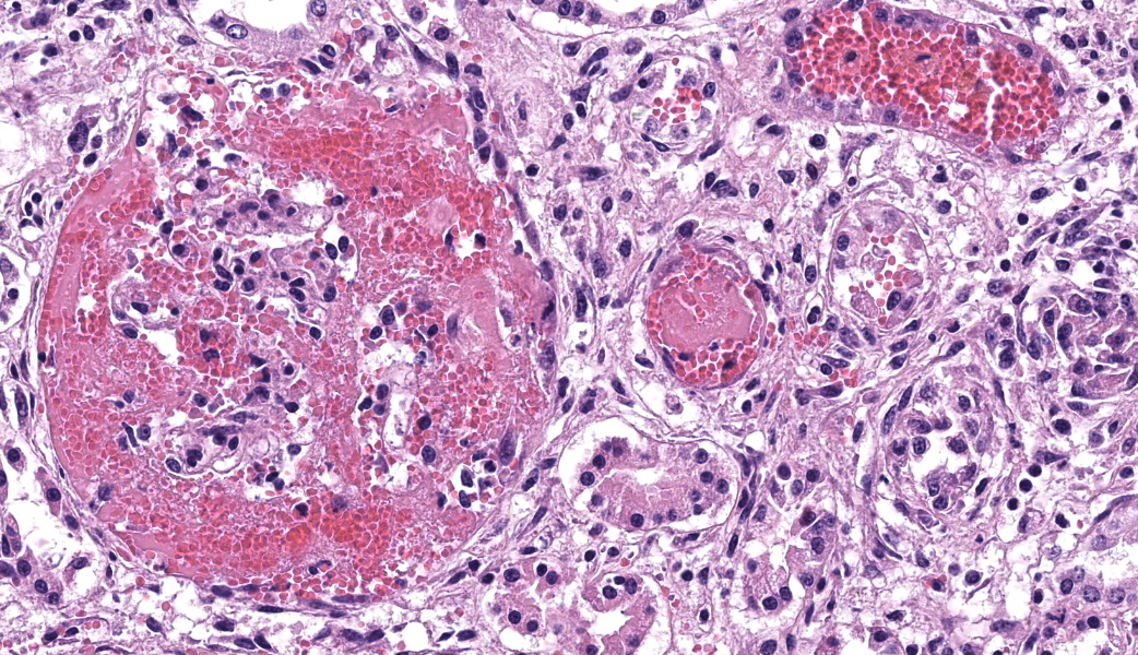
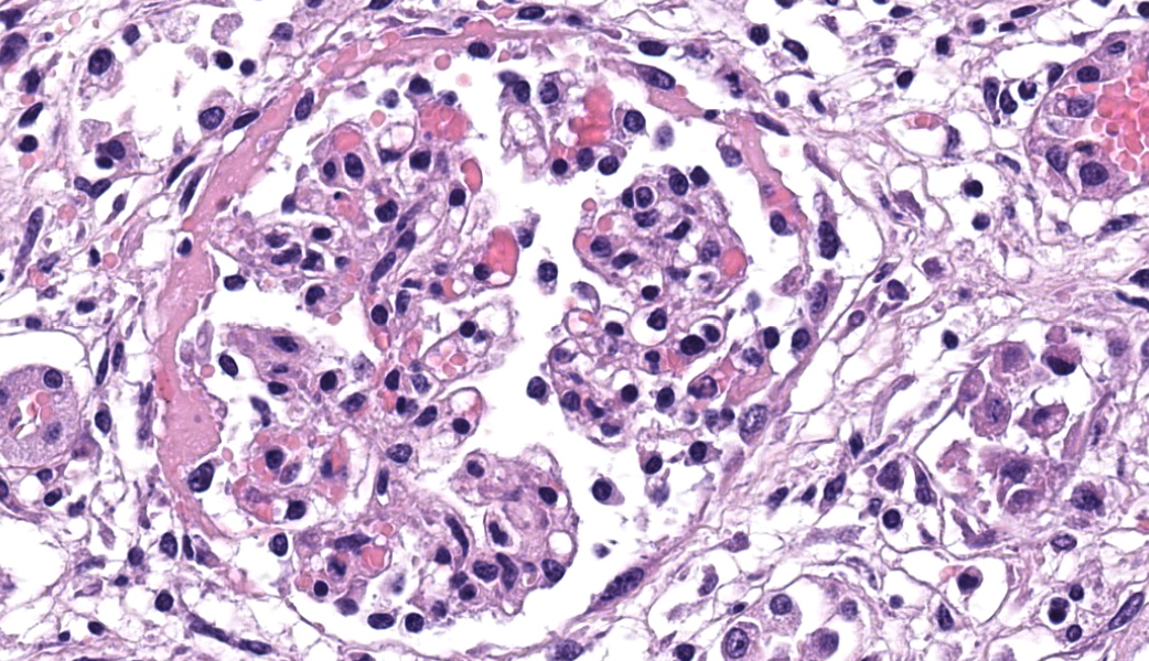
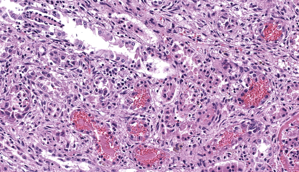
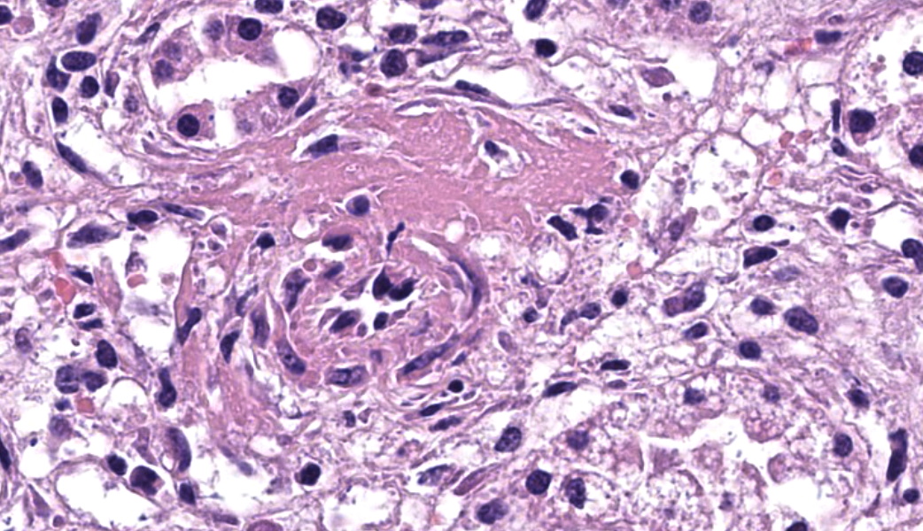
Kidney: Multifocally, up to 70% of the renal corpuscles are affected by a moderate to severe space-occupying accumulation of many extravasated erythrocytes, eosinophilic, fibrillar material (fibrin exudation), and few neutrophils within the urinary space and the mesangium. Few glomeruli are completely replaced by the before mentioned accumulation, partially with loss of continuity of the parietal layer of the Bowman's capsule. Many adjacent tubular structures are mildly dilated and filled with extravasated erythrocytes, eosinophilic, fibrillar material (fibrin), few neutrophils and macrophages. The epithelial cells of some tubules have lost cellular details and exhibit hypereosinophilia, karyorrhexis, and karyolysis (necrosis), with detachment from the basement membrane. Many adjacent blood vessels contain moderate amounts of eosinophilic, fibrillar material, which partially obstruct the lumen (fibrin thrombi). The interstitial space of the cortex is severely infiltrated with neutrophils, macrophages, lymphocytes and plasma cells. Multifocally, there are accumulations of few extravasated erythrocytes (acute hemorrhage). In the medulla, the interstitial inflammatory infiltrate is mild and mainly composed of lymphocytes. In the corticomedullary junction and in the medulla, some tubules contain eosinophilic, homogeneous material (hyaline urinary cast). The kidney is mildly congested.
Contributor's Morphologic Diagnoses:
- Kidney: Severe, multifocal, acute, exudative glomerulonephritis
- Kidney: Severe, acute, neutrophilic and histiocytic, tubulointerstitial nephritis with tubular necrosis, intravascular fibrin thrombi, intratubular urinary casts and interstitial hemorrhage
Contributor's Comment:
Leptospira spp. are obligate aerobic and gram-negative spirochete bacteria divided into distinct serovars,1 Depending on the level of pathogenicity for animals and humans, they are divided into pathogenic, intermediate, or saprophytic.2 One serovar is not specific to a single Leptospira species but is adapted to a specific host, known as the maintenance host. For instance, the serogroup bratislava has been associated with horses.2 The host-adapted serovar may cause disease in its maintenance host or infect incidental hosts, albeit to a lesser extent. The infection may be subclinical, and the host becomes an asymptomatic carrier with persistent spirochetes in the proximal convoluted tubules of the kidneys, or less commonly in the genital tract.1 The spirochetes are then continuously shed within the urine, allowing the transmission of the disease. Outside of the host, the spirochetes can
survive in moist, warm (ideally 28°C), and neutral or mildly alkalinized conditions for weeks or even months.1
Leptospirosis is a spirochetal infection caused by pathogenic Leptospira spp. In farm and domestic animals, the serovars of Leptospira interrogans are an important cause of abortion and stillbirth and might induce an acute or subacute systemic disease (including septicemia, hepatitis, nephritis, meningitis).1
When an animal is exposed to Leptospira spp. in the environment, usually via urine or body fluid of carriers or contaminated water, the bacterium enters the body via mucous membrane or skin lesion, leading to leptospiremia. The spirochetes reach many different organs via the bloodstream, including the renal interstitial capillaries. There, they migrate through the endothelium into the interstitial spaces, where they reach the tubular lumens via migration through intercellular junctions of tubular epithelial cells. The spirochetes associate with the microvilli of the tubular epithelium and are able to persist in the phagosomes of those cells, especially in the proximal and distal convoluted tubules. Usually, the leptospiremic phase lasts up to 7 days, as the organisms are cleared from most organs thanks to the host immune response (clearing via agglutinating and opsonizing antibodies). However, immunologically privileged sites, such as the proximal convoluted tubules, are spared, and the animal becomes a carrier.1
The leptospiremic phase is responsible for the acute or subacute systemic disease, and young animals are more commonly affected. Common clinical and pathological signs include fever, vasculitis (caused by inflammatory cytokine release), hemolytic anemia due to bacterial hemolysins or reaction of antibodies with leptospiral products on erythrocytes, hemoglobinuria, jaundice due to hemolysis or direct liver injury, pulmonary congestion, and occasionally meningitis.1 Because of the localization and persistence of leptospires in the kidneys, acute tubular injury and interstitial nephritis are one of the main findings. The cause of degeneration and necrosis of the tubular epithelium is multifactorial and comprises direct toxic effects of the leptospires, hypoxia, free hemoglobin, and the concomitant interstitial inflammatory reaction1.The inflammatory infiltrate might consist of neutrophils in the acute phase, as in our case, but usually macrophages, lymphocytes and plasma cells predominate (chronic lesion). The inflammatory cells are typically localized at the corticodullary junction. Not only the tubules and interstitium can be affected in acute leptospirosis, but also the glomerular structures.
Exudative glomerulonephritis, characterized by the abundant presence of extravasated erythrocytes, fibrin exudation, as well as fewer neutrophils in the urinary space, should be considered suspicious for leptospiral infection, especially in dogs.3 In the postsepticemic phase, leptospiral infection can manifest itself as abortion, stillbirth or birth of an infected fetus may occur, even weeks to months after infection.1
In horses, serologic evidence of Leptospira spp. is widespread (reservoir hosts) 4 but the occurrence of acute disease is rare. Different serovars (including bratislava,canicola, grippotyphosa, hardjo, icterohaemorrhagiae, and particularly pomona) have been associated with such acute disease, leading to fever, anorexia, acute kidney injury, liver disease accompanied by icterus, epistaxis and acute respiratory distress due to pulmonary hemorrhages.5 Foals are more susceptible and can develop fatal hepatic and renal diseases. The chronic form of leptospirosis is mostly associated with abortion (associated with serovars pomona, grippotyphosa and bratislava), premature foaling and equine recurrent uveitis.1 The latter is a complex, multifactorial syndrome characterized by repeated episodes of uveitis, which has been correlated with Leptospira spp. (especially serovar pomona), even though the exact mechanism remains poorly understood.6
In living animals, the diagnosis mostly relies on the evaluation of antibody titers against Leptospira spp., using the microscopic agglutination test (MAT) as standard serologic assay, in concordance with clinical symptoms.7 Indeed, although the polymerase chain reaction (PCR) is a more rapid and sensitive diagnostic method, it is not always reliable due to the intermittent shedding of the leptospires. In the context of pathological examination, leptospiral nucleic acid can be detected by PCR of infected tissue. However, most PCR are designed to detect only pathogenic leptospires and do not allow the identification of the serogroup.7 Hence, those results should always be interpreted with the clinical and pathological findings. The spirochetes within the cytoplasm and in the lumen of affected tubules can also be identified using a Giemsa or silver stains (low sensitivity), or an immunohistochemistry stain against leptospiral antigens.7
Contributing Institution:
Institute of Animal Pathology Vetsuisse Faculty - University of Bern Laenggassstrasse 122 CH-3012 Bern Switzerland www.itpa.vetsuisse.unibe.ch
JPC Diagnoses:
Kidney: Glomerulonephritis, exudative, subacute, diffuse, marked, with vasculitis and tubular degeneration and necrosis.
JPC Comment:
This week's conference was moderated (in accordance with longstanding JPC/AFIP tradition) by Department chair, COL Sherri Daye. And in line with her own tradition (she is an equestrian extraordinaire among her many talents), horses play a significant role in this week's cases. (See if you can make the connection between Case 4 and equine pathology).
The contributor provides an excellent review of the breadth of disease associated with pathogenic leptospires in horses and other domestic species. While most readers are likely familiar with common forms of glomerulonephritis associated with chronic infectious disease in domestic species, to include membranous, membranoproliferative, and mesangiocapillary (among others), the term "exudative glomerulonephritis" (incorporated into both the contributor's and the JPC's morphologic diagnosis may be unfamiliar and worth a more detailed explanation.
Exudative glomerulonephritis, as the contributor notes, is characterized on light microscopy by the unifying presence of abundant hemorrhage and accompanied fibrin within the urinary space. In acute cases, variable amounts of the following are present and often related to the chronicity and severity of disease: endocapillary capillary hypercellularity (typically by the accumulation of neutrophils), fibrin thrombi within capillary loops, and ultimately segmental to global fibrinoid necrosis of capillary loops. More chronic cases may result in a more generalized hypercellularity of the glomerular tuft, as well as crescent formation.
In both human and veterinary medicine, exudative glomerulonephritis has been most closely associated with various types of infectious disease, and in the human realm, has been referred to as "infection-related glomerulonephritis (IFGN)." The prototypical infection associated with IFGN in humans is post-streptococcal glomerulonephritis which may follow streptococcal pharyngitis or impetigo after a period of 1-4 weeks.8 Post-streptococcal glomerulonephritis is considered the most common cause of glomerulonephritis in children, with up to 475,000 cases diagnosed per year.9 (Interestingly, while Streptococcus equi infection has been incriminated a number of times in IFGN in humans and obviously results in immune-driven systemic vascular lesions associated with thrombocytopenic purpura in the horse, there are apparently no reports in the veterinary literature of S. equi resulting in exudative glomerulonephritis in horses.)
Infectious agents that have been less commonly incriminated in IFGN in humans include Staphylococcus aureus (often associated with intravenous drug use), Bartonella henselae, and Coxiella burnetti. On the other hand, cases of IFGN resulting from spirochetal disease in humans are notably few. Leptospirosis, similar to its effects in most other animal species, results in tubulointerstitial disease in humans (due to its predilection to infect tubular epithelium). Other spirochetes, such as Treponema pallidum and Borrelia burgdorferi only rarely) result in glomerular disease in humans but typically cause immune-complex glomerulonephritis (ICGN) rather than IFGN (but one case of a human IFGN has been reported in association with B. burgdorferi infection.)10
In veterinary medicine, it appears that the dog is the outlier when it comes to the renal effects of spirochetal diseases, as dogs famously develop immune-mediated GN with B. burgdorferi11 and IFGN when infected with leptospires.3
References:
- Jubb K, Kennedy P, Palmer N. Jubb, Kennedy, and Palmer's Pathology of Domestic Animals: Volume 2. Vol 2. Sixth. Elsevier; 2016, pp. 433-439.
- Cilia G, Bertelloni F, Fratini F. Leptospira Infections in Domestic and Wild Animals. Pathogens. 2020 Jul 15;9(7):573. doi: 10.3390/pathogens9070573. PMID: 32679834; PMCID: PMC7400056.
- Hilbe M, Posthaus H, Paternoster G, Schuller S, Imlau M, Jahns H. Exudative glomerulonephritis associated with acute leptospirosis in dogs. Veterinary Pathology. 2023;61(3):453-461. doi:10.1177/03009858231207020
- Hamond C, Adam EN, Stone NE, et al. Identification of equine mares as reservoir hosts for pathogenic species of Leptospira. Front Vet Sci. 2024 May 9;11:1346713. doi: 10.3389/fvets.2024.1346713. PMID: 38784659; PMCID: PMC11112012.
- Ramsay L, Eberhardt C, Schoster A. Acute leptospirosis in horses: A retrospective study of 11 cases (2015-2023). J Vet Intern Med. 2024 Sep-Oct;38(5):2729-2738. doi: 10.1111/jvim.17184. Epub 2024 Aug 28. PMID: 39194176; PMCID: PMC11423456.
- Gerras J, Young K, Roberts D, Waldman G, Salmon JH, Gilger BC. Uveitis and blindness in a closed herd of Equidae following leptospiral infection. Front Vet Sci. 2025 Jan 6;11:1504990. doi: 10.3389/fvets.2024.1504990. PMID: 39834922; PMCID: PMC11743373.
- Sykes JE, Hartmann K, Lunn KF, Moore GE, Stoddard RA, Goldstein RE. 2010 ACVIM small animal consensus statement on leptospirosis: diagnosis, epidemiology, treatment, and prevention. J Vet Intern Med. 2011 Jan-Feb;25(1):1-13. doi: 10.1111/j.1939-1676.2010.0654.x. Epub 2010 Dec 13. PMID: 21155890; PMCID: PMC3040842.
- Khalighi MA, Chang A. Infection-Related Glomerulonephritis. Glomerular Dis. 2021 Apr 28;1(2):82-91. doi: 10.1159/000515461. PMID: 36751423; PMCID: PMC9677723.
- Chamarthi G, Clapp WL, Bejjanki H, Auerbach J, Koratala A. Infection-related Glomerulonephritis and C3 Glomerulonephritis - Similar Yet Dissimilar: A Case Report and Brief Review of Current Literature. Cureus. 2020 Feb 28;12(2):e7127. doi: 10.7759/cureus.7127. PMID: 32140372; PMCID: PMC7047932.
- Rolla D, Conti N, Ansaldo F, Panaro L, Lusenti T. Post-infectious glomerulonephritis presenting as acute renal failure in a patient with Lyme disease. J Renal Inj Prev 2014; 3(1): 17-20. DOI: 10.12861/jrip.2014.07
- Dambach DM, Smith CA, Lewis RM, Van Winkle TJ. Morphologic, immunohistochemical, and ultrastructural characterization of a distinctive renal lesion in dogs putatively associated with Borrelia burgdorferi infection: 49 cases (1987-1992). Vet Pathol. 1997 Mar;34(2):85-96. doi: 10.1177/030098589703400201. PMID: 9066075.