CONFERENCE 10, CASE II:
Signalment:
11 year old, male, Australian shepherd dog, Canis lupus familiaris
History:
The dog’s owner noted asymmetry of the testes with enlargement of the left testis within a short period of time. After clinical examination, neoplasia of one testis was suspected, and the left testis was removed, and submitted for histopathologic examination. Signs of hormonal imbalance or feminization as well as other clinical symptoms were not observed.
Gross Pathology:
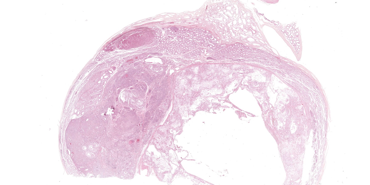
Testis with epididymis (5 x 4 x 3.5 cm) and funiculus spermaticus, testis consisting of approximately 90% neoplastic tissue with a small margin of normal tissue, centrally cystic, multiple confluent whitish and yellowish-brown nodules.
Microscopic Description:
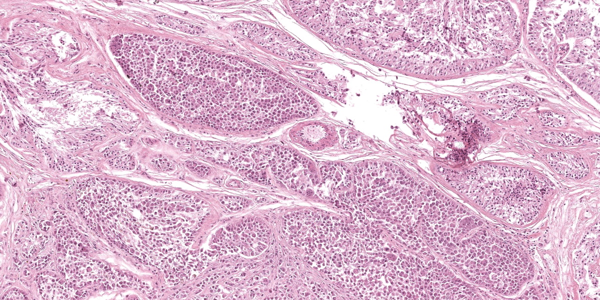
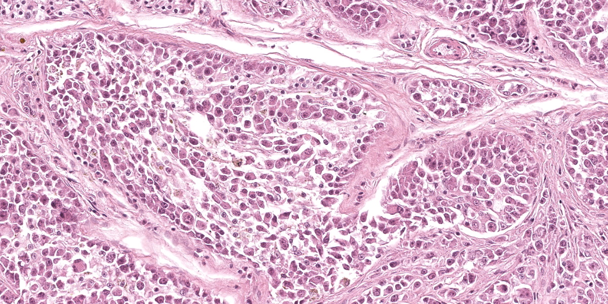
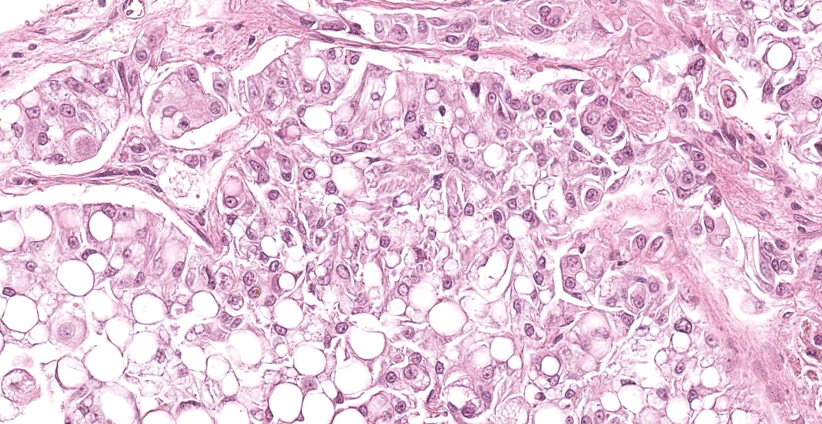
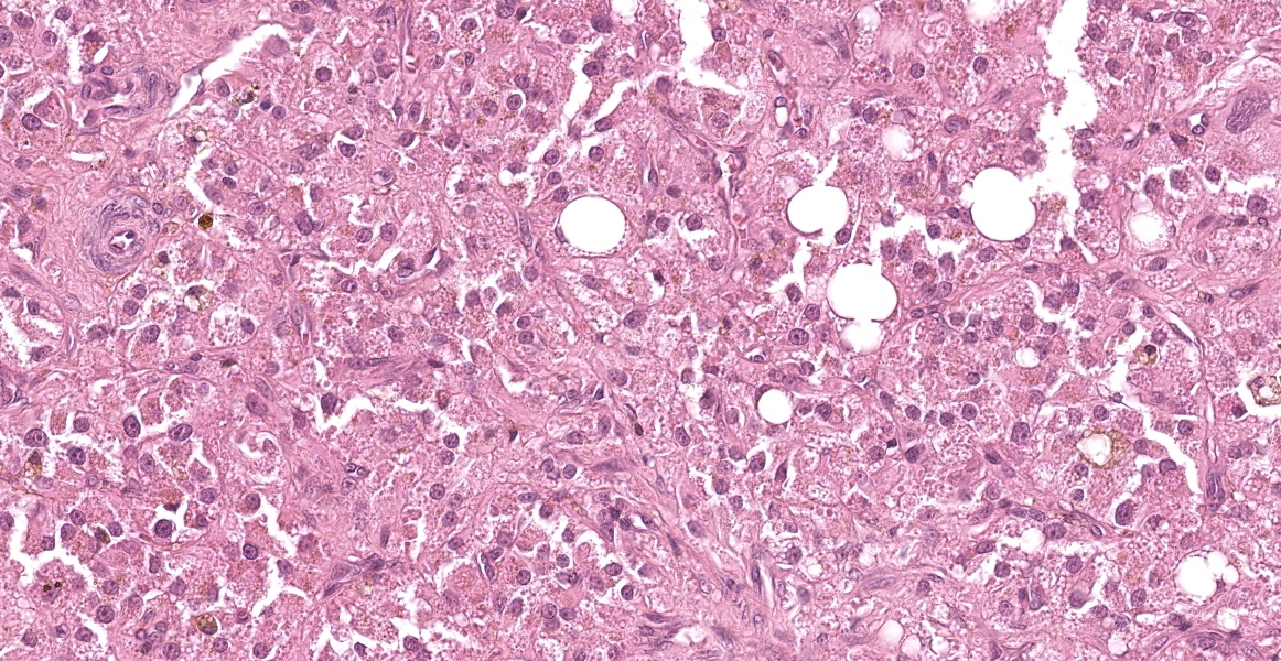
Testis: Infiltrating and compressing the testicular parenchyma and extending to one cut edge is a 2 x 1.5 cm large neoplastic proliferation composed of different cells and components. The first cell population consists of polygonal cells with often distinct cell borders, a moderate amount of eosinophilic, granular, or vacuolated cytoplasm (ranging from finely vacuolated to large vacuoles with compression of cytoplasm), and a round nucleus with finely stippled chromatin and an often prominent magenta nucleolus (interstitial cells). There is moderate anisocytosis and anisokaryosis. The mitotic rate is less than 1 per 2.37 mm2 (10 HPF). These cells form both a 0.5 x 0.3 cm, compact, densely cellular, well-demarcated, partly encapsulated nodule composed of densely packed cords and nest of cells in a fine fibrovascular stroma and a 2 x 1.5 cm moderately cellular, well-demarcated, encapsulated nodule composed of packets and nests of cells with large vacuoles and a central clear space. The second cell population consists of large round cells with distinct cell borders, often a large amount of eosinophilic granular cytoplasm, and large, round, vesicular nuclei with coarse chromatin and 1-2 prominent, magenta nucleoli (germ cells).
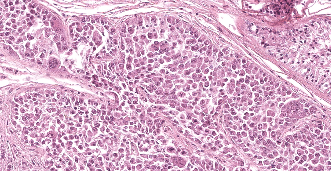
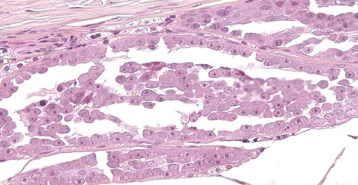
There is moderate anisocytosis and anioskaryosis with frequent multinucleated cells, occasionally forming giant cells with up to 8 nuclei. Some macronuclei are visible. The mitotic rate averages 1-3 mitoses per 0.237 mm2 (1 HPF). These cells are either infiltrate the lumina of the seminiferous tubules, replacing Sertoli and spermatogenic cells, or are intermingled with the third cell population within a 1.5 x 1 cm, moderately cellular, well-demarcated, partly encapsulated, partly infiltrative nodule composed of large nests, packets, and tubules of neoplastic cells, separated and surrounded by bands of fibrous connective tissue. The cells of the second population can sometimes be found within thin walled vessels (vascular invasion). The third population of neoplastic cells is polygonal to columnar with indistinct cell borders , a moderate amount of eosinophilic granular cytoplasm and an oval nucleus with coarsely stippled chromatin and a magenta nucleolus. Palisading of the cells perpendicular to thebasement membrane of the seminiferous tubules is frequently noted. The population exhibits mild to moderate anisocytosis and anisokaryosis. The number of mitoses is less than 1 per 2.37 mm2 (10 HPF). Interspersed within the neoplastic cells are few lymphocytes and plasma cells as well as aggregates of hemosiderin-laden macrophages.
A few seminiferous tubules show remnants of spermatogenesis. Epididymis and ductus deferens contains single macrophages and no spermatids.
Contributor’s Morphologic Diagnosis:
Testis:
- Interstitial cell tumor in two localizations
- Seminoma, intratubular multiple and diffuse
- Mixed germ cell sex-cord stromal tumor (seminoma and Sertoli cell tumor)
- Atrophy of residual seminiferous tubules
Contributor’s Comment:
In intact older male dogs, testicular tumors are one of the most common tumors. The cause for that is unknown. Seminoma, Sertoli cell tumors and Leydig cell tumors are found regularly.1-3 Less common are mixed germ cell sex-cord stromal tumors of the testis,5 embryonal carcinoma, or teratomas. Adenomas or adenocarcinomas of the rete testis are very rare. Secondary testicular tumors (metastases) are extremely rare.
In dogs, cryptorchism is an important predisposing factor for primary testicular tumors. Several types of neoplasms can be found in one testis,1,2 so careful examination and lamellation of the testes is obligatory in preparation for histopathology.
The biologic behavior of testicular neoplasms in dogs is often benign, and metastases are rare, with a higher risk in seminomas and Sertoli cell tumors.3 Leydig cell tumors are considered benign in dogs. Recently, however, two dogs with metastatic Leydig cell tumors have been described. Both cases were found to have a high mitotic count and high Ki-67 index.4 Unfortunately, there are no other obvious cytologic or histopathologic signs of malignancy in testicular neoplasms in dogs. In malignant cases, distant metastases may be suspected in regional lymph nodes or along the spermatic cord.1,2
In the case presented, all three cell types of the common tumor types of canine testicularneoplasms are found, and the examiner must describe the characteristics of the tumor cells and the additional findings in the remaining non-neoplastic tissue. Testicular neoplasms in general and this case in particular are thus very good practice cases for veterinary pathology residents.
Contributing Institution:
Institut fuer Veterinaer-Pathologie, Justus-Liebig-Universitaet Giessen
Frankfurter Str. 96, 35392 Giessen, Germany
http://www.uni-giessen.de/cms/fbz/fb10/ institute_klinikum/institute/pathologie
JPC Diagnosis:
- Testis: Mixed germ cell sex-cord stromal tumor
- Testis, seminiferous tubules: Atrophy, diffuse, moderate, with aspermatogenesis
JPC Comment:
Case 2 presents an interesting diagnostic challenge in that there are 4 potential separate neoplasms in this neoplasm. Our morphologic diagnoses differed from that of the contributor only academically, in that we were unable to fully resolve what was an individual neoplasm versus a component of the larger mixed germ cell sex-cord stromal tumor. Herein,we simply covered all tumor entities under a single banner of mixed germ cell sex-cord stromal tumor. In addition, and with less spirited discussion, conference participants described degenerative changes to the remaining seminiferous tubules, degeneration of spermatids, and lack of sperm within the epididymis.
We performed a full workup for this case in conjunction with Dr. Smedley. Within this section of testis, there were 3 separate, distinct morphologies that participants described. Foremost, there was a seminoma-distinct portion that had intratubular round to polygonal cells with radiating chromatin and occasional multinucleation. Second, there was an interstitial (Leydig) cell-distinct portion that was unencapsulated with markedly eosinophilic polygonal cells with vacuolated cytoplasm. Finally, there was an encapsulated, expansile portion that had polygonal cells palisading along fibrous trabeculae overlapping morphologies of both Sertoli (sustentacular cells) and seminoma.
Conference participants discussed the nomenclature for this case carefully. Seminomas are a testicular germ cell tumor. Conversely, sex-cord stromal tumors represent neoplasms that exhibit features of either (or both) Sertoli cells and Leydig cells. In cases where seminoma (germ cell) and one or more stromal tumor features are overlapping, a diagnosis of mixed germ cell sex-cord stromal tumor is fitting. In some cases, it may be appropriate to diagnosis multiple discrete tumors within one testis when they are clearly distinct. Conference participants opined that the interstitial cell tumor-distinct population was smaller, separated by normal seminiferous tubules, and was morphologically the most distinct. It is possible that this was a separate tumor that arose earlier that was overshadowed by the emergence of the rapidly growing mixed tumor the contributor describes clinically.
Dr. Smedley emphasized that HE diagnoses alone could be misleading for approximately 20% of canine testicular tumors and that IHC was helpful for resolving exact cell type. We performed IHCs for CD117 (C-kit), desmin, vimentin, inhibin, LH, Melan-A, and NSE to characterize these neoplasms. With regard to LH, it should be stressed that LH is not expressed within the gonads (it is produced in the pituitary gland); only its receptor is expressed in the gonads (and neither the JPC laboratory or Dr. Smedley’s laboratory currently performs this assay.) GATA-4 is a transcription factor expressed in sex-cord cells, but not germ cells.6 Dr. Smedley performed this IHC in her laboratory and noted moderate nuclear immunoreactivity within Sertoli cells and select Leydig cells. These patterns confirmed our H&E expectations of this case.
References:
- Agnew DW, MacLachlan NJ. Tumors of the genital system. In: Meuten DJ, ed. Tumors in domestic animals. 5th ed. Ames, IA: Wiley Blackwell; 2017:706-713.
- Foster RA. Male genital system. In: Maxie MG, ed. Jubb, Kennedy, and Palmer’s Pathology of Domestic Animals. 6th ed. Philadelphia, PA: Elsevier; 2016; vol. 3:492-497.
- Kennedy PC, Cullen JM, Edwards JF, Goldschmidt MH, Larsen S, Munson L, Nielsen S. Histological classification of tumors the genital system of domestic animals. WHO, Washington, DC: Armed Forces Institute of Pathology; 1998,second series, vol. 4.
- Kudo T, Kamiie J, Aihara N, Doi M, Sumi A, Omachi T, Shirota K. Malignant Leydig cell tumor in dogs: two cases anda review of the literature. J Vet Diagn Invest. 2019;31(4):557-561.
- Patnaik AK, Mostofi FK. A clinicopathologic, histologic, and immunohistochemical study of mixed germ cell-stromal tumors of the testis in 16 dogs. Vet Pathol. 1993;30:287-295.
- Ramos-Vara JA, Miller MA. Immunohistochemical evaluation of GATA-4 in canine testicular tumors. Vet Pathol. 2009 Sep;46(5):893-6.
- Reineking W, Seehusen F, Lehmbecker A, Wohlsein P. Predominance of granular cell tumours among testicular tumours of rabbits (Oryctolagus cuniculi dom.). J Comp Pathol. 2019;173:24-29.
