Signalment:
9.3-year-old male Indian origin rhesus macaque (Macaca mulatta)
History:
This animal was used in an infectious disease study and had been experimentally infected nine days prior to euthanasia. The animal was otherwise clinically healthy.
Gross Pathology:
The heart was moderately enlarged and flabby (heart and ventricular weights could not be collected). Abundant adipose diffusely expanded the epicardium, obscuring the myocardium. Both ventricles were dilated, and there was multifocal interstitial fibrosis as well as fatty replacement within the myocardium, particularly within the interventricular septum. Other gross lesions were consistent with experimental manipulations.
Laboratory Results:
None available
Microscopic Description:
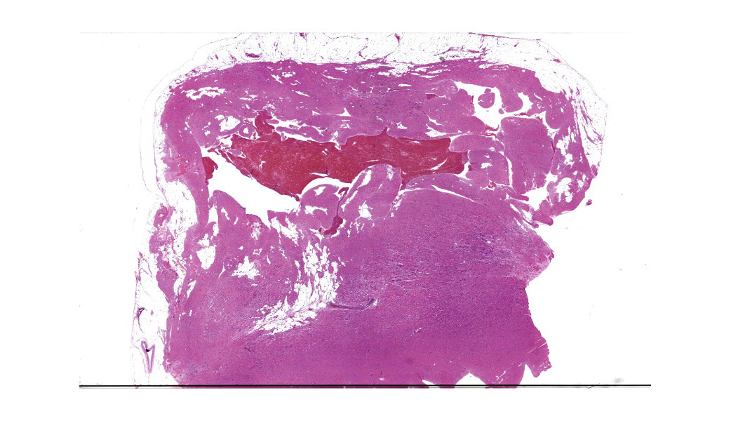
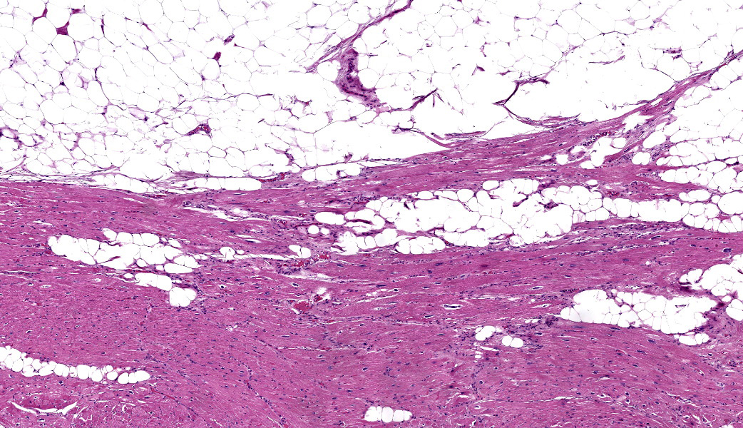
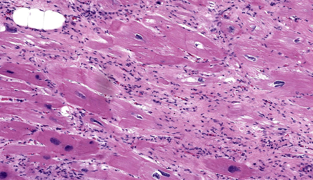
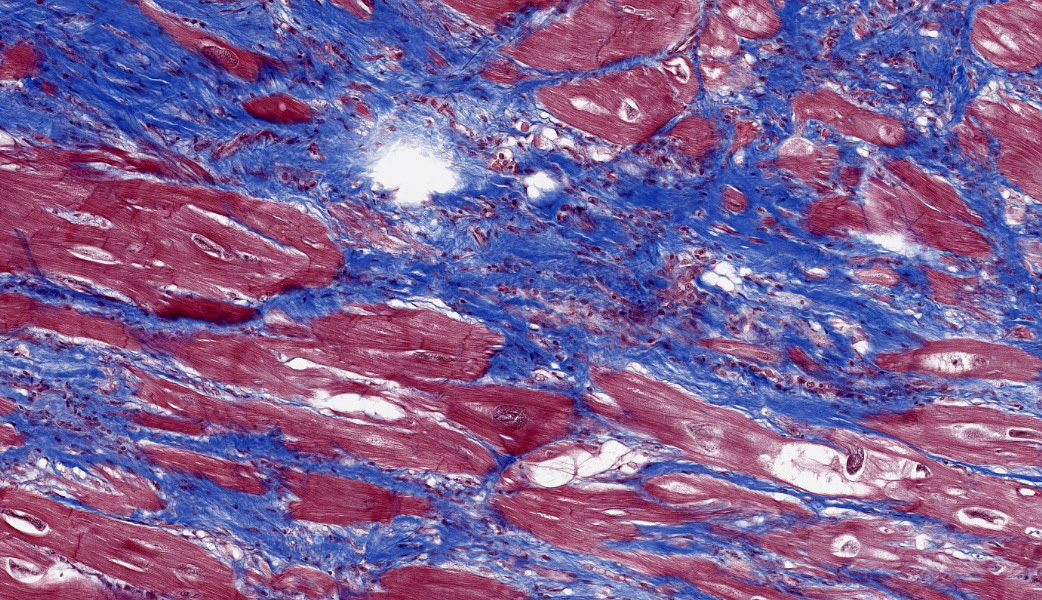
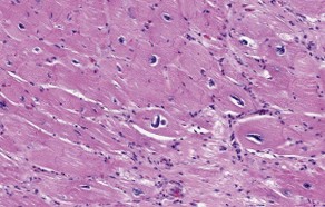
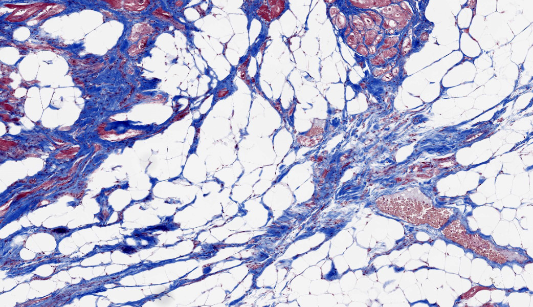
Slide contains a cross section of interventricular septum and right ventricular free wall. There is copious epicardial fat which frequently infiltrates and separates, surrounds, and dissects the cardiomyocytes of the outer myocardium, with occasional individual or small islands of cardiomyocytes encircled by abundant well-differentiated unilocular (white) adipocytes and/or mature fibrosis. There are scattered small foci of individual cardiomyocyte degeneration necrosis with bland histiocytic infiltrates. There is frequent cardiomyocyte hypertrophy with numerous karyomegalic "boxcar" nuclei. There is occasional cardiomyocyte disarray with basketweave formation. There are also hypercontracted cardiomyocytes, particularly at-the cut edges of the section (artifact). There are frequent interstitial perivascular white adipocytes in the myocardium (normal).
Contributor's Morphologic Diagnoses:
Heart, RVFW and IVS: Cardiomyocyte degeneration, necrosis, and loss with fibrofatty replacement, multifocal to coalescing, chronic, moderate to severe, with cardiomyocyte hypertrophy
Contributor's Comment:
The gross and histologic lesions in the heart are consistent with arrhythmogenic cardiomyopathy, formerly arrhythmogenic right ventricular dysplasia.2,4
Although commonly involving the right ventricle, left and biventricular disease may occur. The characteristic features of this entity are progressive epicardial to endocardial loss of cardiomyocytes with fibrofatty replacement, with or without aneurysms of the RVFW, particularly at the RV outflow tract. ACM is a genetic disease, with autosomal dominant with incomplete penetrance and autosomal recessive forms. The vast majority of human cases in which a cause is identified result from mutations in desmosomal genes, most often plakophilin-2 (PKP2). Other mutated desmosomal genes include desmoplakin (DSP), desmoglein-2 (DSG2), plakoglobin (JUP), and desmocollin-2 (DSC2). Implicated non-desmosomal genes include alpha-T-catenin (CTNNA3), N-cadherin (CDH2), transmembrane protein 43 ( TMEM43), lamin A/C (LMNA), desmin (DES), sodium voltage-gated channel alpha subunit 5 (SCN5A), phospholamban (PLN), and ryanodine receptor 2 (RYR2). However, in up to 50% of human cases, no mutation has been identified. Ante-mortem diagnosis can be difficult due to an absence of specific unique diagnostic criteria, variable expressivity, and incomplete pene-trance in relatives. EKG, cardiac imaging, endomyocardial biopsy, and genetic testing are the mainstays of diagnosis. Diagnosis may occur post-mortem, particularly in otherwise healthy young athletes with sudden death. In veterinary medicine, ACM has been reported in cats, dogs (boxers, English bulldogs), horses, a rhesus macaque, two chimpanzees, and a bonobo.1,3,5,7 In boxers, deletions in the intercalated disc gene striatin (STRN) cause AD ACM, with worse disease in homozygotes.
ACM should be differentiated from adipositas cordis, in which there is diffuse fatty infiltration of the ventricular myocardium or interventricular septum without fibrosis or cardiomyocyte abnormalities or destruction.
Contributing Institution:
Pathology Department NIH/NIAID
Integrated Research Facility
JPC Diagnoses:
Heart: Fibrofatty infiltration, chronic, multifocal to coalescing, severe, with myofiber karyomegaly, hypertrophy, degeneration, and loss.
JPC Comment:
This case was selected to provide a contrast between the histologic changes seen in dilated cardiomyopathy (Case 1 in this conference) versus arrhythmogenic right cardiomyopathy this case. It also demonstrates a nice interspecies contrast between the relatively bland degenerating cardiomyocytes in the dog, and the visually striking nuclear changes seen in late-stage cardiomyopathy in primates.
This is not the first time that this condition has been seen in the WSC - it has been previously seen in three different species - WSC 2009 Conference 5, Case 3 (dog), WSC 2018 Conference 13, Case 3 (horse), and WSC 2020 Conference 18, Case 2 (rhesus macaque).
ARVC is a genetic disease (and familial in 50% of cases) linked to defects in over 13 different genes that are vital components of the intercalated disc, particularly the adherens junction. The degeneration of intercalated disks with mutated structural proteins results in weaking of desmosomes, ultimately resulting in the loss of cardiomyocyte adhesion, car-diomyocyte apoptosis and an opportunity for adipocytes to infiltrate between separated and degenerating cardiomyocytes.8 Other mutations in this syndrome more directly affect cardiomyocytes, to include connexin 43, whose mutation results in abnormal gap junctions between cardiomyocytes which are required for the proper movement of ions, nucleotides and protein and sugar molecules needed for prompt repolarization of myocytes following contraction. This particular mutation ultimately impairs the influx of sodium into cardiomyocytes resulting in asynchronous contraction of myocytes and ultimately, the arrhythmias from which this disease gets its name.9 (A more detailed description of the genetic mutations and their fascinating effects on the myocardium is present in WSC 2020 Conference 18, Case 2.). The right ventricular wall is thought to be predisposed to development of this lesion as a result of being thinner and less able to withstand the rigors of exercise due to defects in cardiomyocyte adhesion.10Sudden death is seen in athletes (both animal and human), while progressive heart failure is exhibited by more sedentary individuals (pass the pork rinds and put on the ballgame, please!)
One of the more intriguing changes in the section (and of the other rhesus macaque with ARVC in the WSC, but not the dog or horse case) is the nuclear pleomorphism that is seen within cardiomyocytes adjacent to areas of fibrosis. Studies of this change in human cardi-omyopathy have described it a common change and in affected cases, is seen in proximity to areas of fibrosis.11
While early studies promoted polyploidy as the cause of the nuclear alterations in various forms of cardiomyopathy in humans, recent investigations have focused on more complex interaction between the cardiomyocyte cytoplasm and it nucleus in related process known as mechanosensing and mechanotransduction.12,13 A vast oversimplification of these complex interplay is as follows: actin and des-min filaments as well as microtubules form connections between the sarcomere and the nuclear envelope which result in changes in nuclear morphology and gene expression.12 Mechanical forces are transmitted from the sarcomere via desmin filaments in the Z-disks. Nuclear envelope proteins such as laminin A/C then interact with chromatin at the nuclear periphery, resulting in an increase or decrease in chromatin organization and corresponding changes in gene expression. A much more detailed accounting of these processes and the structures and proteins involved can be found in the references below.12,13
References:
- Celestino-Soper PBS, Lynnes TC, Zhang L, Ouyang K, Wann S, Clyde VL, et al.: Genetic analyses in a bonobo (Pan paniscus) with arrhythmogenic right ventricular cardiomyopathy. Scientific Reports 2018:8(1):4350.
- d'Amati G, Giordano C: Chapter 11 - Cardiomyopathies. In: Buja LM, Butany J, eds. Cardiovascular Pathology (Fourth Edition). San Diego: Academic Press; 2016: 435-483.
- Freel KM, Morrison LR, Thompson H, Else RW: Arrhythmogenic right ventricular cardiomyopathy as a cause of unexpected cardiac death in two horses. Vet Rec 2010: 166(23):718-721.
- Marcus Fl, McKenna WJ, Sherrill D, Basso C, Bauce B, Bluemke DA, et al.: Diagnosis of arrhythmogenic right ventricular cardiomyopathy/dysplasia: proposed modification of the task force criteria. Cir-culation 2010:121 (13): 1533-1541.
- Nakao S, Hirakawa A, Yamamoto S, Kobayashi M, Machida N: Pathological Features of Arrhythmogenic Right Ventricular Cardiomyopathy in Middle-Aged Dogs. Journal of Veterinary Medical Science 2011:73(8):1031-1036.
- Stern JA, Ueda Y: Inherited cardiomyopathies in veterinary medicine. Pflugers Archiv-European Journal of Physiology 2019:471 (5):745-753.
- Tong LJ, Flach EJ, Sheppard MN, Pocknell A, Banerjee AA, Boswood A, et al.: Fatal Arrhythmogenic Right Ventricular Cardiomyopathy in 2 Related Subadult Chimpanzees (Pan troglodytes). Veterinary Pathology 2014:51 (4):858-867.
- Chatterjee D, Fatah M, Akdis D, et al. An autoantibody identifies arrhythmogenic right ventricular cardiomyopathy and participates in its pathogenesis. European Heart Journal. 2018;39:3932-3944.
- Paul M, Wichter T, Gerss J, et al. Connexin expression patterns in arrhythmogenic right ventricular cardiomyopathy. Am J Cardiol. 2013 May 15;111(10):1488-95.
- Basso C, Fox PR, Meurs KM, et al. Arhythmogenic right ventricular cardiomyopathy causing sudden cardiac death in boxer dogs: a new animal model of human disease. Circulation. 2004 Mar 9;109(9):1180-5.
- Mitrut R, Pirici D, Stepan AE, Margaritescu C, Simionescu CE, Kesse AM, Militaru C. Histopathological and
- Morphometric Study of Fibrosis and Nu-clear Pleomorphism in Dilated Cardiomyo-pathy. Curr Health Sci J. 2019 Jan Mar;45(1):73-78. doi:10.12865/CHSJ.45.01.10.
- Coscarella IL, Landim-Vieira M, Rastegarpouyani H, Chase PB, Irianto J, Pinto JR. Nucleus Mechanosensing in Cardiomyocytes. Int J Mol Sci. 2023 Aug 28;24(17):13341.
- Lyon RC, Zanella F, Omens JH, Sheikh F. Mechanotransduction in cardiac hypertrophy and failure. Circ Res. 2015 Apr 10;116(8):1462-1476. doi:10.1161/CIRCRESAHA.116.304937. PMID: 25858069; PMCID: PMC4394185.