Signalment:
Gross Description:
Histopathologic Description:
Morphologic Diagnosis:
Lab Results:
Condition:
Contributor Comment:
Infection of healthy, immunocompetent, non-pregnant cattle by BVD virus usually results in only subclinical to mild clinical disease; however, primary infection by very virulent strains of BVD virus can result in severe clinical disease with mortality which cannot be differentiated from mucosal disease by clinical signs and necropsy findings.(1)
In this particular case, it is uncertain whether this calf was persistently infected and died as a result of mucosal disease, or if this was a healthy calf which became infected by a particularly virulent strain of BVD virus. The low morbidity, high mortality of the group suggests this was a PI calf, which died of fatal mucosal disease. Molecular characterization of the isolated virus may have aided in the differentiation of these two syndromes.
JPC Diagnosis:
Conference Comment:
Virus is shed in body fluids, and primary replication occurs in the tonsils and oropharyngeal lymphoid tissues. Virus then enters circulating monocytes and is transported to lymphoid tissues and the subepithelial connective tissue of the dermis and GI tract, where it spreads locally to overlying epithelial cells. In addition to the subclinical form in immunocompetent adults as mentioned by the contributor, BVD manifests in two other forms. Transplacental infections during days 50-100 of gestation result in fetal death, abortion, or mummification; infection at day 100-150 of gestation results in congenital defects such as microencephaly, cerebellar hypoplasia, hydranencephaly, hydrocephalus, microphthalmia, thymic aplasia, hypotrichosis, alopecia, brachygnathism, growth retardation, and pulmonary hypoplasia. If a calf survives infection prior to 125 days of gestation, it may develop immunotolerance and lifelong, subclinical infection, which is the most pervasive source of infection of other cattle. A second form is mucosal disease, which occurs when a calf is infected with a noncytopathic genotype prior to 125 days of gestation and becomes immunotolerant, and is then infected with a cytopathic strain. Mortality in calves with mucosal disease approaches 100%.(1,2,3)
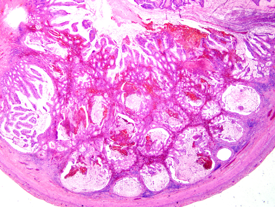
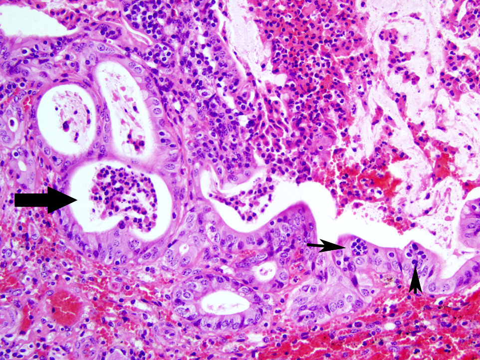
Gross lesions with subclinical BVD are seen as mild erosions or shallow ulcerations of the oral cavity. Mucosal disease manifests with erosions and ulcerations of mouth, tongue, esophagus, oral and ruminal papillae, abomasum, cecum, and colon; linear esophageal ulcerations (tiger-stripe); swollen, necrohemorrhagic Peyer's patches with diphtheritic membranes; and erosive or ulcerative interdigital dermatitis and coronitis. The virus causes widespread vasculitis, epithelial necrosis, and lymphocytolysis.(1,2,3)
References:
2. Gelberg HB. Alimentary system and the peritoneum, omentum, mesentery, and peritoneal cavity. In: McGavin MD, Zachary JF, eds. Pathologic Basis of Veterinary Disease. 5th ed. St. Louis, MO: Mosby Elsevier; 2011:384.
3. Zachary JF. Mechanisms of microbial infections. In: McGavin MD, Zachary JF, eds. Pathologic Basis of Veterinary Disease. 5th ed. St. Louis, MO: Mosby Elsevier; 2011:206-7.

