Wednesday Slide Conference, Conference 17, Case 2
Signalment:
Adult, male, Ezo raccoon dog (Nyctereutes procyonoides albus)
History:
On March 29, 2022, a crow die-off occurred in a public garden in Sapporo, Hokkaido, Japan. Based on RT–qPCR and subsequent gene sequencing analyses, carcasses collected on March 29 and May 18, 2022, were confirmed to be infected with highly pathogenic avian influenza virus (HPAIV). On the same day as the die-off, a diseased raccoon dog was also noticed by the garden workers. Three days thereafter, the raccoon dog showed severe clinical signs of depression and blindness. It was captured for treatment but was considered to be at a humane endpoint from the veterinary viewpoint; thus, it was euthanized via isoflurane inhalation. The raccoon dog was subjected to postmortem examination.
Gross Pathology:
The raccoon dog was severely emaciated and moderately dehydrated. Severe bilateral conjunctivitis with eye discharge was noted, and the lenses were mildly clouded. Nematodes were detected in the stomach. The kidneys were discolored.
Laboratory Results:
HPAIV genomes of the H5 subtype were detected and the virus was isolated from the 10% homogenates of the brain, trachea, and lung of the raccoon dog.2
Microscopic Description:
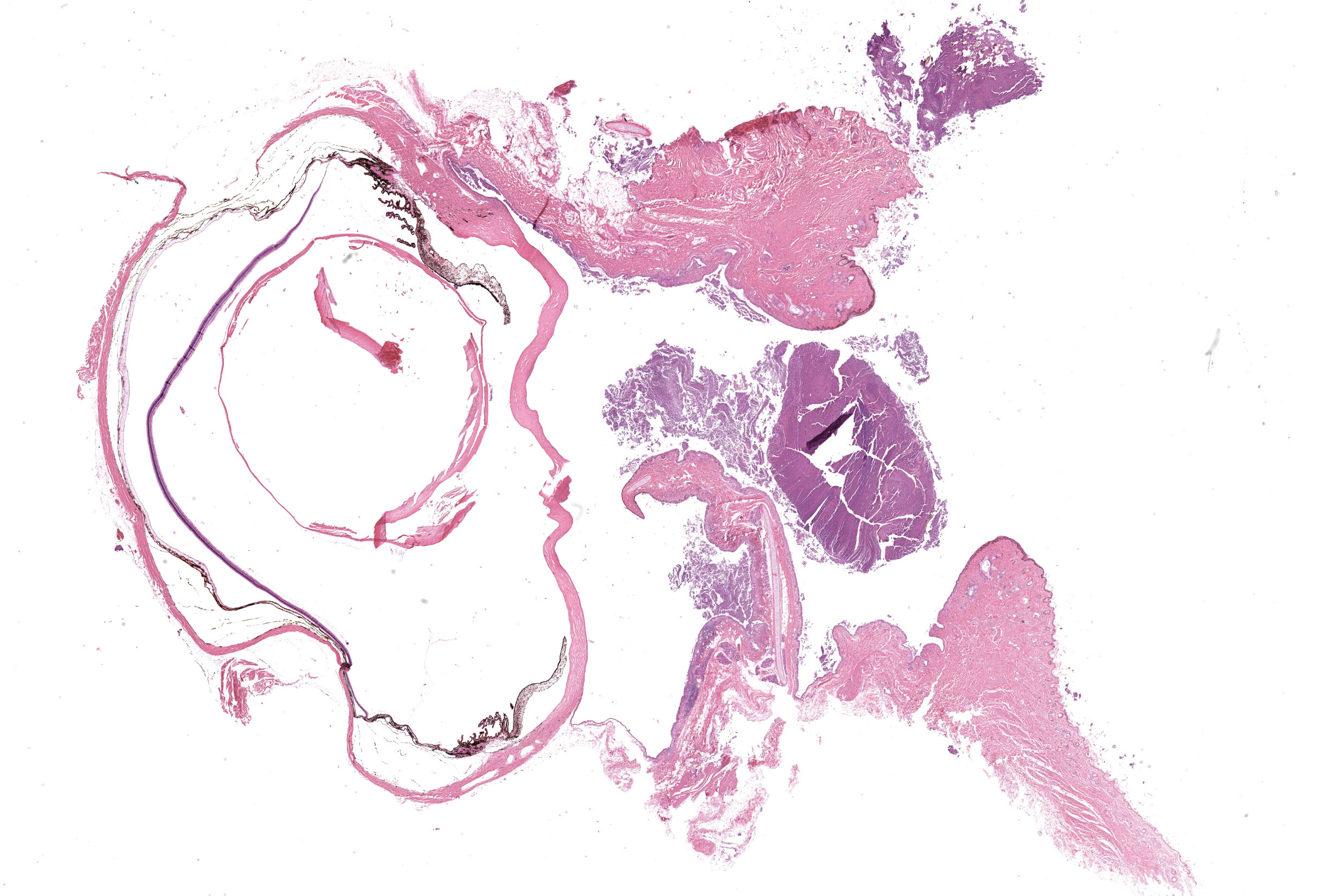
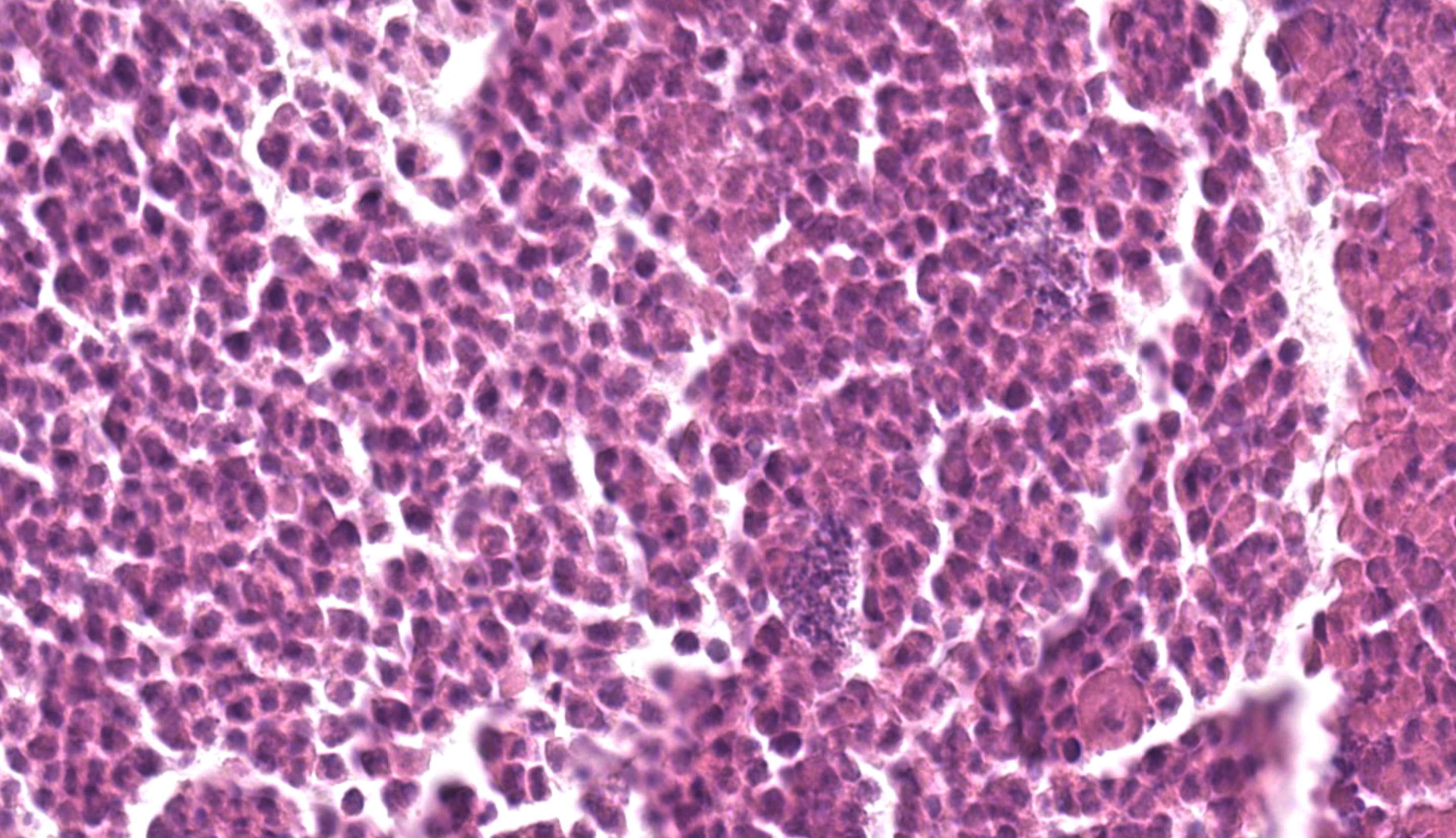
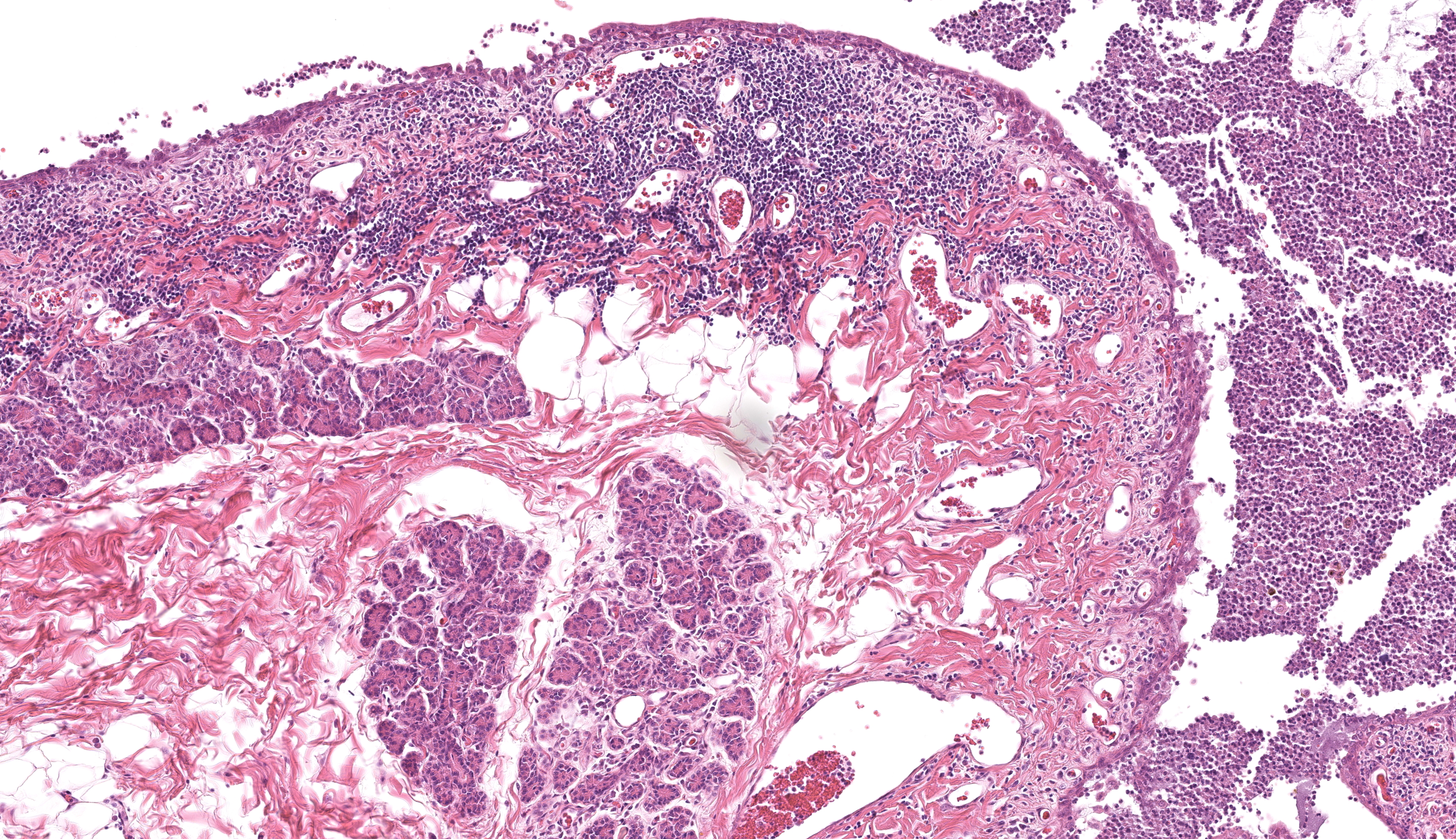
Numerous degenerated and necrotic neutrophils and bacterial colonies are evident in the palpebral fissure. There is a mild infiltration of lymphocytes and neutrophils in the lamina propria of the palpebral and bulbar conjunctiva of the upper eyelid. A moderate to severe lymphoplasmacytic and mild neutrophilic infiltration with mild fibrosis is observed in the lamina propria of the third and lower eyelid. Desquamation of conjunctival epithelial cells is sometimes seen.
Contributor’s Morphologic Diagnosis:
Eye (left): Conjunctivitis, diffuse, chronic, suppurative, severe
Contributor’s Comment:
Immunohistochemistry using anti-influenza virus NP (nucleoprotein) monoclonal antibody (clone 183/5), detected viral antigens in columnar epithelia lining the bulbar and palpebral conjunctiva, most notably of the third eyelid.3 Moreover, viral antigens were detected in the superficial glands under the conjunctiva of the third eyelid.3
This raccoon dog was found emaciated in the same park on the same day that the HPAIV-infected crow was found dead. Raccoon dogs show greater dependency on fruits, plant seeds, and insects, although they also consume animal resources during spring.1 That stated, garden workers did not observe this animal scavenging crow carcasses. Presumably, this raccoon dog was in in close contact with crow carcasses even if it did not ingest them.
In tissues other than conjunctiva, viral antigens were detected only in the tracheal ciliated epithelium and tracheal glands.3 Titers of infectious viruses found in the brain, lung and trachea homogenates were close to the limit of detection,3 which is likely due to clearance of the virus by an immune response. Visual impairment is usually critical for wildlife animals. It was speculated that although the raccoon dog survived the acute phase of HPAIV infection, visual impairment caused by the secondary bacterial infection resulted in malnutrition and dehydration.
Contributing Institution:
Laboratory of Comparative Pathology
Department of Clinical Sciences
Faculty of Veterinary Medicine
Hokkaido University
Kita18, Nishi 9, Kita-ku, Sapporo 060-0818, JAPAN
https://www.vetmed.hokudai.ac.jp/organization/comp-pathol/e/index.html
JPC Diagnosis:
Conjunctiva: Conjunctivitis, neutrophilic and lymphoplasmacytic, chronic-active, diffuse, marked.
JPC Comment:
This second case has an interesting backstory that comes full circle to current events and the ongoing spread of H5N1 HPAI clade 2.3.4.4b viruses worldwide. We previously touched on avian influenza in the context of an experimental case in a pig (Case 3, Conference 14, 2024-2025). Although we discussed the role of viral shedding into milk and zoonotic potential therein, there is still plenty more left to say on this topic.
The slide description for this case is not overly complicated, though conference participants did not immediately offer HPAI as a presumptive cause for the disruption of the conjunctival epithelium and subsequent bacterial infection in this animal. Conjunctivitis is underappreciated as a clinical sign of HPAI, though recent human cases acquired from dairy or poultry workers2,7 have shown that this may be the only overt sign of disease. Similar to the tanuki in this case, workers exposed to HPAI may only be exposed through the conjunctiva because of mask/glove wear or occupational factors such as the proximity of the eyeline of milking parlor workers to the udders of the cows being milked. In comparison to the red fox that scavenged the carcass and developed systemic viral infection3, this animal had evidence of chronic infection and a host adaptive immune response – this may reflect a lower viral load and impact of the route of entry. The role of α2,3 sialic acid receptors on conjunctival epithelium and zoonotic transmission of avian influenza viruses has previously been established and likely played a role in the present case.3,4
Similar to the fox’s outcome in the present case3, domestic cats have fallen ill or died after consumption of raw pet food diets and/or exposure to raw milk contaminated with HPAI.5 Systemic entry of HPAIV has been associated with endothelial cell targeting and vasculitis, a feature which is noticeably absent in this tanuki. Other classic manifestations of HPAI (in cats and beyond) include pancreatic necrosis, encephalitis, myocarditis, and adrenal necrosis.6,8
Finally, the contributor gave us a final puzzle to ponder for this case. As the animal was submitted as an ‘Ezo’ raccoon dog, we explored this aspect further. Accordingly, Ezo is borrowed from the Ainu language (an indigenous people from Northern Japan) and refers to the northern part of Meiji-era Japan, particularly Hokkaido, but also Sakhalin and the Kuril Islands.9 In modern usage, the term has largely disappeared, but is used to refer to several species native to northern Japan such as the Ezo fox (??), Ezo deer ( ????) and Ezo tanuki (?????). Additionally, there is not a consensus on the taxonomy, with Nyctereutes procyonoides albus and Nyctereutes viverrinus both being used to refer to the same species native to Japan. As the contributor of this case is from Hokkaido and refers to the animal as a tanuki,3 we conclude that this is a native Japanese raccoon dog and not a related species that has been widely distributed across Asia and Europe.
References:
- Akihito TS, Teduka M, Kawada S. Long-term Trends in Food Habits of the Raccoon Dog, Nyctereutes viverrinus, in the Imperial Palace, Tokyo. Bull. Natl. Mus. Nat. Sci., Ser. A. 2016;42(3):143–161.
- Drehoff CC, White EB, Frutos AM, et al. Cluster of Influenza A(H5) Cases Associated with Poultry Exposure at Two Facilities - Colorado, July 2024. MMWR Morb Mortal Wkly Rep. 2024 Aug 29;73(34):734-739.
- Hiono T, Kobayashi D, Kobayashi A, et al. Virological, pathological, and glycovirological investigations of an Ezo red fox and a tanuki naturally infected with H5N1 high pathogenicity avian influenza viruses in Hokkaido, Japan. Virology. 2023;578:35-44
- Olofsson S, Kumlin U, Dimock K, Arnberg N. Avian influenza and sialic acid receptors: more than meets the eye? Lancet Infect Dis. 2005 Mar;5(3):184-8.
- Schnirring L. H5N1 confirmed in more cats as probe into raw pet food widens. University of Minnesota Center for Infectious Disease Research and Policy (CIDRAP). 2025 Jan 14; https://www.cidrap.umn.edu/avian-influenza-bird-flu/h5n1-confirmed-more-cats-probe-raw-pet-food-widens.
- Soda K, Tomioka Y, Usui T, et al. Susceptibility of herons (family: Ardeidae) to clade 2.3.2.1 H5N1 subtype high pathogenicity avian influenza virus. Avian Pathol. 2022 Apr;51(2):146-153.
- Uyeki TM, Milton S, Abdul Hamid C, et al. Highly Pathogenic Avian Influenza A(H5N1) Virus Infection in a Dairy Farm Worker. N Engl J Med. 2024 Jun 6;390(21):2028-2029.
- Wünschmann A, Franzen-Klein D, Torchetti M, Confeld M, Carstensen M, Hall V. Lesions and viral antigen distribution in bald eagles, red-tailed hawks, and great horned owls naturally infected with H5N1 clade 2.3.4.4b highly pathogenic avian influenza virus. Vet Pathol. 2024 May;61(3):410-420.
- Tanoshii Japanese. Ezo. https://www.tanoshiijapanese.com/dictionary.