CASE IV:
Signalment:
A 1-year-old female northwest Bornean orangutan (Pongo pygmaeus)
History:
Uneventful clinical history, raised within a family group of orangutans. Without any prior clinical signs, on 22nd May she was found in a semi-comatose state with little muscle tone and carried alternately by different females. She became less responsive and died before veterinary assistance could be provided.
Gross Pathology:
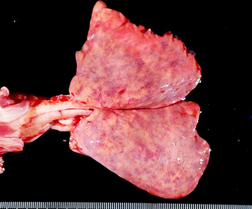
Several subacute to chronic superficial mild wounds on the forehead and face. Within the left hemithorax there is approximately 20 mL of free sero-sanguinolent fluid. Both lungs appear consolidated and present randomly distributed multifocal to coalescing dark red and yellowish patches.
Laboratory Results:
All the previous routine group coprological tests (stool test and cytology) were negative.
Blood drawn shortly post-mortem showed severe, normocytic, normochromic anemia with marked anisocytosis and slight anisochromia.
No significant bacterial or fungal pathogen was isolated from the lung sample.
Microscopic Description:
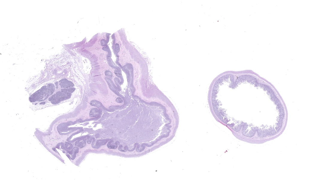
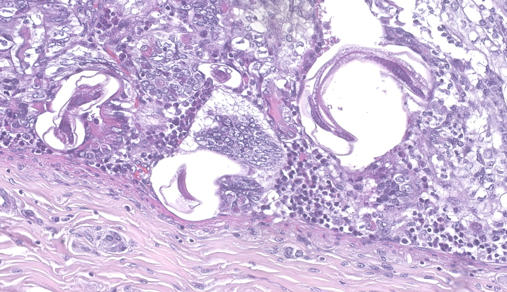
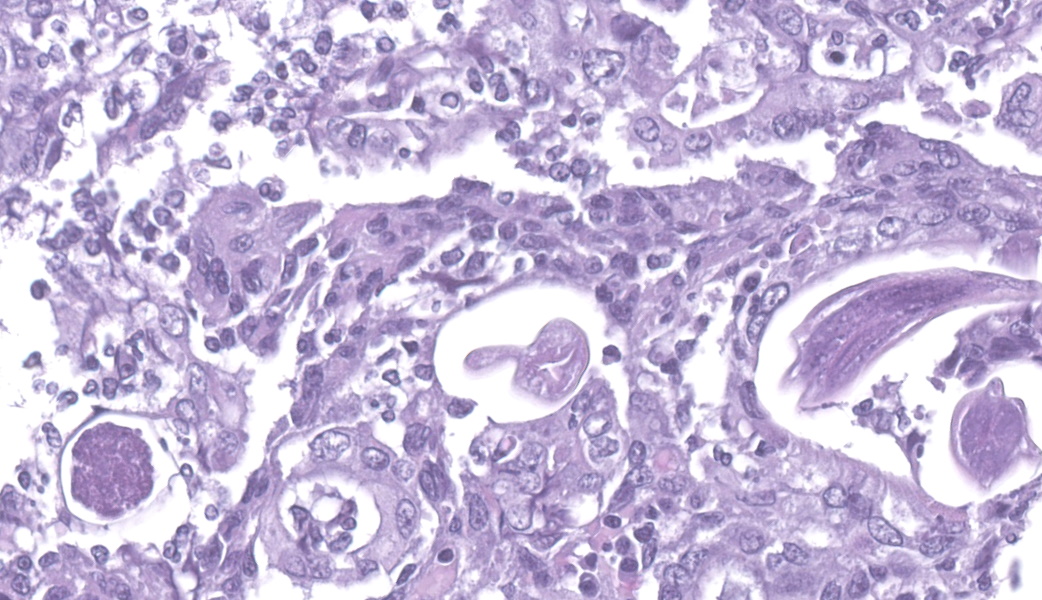
leum (ileocecal valve): effacing 80% of the sample there is a subacute, moderate, inflammatory, and necrotizing process mainly localized in the mucosa of the ileocecal valve. There are multifocal areas of moderate to severe erosions with loss of the mucosa, intraluminal neutrophils, hemorrhages, and bacterial colonies. Intralesional, slender adult nematodes are observed. They are in the lumen of the glands, they have 40-50 µm length, 2-3 µm width, external thin cuticle, hypodermis, pseudocelomen with platymiarian musculature, digestive tract with uninucleate cells and paired genital tract that contain uninucleate eggs. Occasionally ovoid to rounded, eosinophilic eggs 30-50 µm in diameter appear within the epithelium of the mucosa. In the lamina propria macrophages, lymphocytes, plasma cells and low numbers of eosinophils. There is also moderate hyperplasia of intestinal glands.
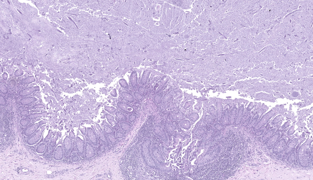
Cecum and regional lymph node: diffusely, the whole section of the cecum shows a diffuse, subacute, moderate inflammatory process that affects mucosa and submucosa. The lumen of the cecum is filled with debris, coccoid bacterial colonies, and high number of nematode larvae (cross and longitudinal sections), that also appear in the lumen of the intestinal glands. Most of the larvae are 200-300 µm in length, 16 ?m width and present a small buccal capsule, rhabditid large esophagus, prominent genital primordium, and straight tail (rhabditiform larvae). There is also another type of larvae, they are larger and thinner, measuring 630 µm and 16 µm (filariform larvae), present in the lumen and invading the mucosa. In the lamina propria, there is a diffuse, moderate inflammation composed of lymphocytes and plasma cells, with multifocal areas of eosinophilic and neutrophilic infiltration. Multifocally, there are numerous filariform larvae in the submucosa with only occasional inflammatory reaction. The GALT is active with slight increased numbers of histiocytes in the germinal centers. Additionally, there is mild dilation of lymphatic vessels, both in the serosa of the intestine and in the lymph node capsule, some of them containing intraluminal bacteria (GRAM positive) and very occasional larvae (cross section). In the lymph node there is mild presence of eosinophils.
Lung (not submitted): all the evaluated section showed a marked widening of the alveolar septa due to hyperplasia and hypertrophy of type II pneumocytes and increased number of mononuclear inflammatory cells in the interstitium. Alveolar lumen was filled with high numbers of foamy macrophages that contain brown intracytoplasmic granules (hemosiderophages) associated with multifocal areas of extravasated erythrocytes (hemorrhage). Multifocally and admixed with the hemorrhage there is moderate numbers of small fibrin thrombi in small blood vessels (DIC). Intraluminal nematode larvae are observed in small blood vessel.
Contributor's Morphologic Diagnoses:
Ileum (ileocecal valve): multifocal to diffuse, subacute, moderate, necrotizing, lymphohystiocytic and eosinophilic ileitis with intralesional eggs and adult female nematodes (Strongyloid spp)
Cecum: multifocal to diffuse, subacute, moderate, necrotizing, lymphohystiocytic and eosinophilic typhlitis with massive strongyloid larvae (rhabditiform, L1 and filariform, L3) infestation.
Lung: severe, generalized fibrino-haemorrhagic and granulomatous pneumonia with intralesional Strongyloid larvae
Contributor's Comment:
The present case was an example of a hyperinfection in a young orangutan by Strongyloides spp. with systemic dissemination. The lesions observed in the lung were severe, and the presence of DIC in alveolar capillaries suggested that the animal suffered septic shock, probably due to bacteria accompanying larval migration. Although the intestinal parasitic load was high, macroscopic lesions were exclusively in the lungs, which is in accordance with the revised bibliography. Environmental factors may have played a role in this case, as the mother showed relaxed maternal behavior, allowing other females to tend to the youngsters, and the group had space restrictions due to construction work. Parasitological examinations of the group, both before and after this incident, have been negative by Baermann?s sedimentation (10+ samplings, tested in-house and at an outside lab).
Strongyloides is a genus of small nematodes belonging to the order Rhabditidia. Other common genera in this group include Rhabdias, Pelodera and Halicephalobus. These worms have platymyarian musculature and an intestine composed of uninucleate cells, and Strongyloides spp. are characterized by paired genitalia.3
Strongyloides nematodes affect millions of people worldwide and have been reported in all great ape species and some Old World primates.5 Of the great apes, orangutans ?5 years old appear to be the most susceptible to severe clinical disease, likely due to their immunologically naive status.5 Infections can be fatal and may represent a major cause of death in captive individuals.11
Most infected animals and humans are asymptomatic shedders, and Strongyloides eggs or larvae are frequently identified in coprological studies of apes in captivity and in the wild.2 Two species can parasitize hominids: Strongyloides fuelleborni (OW monkeys and apes are natural hosts) and S. stercoralis (human natural hosts).7 Strongyloides stercoralis is primarily a human parasite in the Southern Hemisphere, but infections have occasionally also been described in non-human primates, causing severe disease or death.6 S. fuelleborni is found in monkeys and apes in Africa and Asia and comprises two subspecies: S. fuelleborni fuelleborni and S. fuelleborni Kelly. Human infections with this subspecies occur sporadically in Africa.9
Infection occurs by percutaneous penetration or ingestion of third-stage filariform larvae.10 L3 then enter the circulation and travel through the heart to the lungs, where they undergo further development in alveoli. Developing larvae are then coughed up, swallowed, and penetrate the wall of the small intestine, where they develop into parthenogenic adult females that live in the crypts and produce eggs that are shed in the feces (S. fuelleborni) or hatch in the lumen to release larvae that are shed in the feces (S. stercoralis)7. Environmental L1 can develop directly into infectious L3 larvae, or into free-living adult males or females which produce eggs and larvae by sexual reproduction. Likewise, their progeny may either develop into new generations of infectious L3 larvae or noninfectious free-living adults.10
Diagnosis based on coproscopy can be challenging, as it requires Baermann?s sedimentation (funnel technique). In contrast, the flotation technique does not reliably detect the L1. 1 Treatment options for Strongyloides infection include azole drugs and ivermectin.5
Recommended control measures include maintaining exemplary exhibit hygiene and utilization of Baermann or formalin-concentrating fecal screening techniques to identify and treat subclinical infections in all orangutans.5 Crowding, ground dwelling, and bad hygiene, which may occur in populations of captive orangutans, can cause constant reinfection, especially with Strongyloides stercoralis.8 It is therefore important to avoid high densities of animals in the facility if possible. In 2020, the Association of Zoos and Aquariums (ASP) within the "Orangutan Species Survival Plan (SSP)" elaborated a review and recommendations for Strongyloides infection in orangutans, which proposed the prophylactic use of ivermectin in case of clinical suspicion.
Orangutans (Pongo spp.) are threatened in their survival due to habitat loss, hunting, and infections such as Strongyloidiasis, which may represent a severe cause of death in wild and captive individuals.6
Regarding non-primate hosts, S. stercolaris infects cats and dogs, and it has also been reported in arctic foxes.4 Other relevant species are S. papillosus in ruminants, S. westerii in horses, S. ransoomi in swine, and S. felis, S. planiceps, and S. tumefaciens in cats.3
Contributing Institution:
Department of animal health and anatomy,
Autonomous University of Barcelona
Barcelona, Spain
https://www.uab.cat/en/animal-health-anatomy/department
JPC Diagnosis:
Ileocecal junction: Enterotyphlitis, necrotizing, subacute, diffuse, moderate with adult and larval rhabditid nematodes and eggs.
JPC Comment:
The contributor has produced an excellent review of Strongyloides stercoralis in veterinary species, and it parallels the pathogenesis in humans quite closely. One review of 31 isolates of Strongyloides sp. in Bornean orangutans (and one keeper) speciated 30/31 as S. fulleborni, with only one speciated as S. stercoralis.6
Both rhabditiform and filariform larvae may infect the host via ingestion. Still, only filariform larvae may infect the host via skin penetration (Urocanic acid, a histidine metabolite present in high concentration in the skin of mammals, appears to be a potent chemoattractant.)13 Following tissue migration (most commonly in the lungs), filariform larvae become adults in the proximal small intestine, where adult females lay eggs which hatch into rhabditiform larvae. These larvae may be shed in the stool, completing the sexual cycle, or may transform into filariform larvae, which, after exiting and re-entering the gut, may result in reinfection (aka autoinfection). Multiple cycles of autoinfection result in hyperinfection.
Rarely, and most often associated with immunosuppression, Strongyloides stercoralis may leave its favored locations in the body (skin, gut, and lung), resulting in disseminated infection that may be fatal. Because CD4+ TH2 lymphocytes are the primary driver of immunity to S. stercoralis infection, it stands to reason that drugs or diseases that cause immunosuppression (HTLV-1, malnutrition, alcoholism, among others) will promote hyperinfection or disseminated infection.
While Strongyloides sp. in most domestic species involves the proximal small intestinal mucosa, in a few mammalian species, the nematode parasitizes other segments of the GI tract. In macropods, Strongyloides infects the gastric mucosa, particularly in the cardiac region. In cats, a particularly interesting manifestation is seen (WSC 2021-2022, Conference 6, Case 1) in which invasion of the colonic mucosa by adult females results in the formation of proliferative nodules of colonic epithelium within the colonic submucosa. The Strongyloides sp. observed within feline colonic nodules was designated as a separate species, Strongyloides tumefaciens, by the authors who first described it in 1927.13 The species designation was based primarily on the unusual location and lesion, rather than on nematode morphology. Thus, the validity of this species is questionable, and a molecular genetic approach to this parasite's identification would seem warranted.
References:
- Basso W, Grandt L-M, Magnenat A-L, Gottstein B, Campos M. Strongyloides stercoralis infection in imported and local dogs in Switzerland: from clinics to molecular genetics. Parasitol Res. 2019;118:255?66.
- Benirschke K, Adams FD. Gorilla diseases and causes of death. J Reprod Fertil Suppl. 1980;Suppl 28:139-148.
- Gardner CH, Poynton SL. An atlas of metazoan parasites in animal tissues. Washington, DC, Armed Forces Institute of Pathology, American Registry of Pathology; 1999. 2,3,14,16.
- Kapel CMO, Nansen P. Gastrointestinal helminths of arctic foxes (Alopex lagopus) from different bioclimatological regions in Greenland. J Parasitol. 1996;82:17?24.
- Kleinschmidt LM, Kinney ME, Hanley CS. Treatment of disseminated Strongyloides spp. infection in an infant Sumatran orangutan (Pongo abelii). J Med Primatol. 2018 Jun;47(3):201-204.
- Labes EM, Wijanti N, Deplazes P, Mathis A. Genetic characterization of Strongyloides spp. from captive, semi-captive and wild Bornean orangutans (Pongo pygmaeus) in Central and East Kalimantan, Borneo, Indonesia. Parasitology, 2011: 138(11), 1417-1422.
- Linda J. Lowenstine, Rita McManamon, Karen A. Terio, Chapter 15 - Apes, Editor(s): Karen A. Terio, Denise McAloose, Judy St. Leger, Pathology of Wildlife and Zoo Animals, Academic Press,2018, 375-412.
- Mul, Irene & Paembonan, Wardy & Singleton, Ian & Wich, Serge & van Bolhuis, Hester. (2007). Intestinal Parasites of Free-ranging, Semicaptive, and Captive Pongo abelii in Sumatra, Indonesia. Int J Primatol. 28. 407-420.
- Phoo Pwint Ko, Misaki Haraguchi, Takashi Hara, Duong Duc Hieu, Ayaka Ito, Ryusei Tanaka, Mio Tanaka, Takafumi Suzumura, Miya Ueda, Ayako Yoshida, Haruhiko Maruyama, Eiji Nagayasu, Population genetics study of Strongyloides fuelleborni and phylogenetic considerations on primate-infecting species of Strongyloides based on their mitochondrial genome sequences, Parasitology International, Volume 92, 2023
- Thamsborg SM, Ketzis J, Horii Y, Matthews JB. Strongyloides spp. infections of veterinary importance. Parasitology. 2017;144(3):274-284.
- Warren, K. Orang-Utan Conservation - Epidemiological Aspects of Health Management and Population Genetics. Germany: VDM Verlag, 2010.
- Yeh MY, Aggarwal S, Carrig M, Azeem A, Nguyen A, Devries S, Destache C, Nguyen T, Velagapudi M. Strongyloides stercoralis Infection in Humans: A Narrative Review of the Most Neglected Parasitic Disease. Cureus. 2023 Oct 12;15(10):e46908. doi: 10.7759/cureus.46908. PMID: 37954715; PMCID: PMC10639005
- Safer D, Brenes M, Dunipace S, Schad G. Urocanic acid is a major chemoattractant for the skin-penetrating parasitic nematode Strongyloides stercoralis. Proc Natl Acad Sci U S A. 2007 Jan 30;104(5):1627-30.
- Price EW, Dikmans G, et. al. Adenomatous tumors in the large intestine of cats caused by Strongyloides tumefaciens sp. Proc Helminthol Soc Wash 1941; 8.