CASE I:
Signalment:
34-year-old, male gelded, Quarterhorse Paso X, Equus caballus, equine
History:
The horse was used as part of the DVM program teaching herd at Ross University School of Veterinary Medicine and was euthanased due to the presence of large oro-nasal squamous cell carcinoma with metastasis to the submandibular lymph nodes and oesophagus. The tumour was associated with maxillary incisor loss and dysphagia. At autopsy, a clinically silent mass was found in the left thyroid lobe.
Gross Pathology:
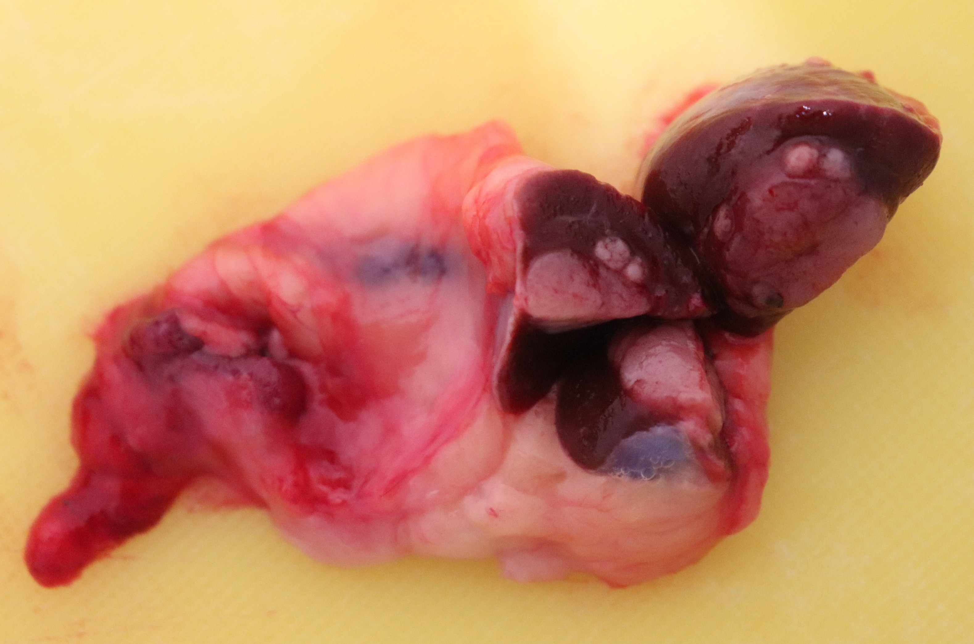
On cut section, the left thyroid lobe contained a well demarcated, pale pink, multinodular, 1.5 - 2 cm diameter mass.
Laboratory Results:
Special Stains
Follicular luminal material (colloid) within and around the thyroid neoplasm were positive for PAS.
Immunohistochemistry
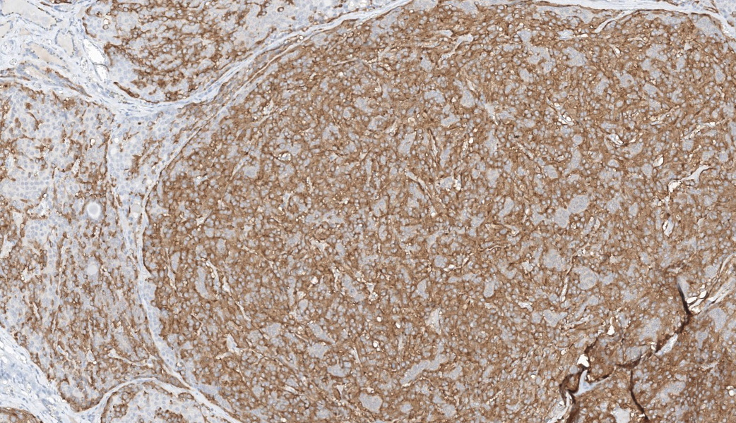
The neoplasm and parafollicular C cells cytoplasm were moderately positive for calcitonin, and faintly positive for chromogranin A, and
negative for thyroglobulin. Follicles entrapped within and around the mass were positive for thyroglobulin.
Microscopic Description:
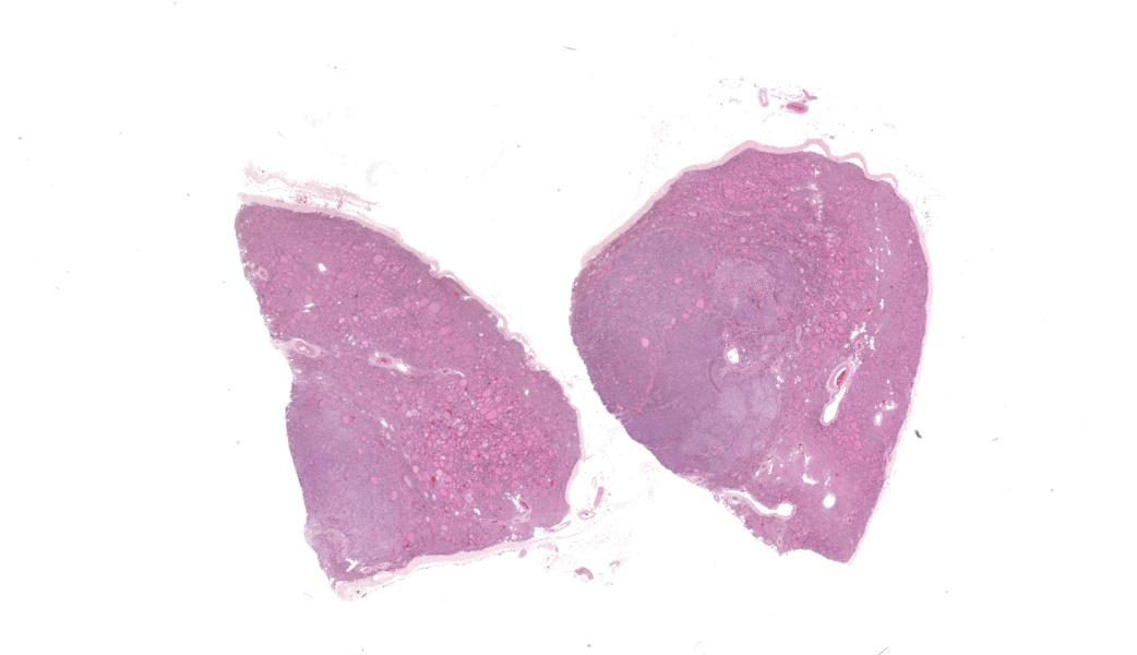
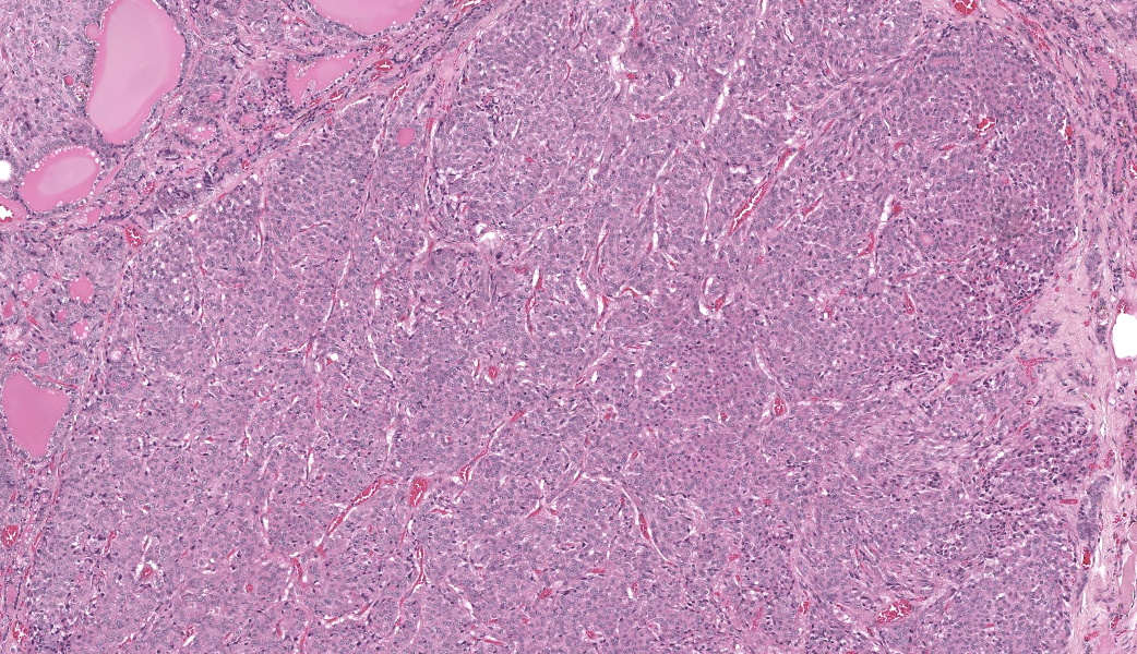
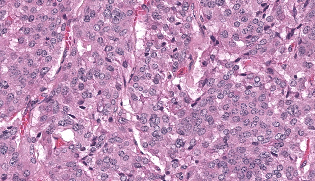
Left thyroid gland: Compressing adjacent thyroid follicles is a multinodular, poorly to moderately demarcated, segmentally infiltrative, unencapsulated mass. It is composed of small sheets and closely packed nests, packets and variably sized follicles (containing homogenous colloid like material) on a fine fibrovascular stroma. The cells are polygonal with indistinct borders and have moderate amounts of finely granulated eosinophilic cytoplasm with a central, round, euchromatic nucleus with 2-3 prominent nucleoli. Anisocytosis is mild and anisokaryosis is moderate with infrequent karyomegaly. There are rare mitoses.
Contributor's Morphologic Diagnoses:
Thyroid C cell carcinoma.
Contributor's Comment:
Primary neoplasms of the thyroid gland have
been histologically classified epithelial, mesenchymal, mixed epithelial-mesenchymal and unclassified. 10 Epithelial tumors can originate from the follicular cells involved in thyroid hormone synthesis or from the calcitonin producing parafollicular C cells (thyroid C cell tumours) although a mixed form is recognised in humans and has been reported in the horse. 5,7 Classically, C cell tumours are described as being more common than thyroid tumours in older horses but non-functional thyroid adenomas and adenocarcinomas were more common in two recent small case series. 8,4
Within the domestic species, thyroid C cell tumours most commonly develop in bulls, dogs and adult to aged horses. 5,3 In horses, they present as unilateral masses and are usually not associated with endocrine disturbances and clinical signs such as dysphagia, oesophageal obstruction (choke), dyspnoea, tracheal compression and stridor 7,4,1 relate to a space occupying lesion in a cranial cervical location.
Surgical excision is the treatment of choice in horses with clinical signs and usually has a good outcome. 4
Grossly, C cell tumours appear as nodular or multinodular masses in one or both thyroid lobes. 5 Smaller masses either represent nodular hyperplasia or adenomas and larger masses with necrosis and haemorrhage are more likely to be malignant with local tissue invasion and potential spread to regional lymph nodes and the lungs, although metastasis is rare in horses with C cell carcinomas. 6,5,1
Histologically, C cell adenomas are larger than a thyroid follicle (in comparison with nodular hyperplasia) and are composed of an unencapsulated expansile mass of polygonal cells with eosinophilic to amphophilic cytoplasm and indistinct neuroendocrine packeting. C cell carcinomas are more pleomorphic with a higher mitotic count and show evidence of local infiltration (as seen in some sections of this case) or lymphovascular invasion. Cytological evaluation of thyroid tumours is also reported to be clinically useful in diagnosing malignant neuroendocrine (and endocrine) tumours. 2
Both carcinomas and adenomas may contain follicular structures that represent entrapped thyroid follicles containing PAS positive colloid, or follicles originating from neoplastic C cells and can therefore mimic a follicular-compact thyroid tumour (as seen in this case). 6 Ambiguous cases may require immunohistochemistry. 6,9,2 C cell tumours are of neural crest origin and should stain positive for calcitonin and chromogranin A and synaptophysin which are markers for neuroendocrine tissue. 5,2 Additionally, C cell tumours should be negative for thyroglobulin, except for any entrapped thyroid follicles and similarly follicles within follicular-compact thyroid tumours are expected to be positive for thyroglobulin. 6
Contributing Institution:
Department of Biomedical Sciences
Ross University School of Veterinary
Medicine, St. Kitts, West Indies
www.veterinary.rossu.edu
JPC Diagnoses:
Thyroid gland: C-cell tumor
JPC Comment:
This week's conference was moderated by JPC alum, Deputy Director of the Biologic Instrumentation Center of the Armed Forces Radiobiology Research Instrumentation (AFRRI) and a member of the Davis-Thompson Foundation's Executive Board and Faculty of Discussants, Dr. Kelsey Fiddes.
It should be noted that in humans, C-cell carcinomas are referred to by the alternate name as medullary thyroid carcinomas; in the older literature, especially in cattle, where this is a fairly common neoplasm, this term will often be encountered.
Whenever a C-cell tumor is mentioned, the prudent pathologist should always consider the possibility of Multiple Endocrine Neoplasia (MEN), as these tumors are a very common constituent of the various syndromes that have been identified in humans and animals.
Multiple endocrine neoplasia is a rare, genetically based condition in humans and animals, which involves the development of concurrent neoplasms in multiple (two or more) endocrine organs. In humans, in which multiple MEN syndromes have been identified, there are four distinct syndromes, all of which are of autosomal dominant inheritance.11
In humans, four syndromes of MEN exist. In MEN1, tumors of the parathyroid gland (95% of affected individuals), pancreatic islets (30-80%) and the anterior pituitary gland (15-50%), and constitutes a total 10% of individuals diagnosed with primary hyperparathyroidism before 30 years of age.11 This syndrome is associated with over a thousand germline inactivating mutations of MEN1, the tumor suppressor gene, which encodes for menin, a protein that regulates transcription and cellular proliferation in endocrine tissues.11
Medullary thyroid carcinoma is commonly associated with MEN2 and MEN3 and is present in 95% of individuals diagnosed with this syndrome.11 Additional tumors in these syndromes include pheochromocytomas (50%) and parathyroid neoplasms (20-30%).11 These syndromes are associated with mutations in the RET (rearranged during transfection) protooncogene, which encodes a membrane-bound tyrosine kinase cell surface receptor.11
MEN4 is the most recently identified (2006) syndrome, with limited numbers of cases being reported. In this syndrome, the tumor palate includes that of MEN1, with additional tumors in the kidneys and reproductive organs. This condition is associated with eight different point mutations in the CDNK1B gene.11
MEN has been reported rarely (accounting for less than 2% of all endocrine tumors) in dogs, cats, cattle, horses, and rats. Isolated cases in a variety of other species have also been reported.12 In animal species, MEN is best described in bulls. The syndrome parallels MEN2/3 in humans, with C-cell tumors, pheochromocytomas, which are often bilateral, and pituitary adenomas.13 Parathyroid tumors have not been reported as part of this syndrome. In most of these cases, genetic mutations have not yet been identified. The high incidence of C-cell tumors in rats has not been associated with mutations in the RET gene.11
The initial report of MEN in horses was published by de Cock et al.13 in 1999. In this report, six individuals (representing 26% of cases in horses with endocrine tumors) possessed both C-cell tumors and pheochromocytomas, similar to MEN2/3 in humans.
The JPC morphologic diagnosis of C-cell tumor is not a typo, but a conscious choice. C-cell tumors, like other neuroendocrine tumors, may be histologically deceptive, as they rarely display obvious signs of malignancy, and this is one of those cases. Criteria of malignancy for these tumors include invasion of the thyroid capsule and extension into surrounding tissue (fixed
tumors), and more obvious signs to include lymphovascular invasion or metastasis to lymph nodes in the region. Unfortunately, none of those are evident in this sample. Like many neuroendocrine tumors, there were karyomegalic cells and some participants felt there was nuclear molding, but those features may be seen in both benign and malignant parafollicular cell neoplasms. Some pathologists might call this C-cell adenoma, but the experience of seeing histologically benign
neoplasms metastasize (especially in the dog), should give a pathologist at least a moment's pause before closing the door on aggressive behavior. For this reason, without the more obvious histologic indicators of malignancy (for which we would happily render a diagnosis of C-cell carcinoma
, we prefer a somewhat less specific diagnosis of C-cell tumor
in this case.
References:
- Elce YA, Ross MW, Davidson EJ, Tulleners EP. Unilateral thyroidectomy in 6 horses. Vet Surg. 2003;32: 187-190.
- Froment R, Allano M, Chiasson A, Dore M, Gara-Boivin C. Pathology in Practice. J Am Vet Med Assoc. 2016;248: 1355-1357.
- Kuwamura M, Shirota A, Yamate J, Kotani T, Ohashi F, Sakuma S. C-cell adenoma containing variously sized thyroid follicles in a horse. J Vet Med Sci. 1998;60: 387-389.
- Marcatili M, Voss SJ, Pollock PJ. Standing thyroidectomy in 10 horses. Vet Surg. 2018;47: 86-92.
- Rosol TJ, Grone A: Endocrine Glands. In: Pathology of domestic animals, ed. Maxie GM, 6 ed. Elsevier, St. Louis, Missouri, 2016
- Rosol TJ, Meuten DJ: Tumors of the Endocrine Glands. In: Tumors in Domestic Animals, ed. Meuten DJ, 5 ed. Wiley & Sons, Ames, Iowa, 2017
- Saulez MN, Viljoen A, Kafka U, Rubio-Martinez L, van Wilpe E, Steyl J. The use of nuclear imaging for a mixed C cell microfollicular carcinoma of the thyroid gland in a mature horse. Equine Veterinary Education. 2013;25: 118-125.
- Troillet A, Bottcher D, Brehm W, Scharner D. Retrospective Evaluation of Hemithyroidectomy in 14 Horses. Vet Surg. 2016;45: 949-954.