Conference 17, Case 4
Signalment:
Four-month-old, male, Oriental magpie robin (Copsychus saularis)
History:
The carcass was submitted from a regional zoo to perform a necropsy of the animal. Carers and veterinarians reported no history of clinical disease. It was found dead inside the house.
Gross Pathology:
The bird was in poor body condition, as assessed by a moderate to severe reduction of the bulk of muscles throughout the body, including both deep and superficial pectoral muscles (muscle atrophy). There were numerous pinpoint white foci in the liver and spleen (moderate, multifocal, acute necrotizing hepatitis and splenitis). Transversal cuts of the left humerus revealed multifocal to coalescing, irregularly shaped, well demarcated, depressed areas filled with a green soft material (severe heterophilic necrotizing osteomyelitis).
Laboratory Results:
The bacteriological results revealed light growths of Yersinia pseudotuberculosis in the lungs and liver by using matrix-assisted laser desorption ionization time-of-flight (MALDI-TOF) mass spectrometry.
Microscopic Description:
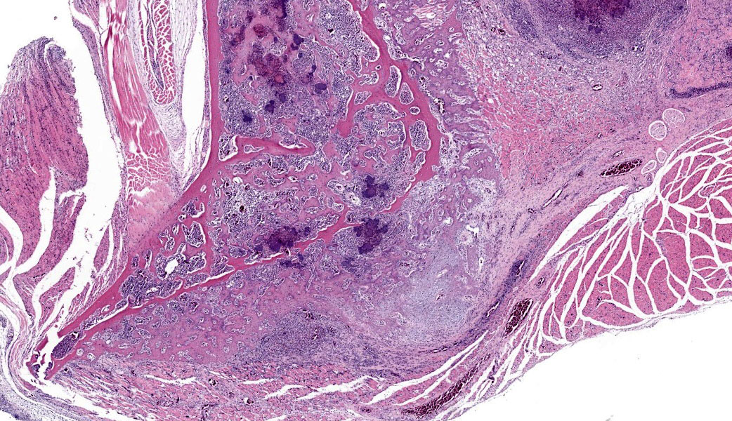
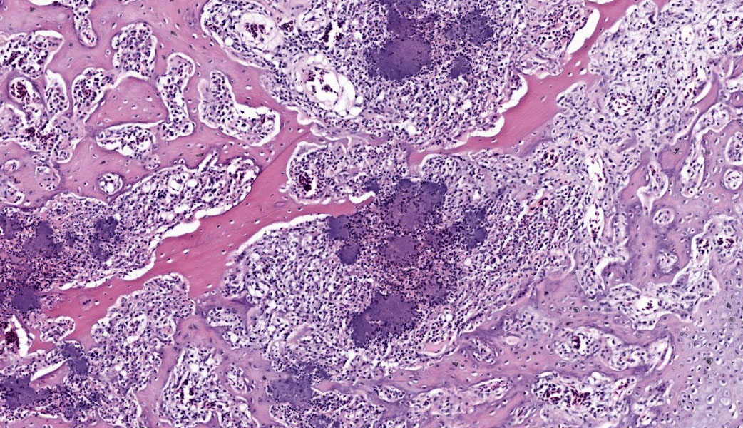
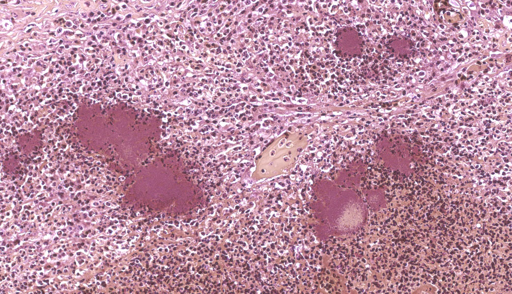
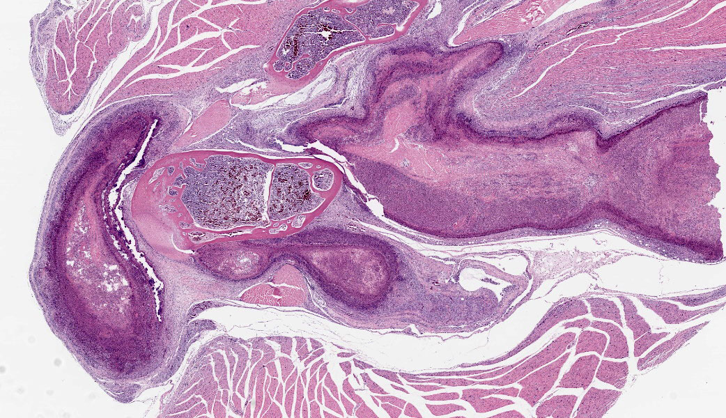
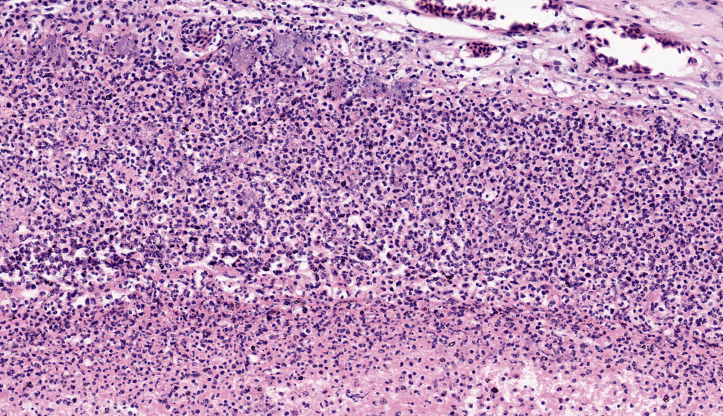
Bones and adjacent soft tissue. Multifocally throughout bone, synovial joint and muscle tissue and affecting approximately 60% of the tissue examined, there are severe, multifocal to coalescing inflammatory infiltrates. The inflammation is chiefly composed of degenerate heterophils, with fewer lymphocytes and plasma cells, embedded in eosinophilic mate-rial showing no cellular outlines (lytic necrosis) with nuclear fragmentation and loss (karyorrhexis and karyolysis); embedded within the necro-inflammatory process are common , up 100 micrometers in diameter, coccobacillary bacterial colonies; in some foci, the necro-inflammatory foci are bordered by macrophages forming multinucleated giant cells containing a 15-50 nuclei with a horse-shoe arrangement with occasional foamy cytoplasm. The inflamed bones show reduction of medullary bony trabeculae (osteolysis), with multifocal scalloping and thinning of the remaining ones with an increased basophilic hue while other show empty lacunae and diffuse eosinophilia (osteonecrosis); the periosteum instead shows a common exophytic proliferation of the cortical bone with cartilage and woven bone formation (periosteal reaction).
The inflammatory infiltrate is also observed infiltrating and filling multifocal synovial joint lumina and capsules (arthrosynovitis), with a layer of fibrin embedding heterophils and covering multifocally the articular carti-age, which shows increase eosinophilia (cartilage degeneration).
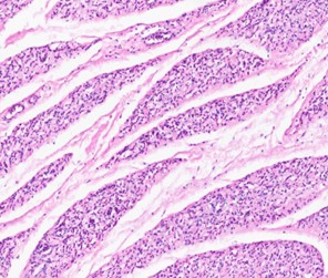
The adjacent rhabdomyocytes exhibit multifocal reduction in size and diameter, with angu-lar edges (muscular atrophy) with the closest one to the inflammation exhibiting an amphophilic sarcoplasm and linear arranged central nuclei (myotubes) and rare areas of endomysial fibrosis.
Contributor’s Morphologic Diagnoses:
- Bone: multifocal to coalescing, severe, subacute, heterophilic and necrotizing osteomyelitis, with osteolysis, perio-teal reaction, osteonecrosis and with intralesional bacterial colonies.
- Synovial joints: diffuse, severe, sub-acute, fibrinous and heterophilic ar-thro-synovitis with intralesional bacte-rial colonies.
- Skeletal muscle: Severe, multifocal, subacute, heterophilic and necrotizing myositis with intralesional bacterial
Contributor’s Comment:
Yersiniosis has been frequently reported in more than a hundred different species world-wide, including avian, mammals and reptiles, and it may act as a zoonotic pathogen.3 Yersiniosis can be caused by Yersinia pseudotuberculosis and Y.enterocolitica, although the latter is more frequently involved in humans and primates cases. Y. pseudotuberculosis can be commonly isolated from wild animals and birds, but there are very few reports of cases of avian osteomyelitis sustained by Y. pseudotuberculosis.4 A recent outbreak in a slaughter turkey flock was reported in Finland, while other older outbreaks were documented in the UK and California, still in turkeys.1,6,8 In mammals, a Y. pseudotuberculosis osteomyelitis case was reported in a ring-tailed lemur in UK.7
We decided to submit this case due to the paucity of Y. pseudotuberculosis induced osteo-myelitis in birds reported in literature. Such case is, to the best of our knowledge, the first pathological description of a Y. pseudotuberculosis induced osteomyelitis in an Oriental Magpie Robin. The present case was characterized by extensive inflammation with large bacterial colonies also spotted in other organs (spleen, liver, lung and large intestine), and therefore suggesting a systemic infection.
Yersinia pseudotuberculosis is a ubiquitous gram-negative anaerobic coccobacillus bacterium from the Enterobacter family. The most common route of infection is oro-fecal thought contaminated food or water, although the epidemiology of the disease is considered multifactorial and highly complex. Therefore, the severity of the clinical signs and lesions would be highly variable and dependent on numerous environmental (e.g. management, temperature, etc.) and host (e.g. age, stress, parasitism and / or concomitant infections, etc.), conditions. No predisposing conditions to such infection were detected here.
Although the bacteriological culture isolated Yersinia pseudotuberculosis, other differential diagnosis for large colonies of bacteria would be: Actinomyces sp., Actinobacillus sp. Arcanobacter sp., Corynebacterium sp., Staphylococcus sp. or Streptococcus sp. (YACS acronym).
Contributing Institution:
University of Liverpool, Institute of Infection, Veterinary and Ecological sciences, Department of Veterinary anatomy, Physiology and Pathology.
JPC Diagnoses:
- Wing: Osteomyelitis, tenosynovitis, myositis, and cellulitis, necrotizing and heterophilic, chronic, multifocal, severe, with pannus and large colonies of bacteria.
- Skeletal muscle, adipose tissue: Atrophy, chronic, diffuse, severe.
JPC Comment:
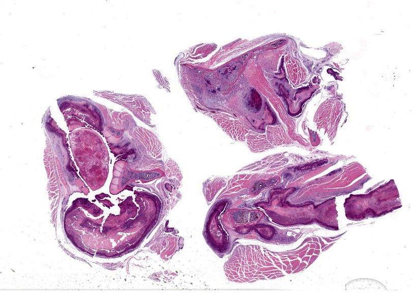
The avian skeletal system has several unique functional adaptations. Birds have three types of bone: pneumatic, hematopoietic, and medullary.5 Pneumatic bones have air sacs in the endosteum in place of bone marrow. The number and distribution of pneumatic bones varies by species, but most birds have pneumatic hu-meri, femurs, sternums, and skulls.5 The remainder of the bones are hematopoietic and have bone marrow. Medullary bone is present in female birds prior to laying eggs when they deposit calcium on the endosteum, with de-posits filling the marrow cavity of long bones.5 Additionally, many bones are fused(e.g., tibiotarsus, synsacrum, etc.). The slide in this case had a pneumatic bone that articulated with two hematopoietic long bones sur-rounded by abundant skeletal muscle; this consistent with wing where the humerus (pneumatic bone) articulates with the radius and ulna (hematopoietic bones). The leg (at the articular of the femur and tibiotarsus) was also considered, but the amount of skeletal muscle was deemed to be excessive for this lo-cation. Foci of inflammation and necrosis were present in both pneumatic and hemato-poietic bone, suggestive of systemic bacterial sepsis.
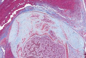
Many classic features of bone and joint inflammation and healing were demonstrated in this case, including osteonecrosis and periosteal new bone formation (characterized by lattices of woven bone oriented perpendicularly to the bone’s long axis). In addition to the excellent description provided by the contributor, conference participants also noted de-creased basophilia of the articular cartilage. This is a common finding of arthritis and correlates to loss of proteoglycans in the chondroid matrix.2 Additionally, one of the joints was overlain by a thin layer of fibrovascular tissue, consistent with pannus. Pannus is caused by proliferation of stromal cells in chronic arthritis and was nicely highlighted with a Masson’s trichrome stain.2
Conference participants also discussed the cause of the severe skeletal muscle atrophy in this case. Skeletal muscle atrophy can be caused by disuse atrophy, which was presumably present in this animal. However, negative energy balance can also lead to primary skeletal muscle atrophy due to birds’ high metabolic demands. Examination of body condition score in birds often involves palpation of the breast muscle, as muscling has direct correlation to overall nutritional status. This animal had severe adipose atrophy as well, indicative of negative energy balance, which likely also contributed to muscular atrophy.
References:
- Blomvall L, et al. Osteomyelitis in a slaughter turkey flock caused by Yersinia pseudotuberculosis sequence type ST42. Veterinary Microbiology. 2022;269:109424.
- Craig LE, Dittmer KE, Thompson KG. Bones and In: Maxie MG, ed. Jubb, Kennedy & Palmer’s Pathology of Do-mestic Animals. Vol 1. 7th ed. Saunders Ltd; 2026:16-163.
- Fredriksson-Ahomaa, M, Joutsen S, Lauk-kanen-Ninios Identification of Yersinia at the species and subspecies levels is chal-lenging. Curr Clin Microbiol. 2018;5:135–142.
- Le Guern AS, Martin L, Savin C, Carniel
- Yersiniosis in France: overview and potential sources of infection. Int J Infect Dis. 2016;46:1-7.
- Smith Anatomy and postmortem examination. In: Schmidt RE, Struthers JD, Phalen DN, eds. Pathology of Pet and Aviary Birds. 3rd ed. John Wiley & Sons; 2024:1-18.
- Wallner-Pendleton E, Cooper, G. Several outbreaks of Yersinia pseudotuberculosis in California turkey flocks. Avian Dis. 1983;27:524–526.
- Walker D, Gibbons J, Harris JD, et Systemic Yersinia pseudotuberculosis as a cause of osteomyelitis in a captive ring-tailed lemur (Lemur catta). J Comp Pathol. 2018;164:27-31.
- Wise DR, Uppal Osteomyelitis in turkeys caused by Yersinia pseudotuberculosis. J Med Microbiol.1972;5:128–130.