CASE II:
Signalment:
12 year old female Cynomolgus macaque (Macaca fascicularis)
History:
This 12-year old female cynomolgus macaque had a caudal abdominal mass discovered on annual physical examination; endometriosis was suspected. The NHP (nonhuman primate) was treated on 17 July 20 with medroxyprogesterone acetate. The mass reduced in size but did not resolve and the clinicians elected to euthanize the animal.
Gross Pathology:
Within the abdomen there were many adhesions between the urinary bladder, uterus, ovaries and descending colon, and red serosal nodules on the surface of the urinary bladder. Additionally, there were also adhesions between the omentum, mesentery of the large intestine and the uterus/mesometrium. There were multiple thin walled, brown cysts that ranged in size from 3-6 mm on and around the adhesed uterine serosa and nodular thickening in the wall and serosal surface of the uterus. Both ovaries appeared enlarged, up to 2.5 cm, including the presence of thin wall cysts that contained brown gelatinous debris.
Microscopic Description:
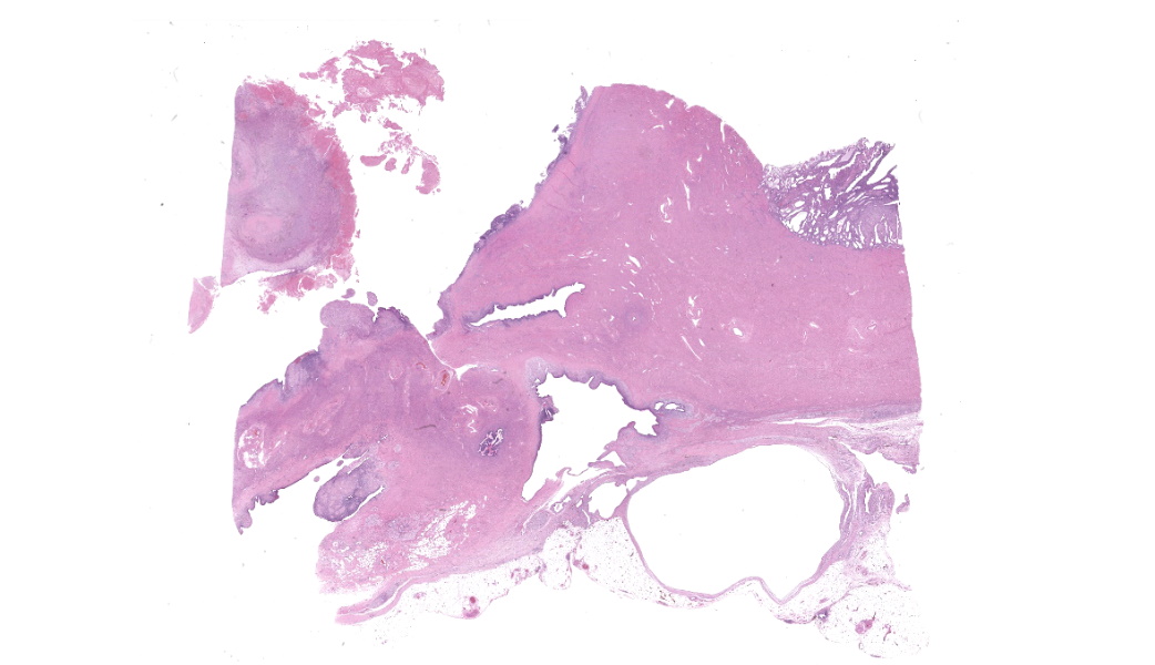
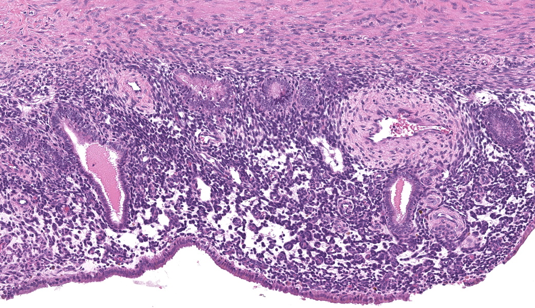
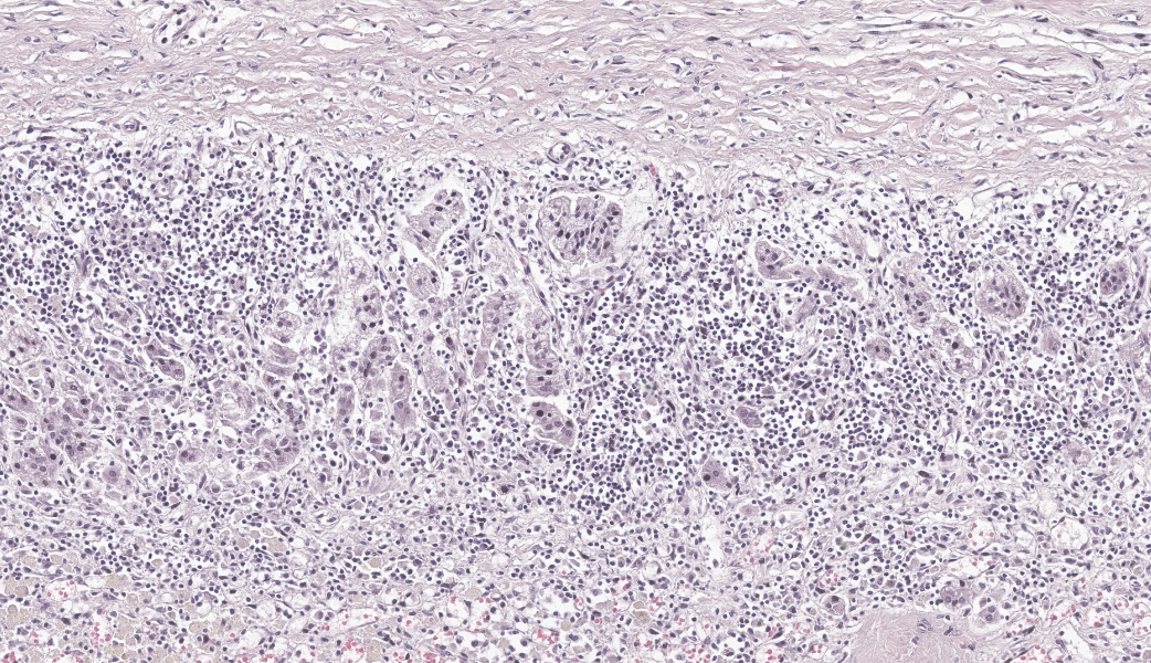
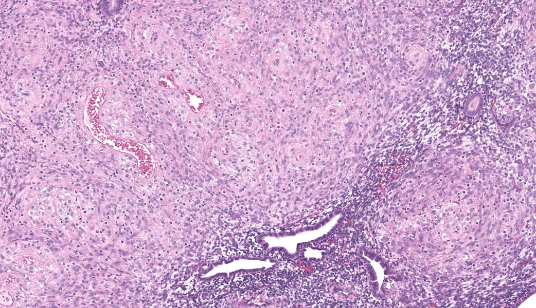
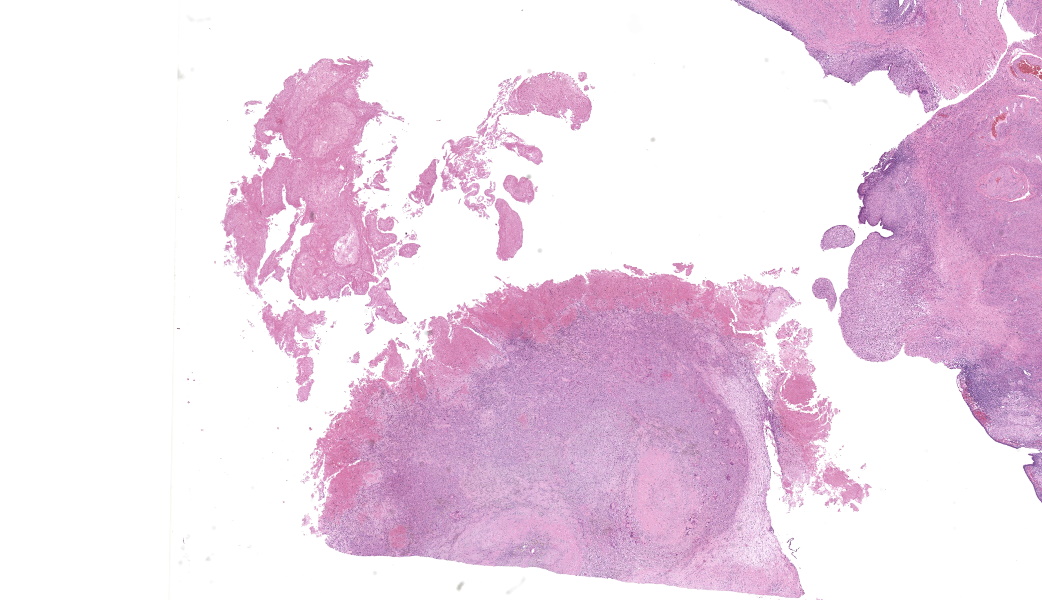
Uterus and mesometrium: Expanding and disrupting the serosal surface of the uterus and adjacent mesometrium is a vigorous endometrial lining with a variably thick underlying stroma, occasionally forming tortuous uterine glands, and large 2-6 mm fibrovascular polypoid nodules that blend inconspicuously with the incongruous uterine stroma. The fibrovascular nodules contain dense whorls of convoluted, hyperplastic and occasionally vacuolated spindle cells, variable amounts of dense sclerotic collagen and low to moderate numbers of macrophages and multinucleate giant cells which often contain either brown pigment (hemosiderin), pale eosinophilic to amphophilic material (secretory product) or clear vacuoles (lipid). There is a moderate amount of a coagulum containing necrotic debris, eosinophilic secretory product and hemorrhage ("chocolate cyst" contents) adhering to the outer surface of said nodules. There are small amounts of hemorrhage and low numbers of lymphocytes underlying and/or sporadically within the disarranged endometrial epithelium. There is a 7 mm empty, thin walled cyst on the surface of the uterine serosa lined by a flattened, attenuated epithelium and surrounded by fibrous tissue containing low numbers of the aforementioned macrophages and multinucleated giant cells. Rarely within the myometrium there are uterine glands surrounded by low numbers of lymphocytes. The endometrium is mildly hyperplastic containing enlarged, tortuous uterine glands on a thickened stroma.
Contributor's Morphologic Diagnoses:
Uterus and mesometrium:
- Endometriosis, cynomolgus macaque (Macaca fascicularis), nonhuman primate.
- Uterus, serosa: Cyst, focal.
- Uterus, myometrium: Adenomyosis, multifocal, minimal.
Contributor's Comment:
Endometriosis is the presence of "ectopic" endometrial tissue at a site outside of the uterus and it is the most common reproductive disorder in menstruating Old World primates. The prevalence of endometriosis increases with increasing age in rhesus macaques and there is evidence that endometriosis may have a genetic basis in humans and rhesus macaques.8 Besides age and familial association, other risk factors include previous hysterotomy and long term estrogen treatment. Dioxin exposure has also been implicated in the development of endometriosis and exposure to dioxin has been shown to facilitate the growth of endometrial explants.3
While the precise etiology of endometriosis is unclear, one of the most widely accepted theories includes retrograde menstruation of endometrial material into the peritoneal cavity.5 A second theory includes the development of endometriosis from metaplasia of cells lining serosal surfaces, including the abdominal and thoracic structures. Finally, a third theory referred to as the induction theory proposes retrograde menstruation produces substances that induce metaplasia of peritoneal serosa.1
Clinically, presenting signs vary depending on the location of the endometriosis tissue implant. The disease may often be clinically silent and diagnosed as an incidental finding at necropsy. In symptomatic cases, animals may exhibit apparent abdominal discomfort or prostration. Other clinical signs that have been reported include: cyclical anorexia, depression, irregular menses and possibly constipation lasting several days. On physical examination, abdominal masses may be palpated.3
While the precise etiology of endometriosis is unclear, one of the most widely accepted theories includes retrograde menstruation of endometrial material into the peritoneal cavity.5 A second theory includes the development of endometriosis from metaplasia of cells lining serosal surfaces, including the abdominal and thoracic structures. Finally, a third theory referred to as the induction theory proposes retrograde menstruation produces substances that induce metaplasia of peritoneal serosa.1
Clinically, presenting signs vary depending on the location of the endometriosis tissue implant. The disease may often be clinically silent and diagnosed as an incidental finding at necropsy. In symptomatic cases, animals may exhibit apparent abdominal discomfort or prostration. Other clinical signs that have been reported include: cyclical anorexia, depression, irregular menses and possibly constipation lasting several days. On physical examination, abdominal masses may be palpated.3
The condition can be quite heterogeneous, varying in degree and level of invasiveness between patients. While it is most commonly found in the pelvic region, it may be located anywhere throughout the abdomen and rarely in extra-abdominal locations such as the thorasic cavity.1 At necropsy, abdominal distention may be evident with the presence of endometrial explants and discrete cysts. Secondary lesions of the urinary tract such as hydroureter or hydronephrosis may occur due to fibrosis and adhesions.6 In this case, the endometriosis appeared to be quite invasive with the formation of exophytic masses as well as development of invasive islands of endometrial stroma and glands and the formation of numerous cysts and reactive fibrosis. This case also had endometrial glands within the myometrium (adenomyosis) which is also considered to be part of endometriosis.4 Decidualization of foci of endometriosis in response to endogenous or exogenous progestins has also been reported in multiple nonhuman primate species but was not observed in this case.2
Spontaneous endometriosis in non-human primates has been studied as an animal model for the disease in women. Both cynomolgus macaques and baboons have similar menstrual cycles as women and develop endometriosis spontaneously. Laparoscopic or open surgery has been used to diagnose and monitor the condition in non-human primates. Levels of CA125 had also been measured in cynomolgus macaques with endometriosis and elevated levels were found to correlate with the presence of chocolate cysts.7
Contributing Institution:
USAMRIIDAttn: Pathology Division1425 Porter Street, Fort Detrick, MD 21702 https://www.usamriid.army.mil/
JPC Diagnoses:
- Uterus, mesometrium: Endometriosis with multifocal decidualization.
- Uterus: Endometrial hyperplasia, chronic, diffuse, moderate with adenomyosis and multifocal decidualization.
JPC Comment:
The contributor has reviewed the basics of endometriosis in humans and non-human primates, but because maintaining non-human primates for laboratory research and drug development is difficult, various smaller animal models, particularly rodents, have received significant focus in the last decade. Researchers have developed a variety of rodent (primarily mouse) models. The intraperitoneal injection, laparoscopic implantation, or peritoneal suturing of uterine endometrial tissue
from donor mice creates autologous (homologous) mouse models. Heterologous models are created by injecting human endometrial tissue into the peritoneal cavity of immune-deficient mice. Other mouse models include transgenic and knockout mice, which allow determination of the effects of particular hormones or cytokines on disease progression, and mouse models expressing green fluorescent proteins that help monitor disease progression in immune-deficient mice in a less invasive manner.9,10
There are also numerous advances in the etiopathogenesis of the human disease - too many to recount here; one interesting development is the discovery of the roles of microRNAs (miRNAs) in the pathogenesis of endometriosis.11 MicroRNAs are small noncoding RNA fragments consisting of 20 or 20 base pairs that do not produce protein, but regulate gene expression. Researchers have identified these miRNAs in both human patients and baboons with endometriosis. Upregulated MiR-29c has been shown to increase progesterone resistance, contributing to endometriosis-associated infertility. MiR-451 has been shown to downregulate apoptosis and enhance proliferation and invasion of endometriotic lesions. MiR-210 has been shown to enhance cell proliferation and migration in ectopic endometriosis lesions, and MiR-15a-5p, when suppressed, is involved in angiogenesis. This is only one facet of the fascinating research into endometriosis today.
While we agree with the contributor's diagnosis in this case, we found areas of decidualization in the submitted sections. (It is not unusual that sections that the contributor evaluated are not necessarily those submitted for a conference.) Atkins et al. (2016) documented decidualization of endometrial lesions.2 In the normal animal, decidualization of endometrial stromal cells is a normal progesterone-driven phenomenon that transforms endometrial stromal cells in order to support early embryonic development and ultimately help to maintain pregnancy. In primates during pregnancy, this tissue is also commonly found in ectopic locations, primarily on the exterior of the ovary and uterine tube, but may explant to other, predominantly intra-abdominal, locations.2
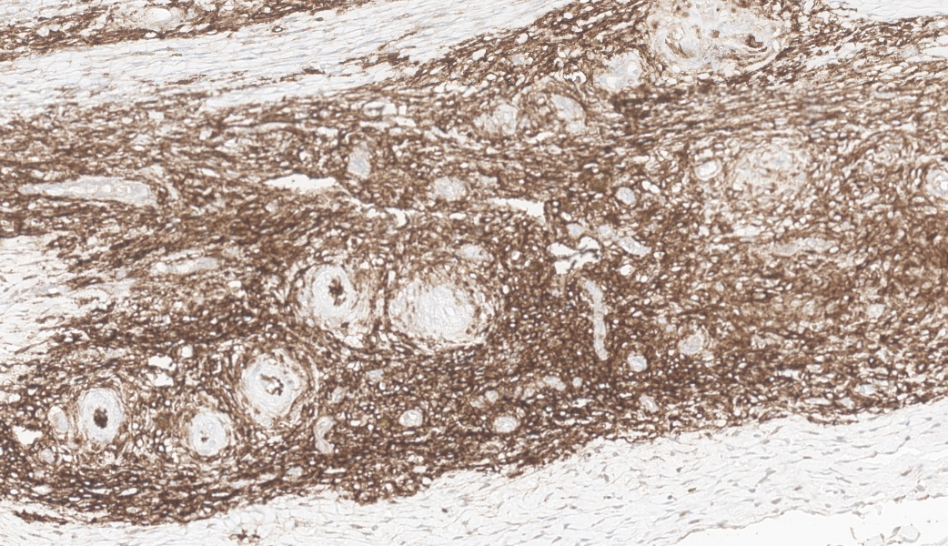
A similar process will occur in primates with endometriosis. In primates with endometriosis, endometrial stromal cells surrounding some or all of the endometrial explants demonstrate a characteristic morphologic change with marked increase in lightly eosinophilic cytoplasm. These foci may be infiltrated by endometrial granular leukocytes (a form of gamma/delta T-cells) which possess numerous cytoplasmic red granules (seen in low numbers in decidualized foci in this slide).2 In this case, decidualized tissue is also strongly positive for CD10 and vimentin.2 (Both of these immunomarkers stain both normal and neoplastic endometrial stroma, so they are not specific for the decidualization process, but merely identify these cells as endometrial stroma.
References:
- Assaf BT and Miller AD. Pleural endometriosis in an aged rhesus macaque (Macaca mulatta): a histopathologic and immunohistochemical study. Vet Pathol. 2011;49(4):636-641.
- Atkins HM, Lombardini ED, Caudell DL, Appt SE, et al. Decidualization of endometriosis in macaques. Vet Pathol. 2016; 53(6):1252-1258.
- Cline JM, Brignolo L, Ford EW. Urogenital System. In: Abee CR, et al. ed. Nonhuman Primates in Biomedical Research:Diseases, 2nd Vol. 2. Elsevier; 2012: 510-514.
- Foster RA. Female Reproductive System and Mammae. In: Zachary JF, ed. Pathologic Basis of Veterinary Disease, 6th Elsevier; 2017: 1167.
- Gruber-Dujardin E, Bleyer M, Matz-Rensing K. Morphological and immunohistochemical characterization of spontaneous endometriosis in rhesus macaques (Macaca mulatta). Primate Biol. 2017; 4:77-91.
- Kirejczyk S, Pinelli C, Gonzalez O, Kumar S, et al. Urogenital lesions in nonhuman primates at 2 national primate research centers. Vet Pathol. 2021; 58(1):147-160.
- Nishimoto-Kakiuchi A, Netsu S, Okabayashi S, Taniguchi K, et al. Spontaneous endometriosis in cynomolgus monkeys as a clinically relevant experimental model. Human Repro. 2018; 33(7):1228-1236.
- Simmons HA. Age-associated pathology in rhesus macaques (Macaca mulatta). Vet Pathol. 2016; 53(2):399-416.