Wednesday Slide Conference, Conference 15, Case 4
Signalment:
16 weeks, male, domestic white turkey (Meleagris gallopavo).
History:
A flock of 7,500 domestic white male turkeys was experiencing elevated mortality as a result of both aortic rupture and culling of lame, recumbent birds. Recumbent birds hadswollen intertarsal (hock) joints with bruising of the nonfeathered skin of the hock. Five affected legs were removed at the coxofemoral joint from culled carcasses and shipped to the Minnesota Veterinary Diagnostic Laboratory.
Gross Pathology:
The submitted turkey legs had skin bruising of the caudal aspect of the hock. The hocks were swollen as a result of periarticular subcutaneous edema (serosanguinous fluid) and increased volume of synovial fluid within the hock joints and sheaths of the gastrocnemius tendon and digital flexor sheaths. In two legs, the gastrocnemius tendon was partially ruptured proximal to the hock joint. Longitudinal sections of the femur, tibiotarsus and tarsometatarsus showed no overt lesions (no evidence of osteomyelitis or chondrodysplasia) in bone or growth plate.
Laboratory Results:
-Aerobic culture: Hock joint fluid- no significant growth
-Molecular diagnostics: Gastrocnemius tendon was positive (CT 32.5) for avian reovirus by universal avian reovirus PCR
Synovial fluid was negative for Mycoplasma gallisepticum and Mycoplasma synoviae by PCR
-Virology: Reovirus was isolated after one pass on embryonated eggs (yolk sac inoculation) and after two passes on QT-35 (quail fibroblast) cell line
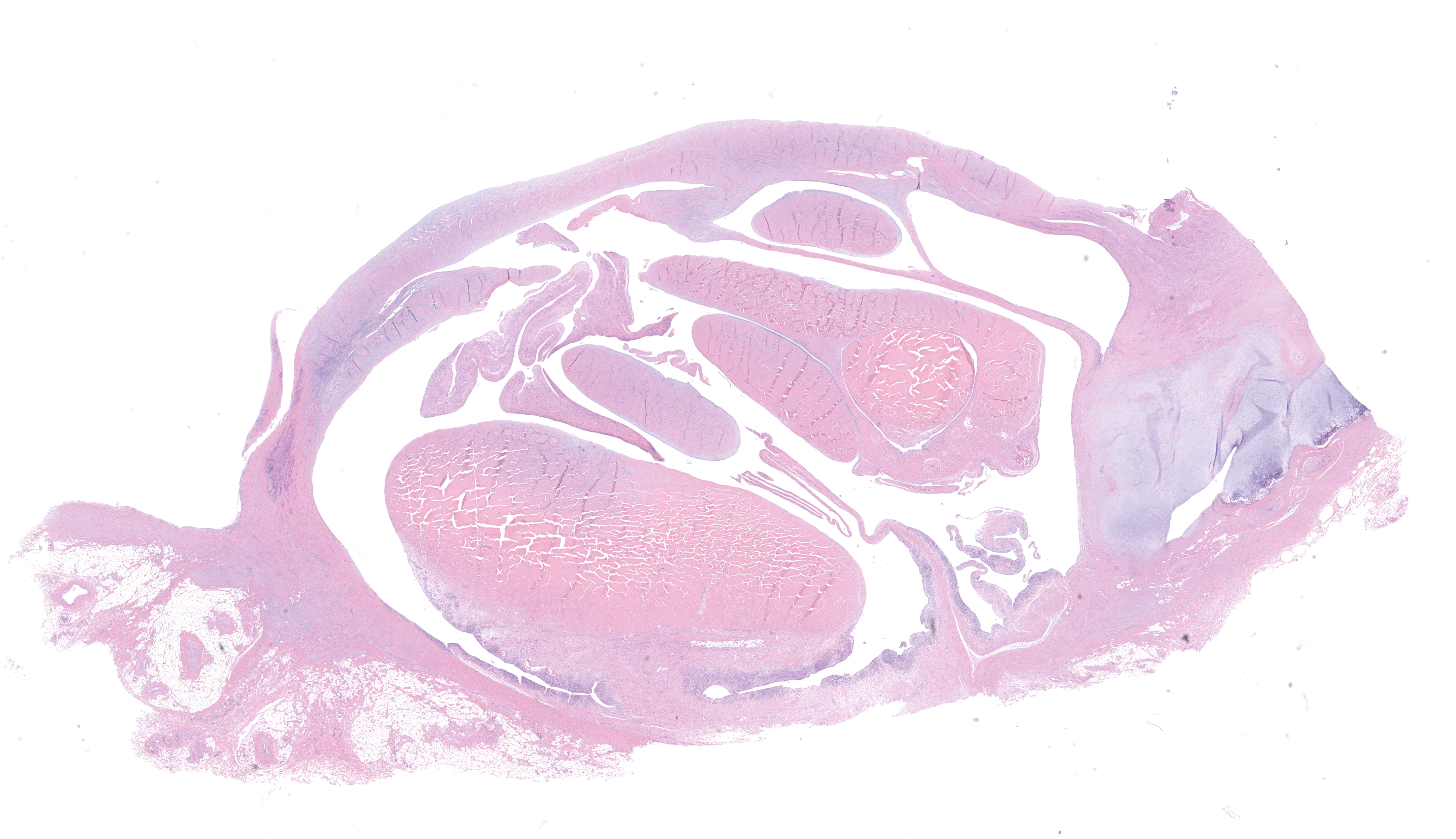
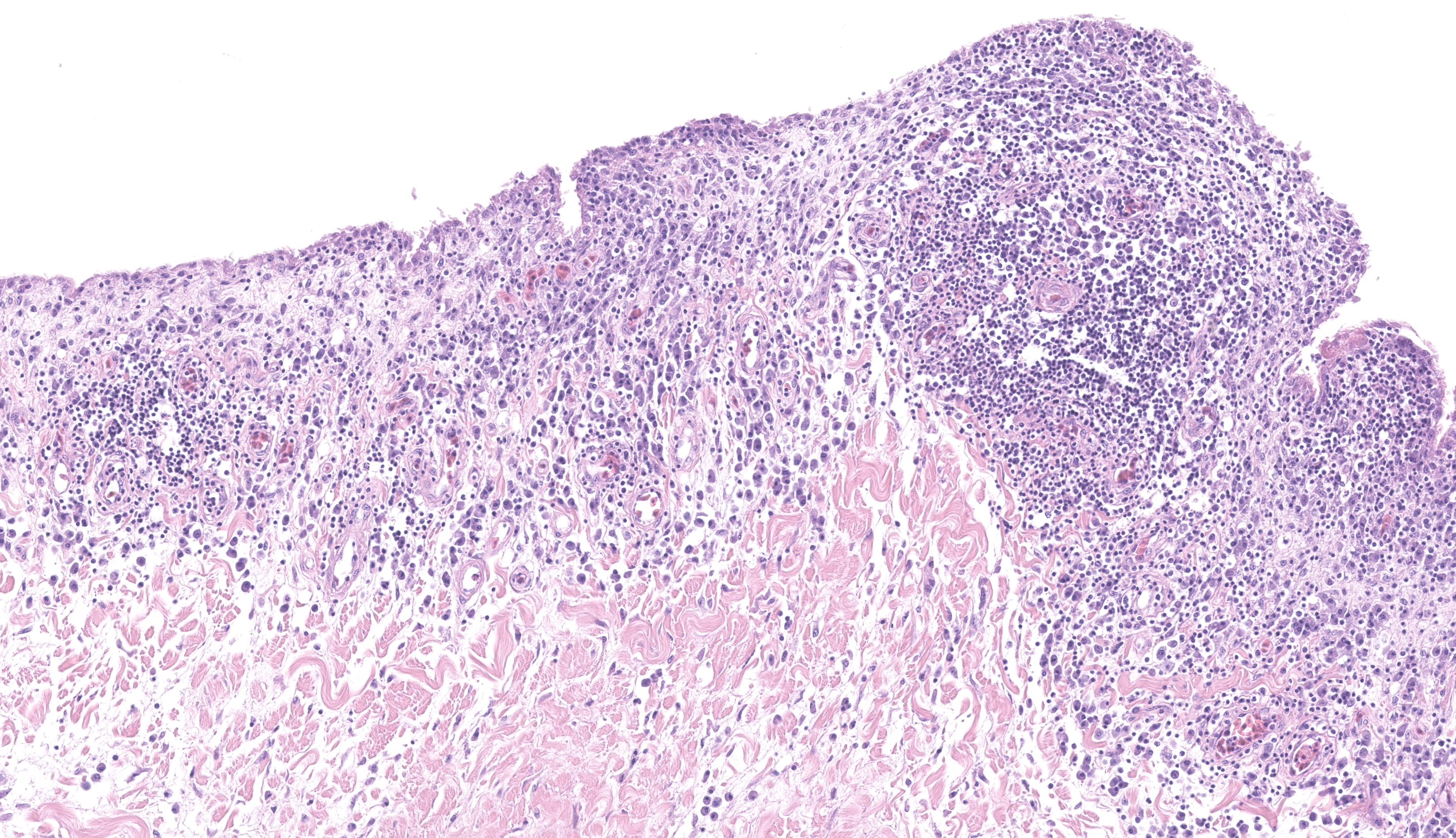
Microscopic Description:
The tissue is a cross-section of gastrocnemius tendon complex, composed of the tendon and sheaths of the primary gastrocnemius tendon and multiple smaller digital flexor tendons and sheaths. Inflammation largely affects the tendon sheaths and consists of multifocal, moderate to marked infiltrates of lymphocytes and plasma cells either scattered or arranged in perivascular fashion within the edematous subsynovium. Rare heterophils are also observed. Adjacent synoviocytes are hypertrophic. No microorganisms are observed.
Contributor’s Morphologic Diagnosis:
Gastrocnemius and digital flexor tendons: tenosynovitis, lymphoplasmacytic with subsynovial edema and synoviocyte hypertrophy
Contributor’s Comment:
Turkey arthritis reovirus (TARV) causes lameness in domestic turkeys, including both turkey breeders and meat-type turkeys, and affecting both sexes by 12-17 weeks of age. Males generally show clinical signs more often than females, likely because of the greater male body weight. In the 1980s, there were two reports of reovirus isolated from the gastrocnemius tendons of domestic turkeys affected with arthritis/tenosynovitis, but this condition was not experimentally reproduced at the time and was not observed again for nearly 25 years when it was reported in Minnesota.2,4,5 Thereafter, there were multiple reports of TARV-associated outbreaks of lameness in market age turkeys,3,10 resulting in substantial economic losses in the form of increased culling, mortality, poor feed efficiency, low rates of weight gain, aortic ruptures and increased condemnations at the processing plant. 3,6,10 The disease has been experimentally reproduced to confirm the involvement of reovirus and the infection has consistently been associated with uni- or bilateral lameness due to swelling of the intertarsal (hock joints), periarticular fibrosis, tenosynovitis, occasional erosion of articular cartilage of the hock joints and rupture of the gastrocnemius tendon or digital flexor tendons. 4,6,7,8 More recently, reoviruses that are genetically identical to turkey arthritis reovirus have been shown to cause or have been associated with hepatitis and meningoencephalomyelitis in turkey poults.1 Avian reoviruses of turkeys are members of the genus Orthoreovirus in the family Reoviridae containing a double-stranded, segmented RNA genome in a double-shelled capsid. The ten genome segments are classified as L class (L1-L3), M class (M1-M3) and S class (S1-S4) based on their electrophoretic mobility.9 A similar condition of reoviral tenosynovitis in chickens (“viral arthritis”) has been recognized for many years; however, gene segments of the chicken arthritis reoviruses bear only 80-85 % homology with the turkey arthritis reoviruses. It is likely that the turkey reoviruses represent a more recent mutation of the chicken reoviruses.
Contributing Institution:
University of Minnesota Veterinary Diagnostic Laboratory
https://vdl.umn.edu/
JPC Diagnosis:
Gastrocnemius and digital flexor tendons (presumptive): Tenosynovitis, lymphoplasmacytic, chronic, multifocal to coalescing, moderate.
JPC Comment:
The final case of this conference is a cross-section of multiple large tendons. From the H&E alone, we speculated that this likely represented the gastrocnemius and digital flexor tendons (given the approximate size/width presented) and adjusted our morphologic diagnosis accordingly. The section is nicely presented, with the distribution of lymphoplasmacytic inflammation being readily apparent even from low magnification. Conference participants also considered Enterococcus and Mycoplasma synoviae as less likely differential diagnoses for this case.
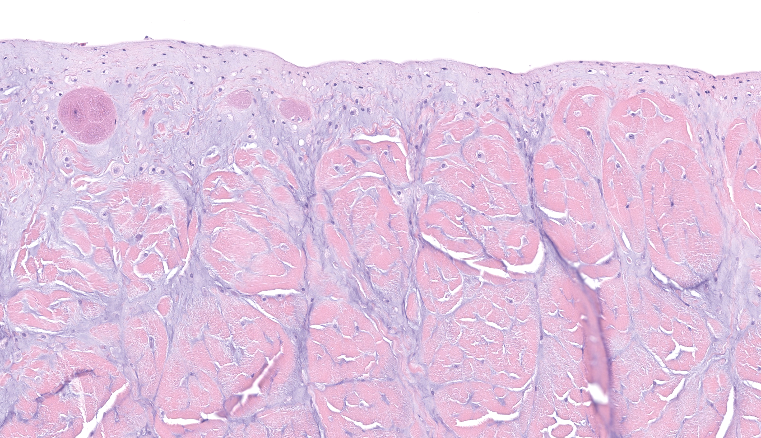
An interesting finding in this slide is the large amount of amphophilic to basophilic substance present in the affected tendon reflects notable deposition of ground substance (including proteoglycans, glycosaminoglycans;) and even cartilaginous metaplasia (Fig 4-3). We debated its origin carefully, weighing the natural progression of ‘turkeydom’ and rapid weight gains effect on the tendons of the legs versus virally-induced changes. The bulk of participants felt that the deposition of anastomosing ground substance was most attributable to the rapid weight gain (and increased shear and concussive force in this area.) Though it is possible that reoviral changes could weaken collagen and augment rupture of tendons, we did not have clear evidence of inflammatory cells associated with this material (i.e. tendonitis). As such, we probably could (should?) have two morphologic diagnoses for this case, but we could not agree on what to call this process with some participants unsatisfied with the vagueness of ‘tendinopathy’ or ‘tendinosis’ or if (in true WSC fashion) an expected lesion in a heavy meat bird merited an entire paragraph to explain it (it appears it does)! It is some consolation perhaps to consider a comparison to human sports-induced tendinopathies,11 which have analogous gross and histologic appearance – something worth considering for those that enjoy running like the JPC residents do. Careful description of disorganized collagen, ground substance, the presence or absence of inflammatory cells, and changes in supporting stroma are helpful in categorizing these conditions.11
References:
- Kumar R, Sharafeldin TA, Nader MS, et al. Comparative pathogenesis of turkey reoviruses. Avian Pathol. 2022;51:435-444.
- Levisohn A, Gur-Lavie A, Weisman, J. Infectious synovitis in turkeys: Isolation of tenosynovitis virus-like agent. Avian Pathol. 1980;9:1-4.
- Lu HY, Dunn PA, Wallner-Pendleton EA, et al. Isolation and molecular characterization of newly emerging avian reoviruse variants and novel strains in Pennsylvania, U.S.A., 2011-2-14. Sci Rep. 2015;5:14727.
- Mor SK, Sharafeldin TA, Porter RE, et al. Isolation and characterization of a turkey arthritis reovirus. Avian Dis. 2013;57:97-103.
- Page RK, Fletcher Jr OJ, Villegas P. Infectious tenosynovitis in young turkeys. Avian Dis. 1982;26:924-927.
- Porter R. Turkey Reoviral Arthritis: A Novel Condition in the United States. In: Proceedings International Turkey Production and Science Meeting, Chester, England. March 22, 2018. Proceedings pp. 31-35.
- Sharafeldin TA, Mor SK, Bekele, AZ, et al. The role of avian reoviruses in turkey tenosynovitis/arthritis. Avian Pathol. 2014;43:371-378.
- Sharafeldin TA, Mor SK, Bekele AZ, et al. Experimentally induced lameness in turkeys inoculated with a newly emergent turkey reovirus. Vet Res. 2015;46:11-16.
- Spanididos DA, Graham AF. Physical and chemical characterization of an avian reovirus. J Virol. 1976;3:968-976.
- Tang Y, Sebastian A, Yeh YT, et al. Genomic characterization of a turkey reovirus field strain by Next-Generation Sequencing. Infect. Genetic. Evol. 2015;32;313-321.
- Khan KM, Cook JL, Bonar F, Harcourt P, Astrom M. Histopathology of common tendinopathies. Update and implications for clinical management. Sports Med. 1999 Jun;27(6):393-408.