Wednesday Slide Conference, 2025-2026, Conference 9, Case 1
Signalment:
3 months-old, female, Piétrain x Large White, pig, Sus scrofa domesticusHistory:
Gross Pathology: Multiple, 1-2 mm in diameter, non-raised, red to blue, irregular lesions were found diffusely distributed through the skin of the ventral and inguinal regionas well as the head, nose and ears. Upon incision, lesions were localized at the dermal/subcutaneous junction.Laboratory Results:
Cells lining cystic cavities immunohistochemically stained positive with pan-cytokeratin and negative with CD31. Immunohistochemistry with Ki67 and alpha-SMA revealed a low proliferative index in the apocrine epithelial cells and strong positivity for alpha-SMA in the basal area of the cystic lesions.Microscopic Description:
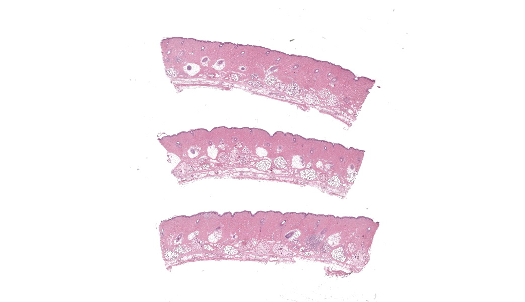
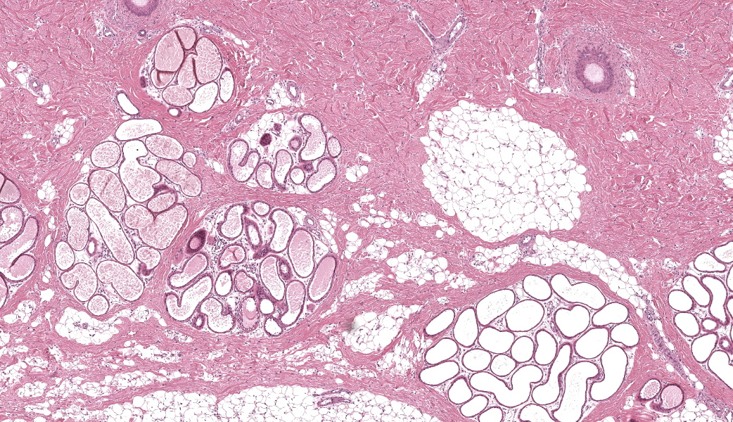
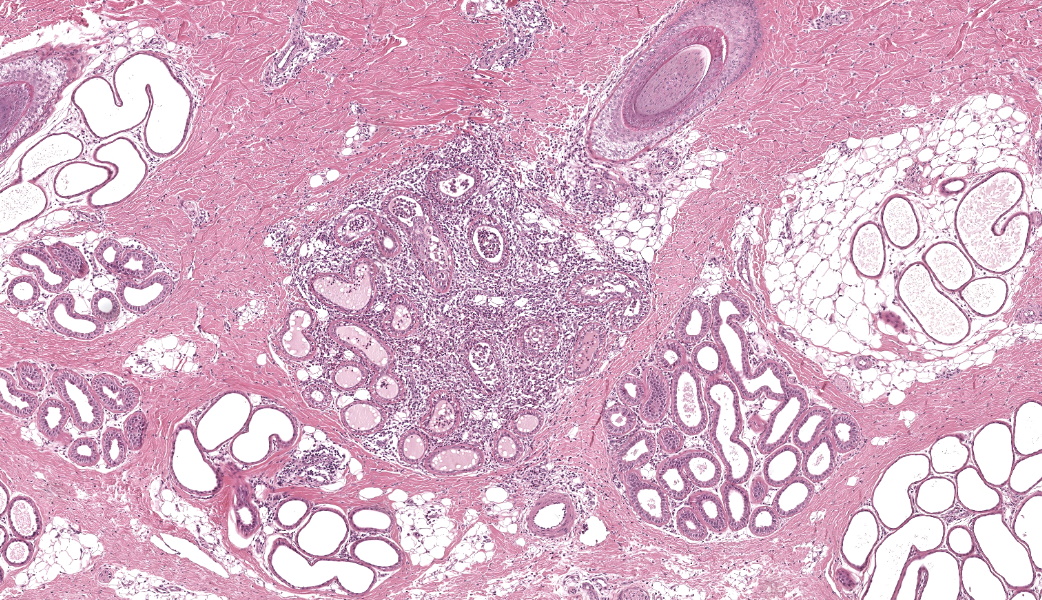
Haired skin (dorsal nose, three sections): Within the deep dermis and subcutis there are multiple well delineated clusters of variably distended apocrine glands with sinuous cystic cavities and partial tubular folding, which expand and replace dermal adipose tissue and muscle . Most of the clustered cysts are lined by a single layer of flattened columnar epithelial cells without apical blebbing. Adjacent smaller clusters of lesser distended glands exhibit one to several layers of more cuboidal epithelium. The outer layer of the cyst wall consists of thin layers of myoepithelial cells. Many gland lumina contain pale eosinophilic secretory product. Surrounding tissue and stroma comprises mild lymphoplasmacytic infiltrates.Focally in the mid dermis and associated with a hair follicle there is a localized, well demarcated focus of moderate mixed cellular inflammation composed of macrophages, neutrophils and lesser lymphocytes and plasma cells. These encase numerous distorted and/or ruptured apocrine glands, infiltrate their epithelium and are present within the lumen of remaining glandular structures (foreign body type reaction). Macrophages frequently contain cellular debris.
The remaining epidermis and dermis are unremarkable.
Contributor's Morphologic Diagnoses:
Skin: cystic distention of apocrine glands, diffuse, severe, with compression of adjacent muscle tissue consistent with cutaneous apocrine cystomatosis.Skin: hidradenitis and dermatitis, focally extensive, moderate, chronic, mixed cellular with apocrine gland rupture (foreign body type reaction).
Contributor's Comment:
Cutaneous apocrine cystomatosis is an uncommon non-neoplastic condition, which has been first reported in three pigs from three different abattoirs in Spain in 2020.6 There, the lesions appeared as brown, non-prominent nodules within the subcutaneous fat distributed over the entire dorsal region. Microscopically, the authors’ findings correspond to results reported here. Ki67 immunohistochemistry showed only few glands with single cells of positive stained epithelium indicating a low proliferative index. In addition to the findings of Lopez-Figueroa et al. an inflammatory site was present in the case described here. This possibly occurs due to rupture of an adjacent dilated gland.Cutaneous apocrine cystomatosis has also been reported in dogs and cats4,9,10. In dogs, it is mainly distributed over the head and neck but was also described at the dorsal trunk and flanks as well as solitary at the cheeks. Microscopically, the lesions are described as multiple clusters of cystically dilated sweat glands, but, in contrast to the lesions in the pigs, show the same morphology as normal apocrine glands with a single layer of cuboidal to columnar epithelium with dome-shaped apical surface. In addition, the epithelial cells of the cyst wall in dogs show some pseudo-papillary projections protruding into the lumen. Inflammation and fibrosis in the surrounding tissue is rare and mild. In case of cyst rupture, moderate inflammation can occur comprising plasma cells, neutrophils and lymphocytes. Pale siderophages, which contain pigment from the iron content of apocrine secretions, may be found.
Feline ceruminous cystomatosis is the corresponding disorder described in cats.4 The lesion develops from the ceruminous glands and occurs in the external ear and the inner pinna.5 Microscopically, the cysts are lined by a single layer epithelium that varies from attenuated to cuboidal to columnar. In contrast to dogs and pigs, the surrounding dermis often shows mild fibrosis with detection of inflammatory cells like lymphocytes, neutrophils, mast cells and pigmented macrophages.
The etiology of cutaneous apocrine cystomatosis is uncertain. For dogs, a senile degenerative change has been discussed because affected animals were middle-aged or older. The average age in affected cats has been reported between 8 and 9.5 years, but animals as young as one year can be affected.4 As all described cases in pigs were from abattoirs or, in our case, from experimental studies, and therefore comparatively young (3 to 6 months), the disease in pigs seems to be unrelated to the aging process. A congenital origin is also discussed as normal apocrine glands are absent and no other alterations in the surrounding tissue are detectable.6 Whether there is a genetic origin in dogs and cats is unknown.
In cats, there seems to be a breeding predisposition in Persian and Abyssinian cats with a slight male predominance, whereas in dogs and pigs no over-representation in certain breeds or sex predilection is described to date.3
The differential diagnosis of cutaneous apocrine cystomatosis includes apocrine hidrocystoma/cystadenoma, particularly described in cats, where it is predominantly located in the eyelid.1,2 It is a benign neoplasm characterized by cysts with a single or multilayered cuboidal to columnar epithelium and true papillary projections into the cavity. These neoplasms usually show a higher proliferative index.
In humans, cutaneous apocrine cystomatosis has not been described. The related benign neoplasm is, corresponding to the differential diagnosis in animals, the apocrine hidrocystoma/cystadenoma, which includes a spectrum of benign cystic lesions of ductal sweat gland origin, with architecture ranging from simple cystic (hidrocystoma) to more complex (cystadenoma).7
Contributing Institution:
Ludwig-Maximilians-Universität München (LMU), Institute of Veterinary Pathology, Veterinärstr. 13, 80539 Munich, Germany www.patho.vetmed.uni-muenchen.dein cooperation with:
Technical University of Munich (TUM), Institute of Pathology, Comparative Experimental Pathology (CEP), Trogerstr. 18, 81675 Munich, Germany web.med.tum.de/path/vergleichende-experimentelle-pathologie
JPC Diagnoses:
Haired skin, apocrine glands: Dilation, chronic, diffuse, marked.JPC Comment:
The JPC was fortunate enough to host Dr. Charles Bradley again this year for an excellent dermatopathology-focused conference that was preceded by his highly educational and forever crowd-pleasing “Approach to a Case Using Pattern Analysis” lecture. This first case was a great exercise in reminding the pathologist to not overinterpret histological findings. Participants are, rightfully so, primed to expect description-heavy cases in conferences. Every once in a while, though, a moderator selects a case that is on the leaner descriptive side to make this point: don’t make a mountain out of a molehill and only describe what you see, not what you want to see. Participants did an excellent job adhering to this principle, and most were able to reach the correct diagnosis. The contributor in this case provided an excellent write-up of this lesion across species and it is well-worth the read.Conference discussion for this case covered numerous topics, including the specific histologic findings that can assist the pathologist in species identification based on skin. Dr. Bradley insists on this as part of his dermatopathology diagnostic process: look at the slide first, try to identify the species you have, and then check the case information. If they don’t seem to match up, a call to the submitting clinician might be in order. For most porcine skin, the epidermis is thicker than other common species, usually 5-7 nucleated cells thick, and forms papillary, shallow rete pegs that project into and interdigitate with the superficial dermis.8 This papillary epidermis is also a feature of human skin. Porcine skin is especially thick on the dorsum. Additionally, pig sebaceous glands are sparse and typically associated with hair follicles. Porcine hair follicles are, generally, large and far apart from one another.
Participants were asked to describe the difference between apocrine and eccrine glands and why they chose apocrine cystomatosis over eccrine cystomatosis. Histologically, apocrine and eccrine glands are indistinguishable, but location, which varies by species, can help with differentiation. In humans, eccrine glands are all over the body and responsible for sweat production, which aids in thermoregulation, whereas apocrine glands are restricted to the axilla and groin regions. In pigs, eccrine glands are restricted to the tip of the snout and to the carpal regions and serve no thermoregulatory function.5 In dogs, eccrine glands are restricted to the paw pads and nose, and have limited sweating function.5 Apocrine glands however, are everywhere in pig skin and are, like the sebaceous glands, associated with hair follicles. The skin samples in this case were from the abdomen and dorsal nose, neither of which is an eccrine gland location in pigs. Differentiating between the two can be diagnostically important, especially in cases of neoplasia, as eccrine adenocarcinomas are considered more aggressive than apocrine adenocarcinomas.
There was brief discussion on the inflammatory component in this case. Mild inflammation of apocrine glands, known as hidradenitis, is a common background lesion in many species that is usually associated with folliculitis. In this case, the focus of inflammation was severe enough that it was unanimously thought by conference participants to be due to a ruptured apocrine gland secondary to dilation. There was a question posed to participants regarding possible other differentials based on gross images in other species, and included apocrine cystadenoma, basal cell adenoma, sebaceous adenoma, or melanocytic neoplasms. Of these, apocrine cystadenoma vs. apocrine cystomatosis was discussed most heavily for educational purposes, the main differences being that this lesion was diffuse throughout the skin, which is more common with cystomatosis, and there are no papillary projections into the lumens of affected glands. Papillary projections of epithelial cells into the cystic glandular lumen are a common finding in apocrine cystadenomas, but not in cystomatosis.7 Wrapping up conference discussion of this case was a brief review of apocrine cystomatosis lesions in other species, which are discussed by the contributor in their comment.
Apocrine gland cystomatosis is also known as epitrichial sweat gland cystomatosis, apocrine cystic hyperplasia, and cystic hyperplasia of apocrine sweat glands (human and canine term).6,10 When initially described in the medical literature, it was proposed that cyst formation might be due to the occlusion of excretory ducts.7,9 Later, it was suggested that apocrine cysts result from episodes of glandular hyperplasia followed by an involution phase and inability of the body to reabsorb the contents of the dilated glands.7,9 More recently, though, single or multiple apocrine cysts have been described as either benign sweat gland tumors, as idiopathic, non-neoplastic, senile degenerative changes or, such as in this pig’s case, as a congenital condition.6,7,9
References:
- Cantaloube B, Raymond-Letron I, Regnier A. Multiple eyelid apocrine hidrocystomas in two Persian cats. Vet Ophthalmol. 2004;7:121-125.
- Chaitman J, van der Woerdt A, Bartick TE. Multiple eyelid cysts resembling apocrine hidrocystomas in three Persian cats and one Himalayan cat. Vet Pathol. 1999;36:474-476.
- Goldschmidt MH, Shofer FS. Skin Tumors of the Dog and Cat. Pp 280-283. Oxford: Pergamon Press;1992.
- Gross TL. Sweat gland tumors. In: Gross TL, Ihrke PJ, Walder EJ. Skin Diseases of the Dog and Cat: Cinical and Histopathologic Diagnosis. 2nd ed., pp. 666-674. Oxford: Wiley-Blackwell 2008.
- Jubb KVF, Kennedy PC, Palmer NC. Pathology of Domestic Animals. 6th ed., vol. 1, p718. Elsevier;2015.
- Lopez-Figueroa C, Domingo M, Marti B, Vidal E, Segales J. Cutaneous apocrine cystomatosis in three slaughter-aged pigs. J Vet Diagn Invest. 2020;32:159-161.
- Sangüeza OP, Cassarino DS, Glusac EJ. Benign tumours with apocrine and eccrine differentiation: Hidrocystoma/cystadenoma. In: Elder DE, Massi D, Scolyer RA. WHO Classification of Tumours: Skin Tumours. 4th ed.;2018.
- Skydsgaard M, Dincer Z, Haschek WM, Helke K, Jacob B, Jacobsen B, Jeppesen G, Kato A, Kawaguchi H, McKeag S, Nelson K, Rittinghausen S, Schaudien D, Vemireddi V, Wojcinski ZW. International Harmonization of Nomenclature and Diagnostic Criteria (INHAND): Nonproliferative and Proliferative Lesions of the Minipig. Toxicol Pathol. 2021;49(1):110-228.
- Stazi M, Silvestri S, Mechelli L, Brachelente C. A case of generalised cutaneous apocrine cystomatosis in a Pekingese dog. Vet Med Sci. 2022;8:450-453.
- Vilafranca M, Domingo M , Roura X. Generalized Apocrine Cystomatosis in an Old English Sheepdog. Veterinary Dermatology. 1994;5:83-87.