Wednesday Slide Conference, Conference 8, Case 4
Signalment:
10-week-old, female, C57BL/6 mice (Mus musculus)
History:
Mice arrived from vendor with no abnormalities observed upon entrance. Lab members shaved animals to prepare for subcutaneous tumor injection 2-week post-arrival and noted abnormal skin (bumpy, thick with patchy fur growth). No other health concerns. Lab stated this condition has only been observed with mice from this vendor. Three mice were submitted for skin histopathology.
Gross Pathology:
All mice from this cohort had skin lesions consisting of patchy alopecia and irregular/bumpy skin foci in the dorsal flank area with extension to the limbs and (rarely) abdomen.
Microscopic Description:
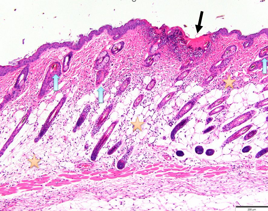
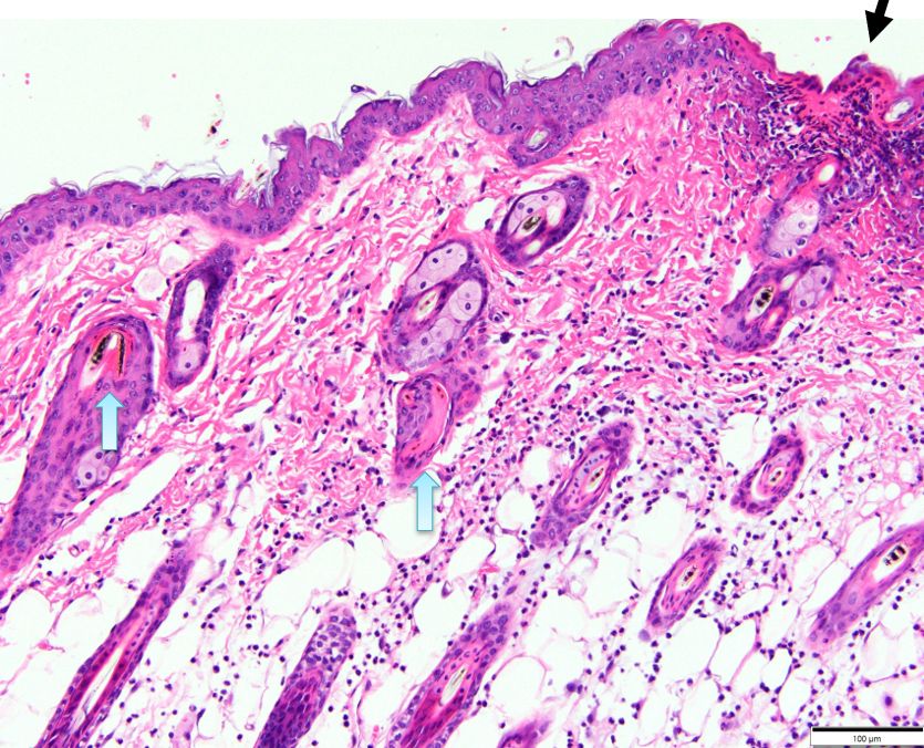
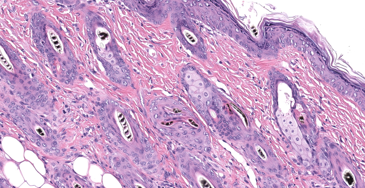
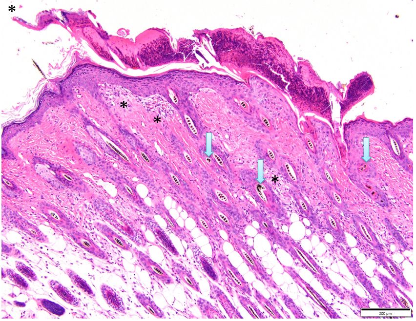
Haired skin: Three skin sections from different mice in the cohort are examined. In all sections, there are focal ulcers and erosions. Multifocally, longitudinal follicle sections show hair shafts with twist severely in the infundibulum and/or disintegrating of hair in the superficial dermis and as it emerges on the surface. Rarely, hair shafts can be seen penetrating the follicle wall with fragments free in the dermis and hypodermal fat layer surrounded by a mixed inflammatory cell infiltrate consisting of neutrophils, macrophages, rare multinucleated giant cells, and occasional lymphocytes forming a foreign body granuloma (trichogranuloma). Other areas have a more diffuse chronic lymphoplasmacytic infiltrate in the dermis and hypodermal fat. The overlying epidermis exhibits mild to moderate acanthosis, hypergranulosis, and orthokeratotic hyperkeratosis. Resolved ulcers often contain underlying superficial dermal granulation tissue proliferation, fibroplasia, and increased connective tissue.
Contributor’s Morphologic Diagnosis:
Haired skin: Dermatitis, chronic, hyperplastic, ulcerative, with hair shaft twisting, fragmentation, trichogranuloma formation, and superficial dermal scarring.
Contributor’s Comment:
Mice of the strain C57BL/6 and those on a C57BL/6 background develop ulcerative dermatitis (UD), a disease of unknown etiology that leads to significant morbidity.4,10 In this entity, ulcerations present on the dorsal scapulae, torso, shoulder, and face from pruritis-induced self-trauma, and may be single or multifocal in distribution.1,4,10 A recent large-scale studies in mice reveal that chronic ulcerative dermatitis is still the primary non-tumoral cause of euthanasia in both sexes (39.1% in males and 35.4% in females).2 In the case of these mice, the lesions did not fit the typical clinical, gross, or histopathology lesions characteristic of UD. Specifically, no pruritis was noted in any of the cohort clinically, and lesions were initially noted when fur was removed. Histopathologically, small ulcerations were noted, however, the changes to the hair follicles, shafts, and secondary inflammation of the dermis were the most characteristic findings.
Many of the scarring alopecias in humans and other species, are based on primary sebaceous gland pathological features.7 Although sebaceous gland loss can be seen in some areas with chronic scarring in these mice, the primary lesions are centered on the follicles. An entity was characterized in 2011 in C57BL/6J mouse substrains describing a primary follicular dystrophy (PFD) which leads to trichogranulomas, chronic scarring of the dermis, and alopecia.8 The changes in the primary follicles include fragmentation and twisting of the hair shaft as we saw in these mice. In 2017, a large scale screening study of knockout mice revealed sporadic trichogranulomas are common in mutant mice, however, these are not consistently seen in all mice of a specific line. PFD was noted at a much lower frequency.9 Although the etiology of PFD remains unclear, C57BL/6J mice were found to have defects in vitamin A metabolism.9 The skin takes up circulating retinol and can either store it in the form of retinyl esters or metabolize it to retinoic acid.5,8 Two enzymes are present in the skin that can oxidize retinol to retinal. These include the medium chain alcohol dehydrogenase type 4 (ADH4) and the short chain dehydrogenase/reductases epithelial retinol dehydrogenase (DHRS9).3,8 DHRS9 is microsomal and can oxidize both free and CRBP-bound retinol.3,8 Upregulation of DHRS9 in C57BL/6J and C57BL/6Tac but not C57BL/6NCr or C57BL/6Crl mice provides a potential explanation for why the first two strains have a much higher frequency of dorsal skin alopecia.8
The histologic lesions seen in mice with primary follicular dystrophy resemble the human disease currently termed central centrifugal cicatrical alopecia (CCCA).8 Premature desquamation of the inner root sheath occurs in CCCA leading to marked thinning of the outer root sheaths with reactive perifollicular inflammation, and eventually entry of the hair fiber into the dermis with resulting destructive chronic granulomatous inflammation.6,8,9 Although this entity does not have all features of CCCA in people, the degenerative features of the inner root sheath in PFD mirror those changes in CCCA.8
Contributing Institution:
Division of Laboratory Animal Resources
University of Pittsburgh
S1040 Thomas E. Starzl Biomedical Science Tower
200 Lothrop Street
Pittsburgh, PA 15261
http://www.dlar.pitt.edu/
JPC Diagnosis:
Haired skin: Follicular dysplasia, multifocal, moderate with ulcerative dermatitis, trichogranulomas, and superficial fibrosis.
JPC Comment:
The final case of this conference proved tricky for participants. Although these sections of skin featured dermatitis that was ulcerative, the cause was follicular dysplasia with ulcerative dermatitis developing similarly to the CCCA that the contributor describes. From low magnification, the twisting of hair shafts and abnormal orientation of hair follicles relative to the epidermis (figure #) are key details. Under higher magnification, dystrophic hairs are hypereosinophilic due to poor cuticle maturation as well. Although subtle, these sections of skin also have increased space (widening) between abnormal hair follicles as well. In contrast, UD is characterized by marked lymphohistiocytic and neutrophilic inflammation of the dermis, epidermis, and deeper tissues (adipose, muscle, nerves) that can progress to fibrosis in chronic cases.2 The overlying epidermis is also often hyperplastic. These features of UD are notably absent in this case however. Other causes of ulcerative skin disease and alopecia in mice include barbering, feeder/waterer-associated dermatitis, ectoparasites (e.g. Myobia musculi), self-trauma, fighting, and chemotherapy agents. Primary deficiencies of genes associated with hair development as was likely in this case should also be considered.
Lastly, conference participants discussed some of the unique features of mouse skin. Mice lack apocrine glands and rete ridge formation. They also have synchronized hair cycles such that growth occurs in waves (i.e. adjacent follicles in section are in the same phase) though overall mice tend to be anagen-heavy. In this case, there is disruption of this synchronization of hair development due to alopecia and inflammation.
References:
- Andrews AG, Dysko RC, Spilman SC, Kunkel RG, Brammer DW, Johnson KJ. 1994. Immune complex vasculitis with secondary ulcerative dermatitis in aged C57BL/6NNia mice. Vet Pathol 31:293–300.
- Elies L, Guillaume E, Gorieu M, Neves P, Schorsch F. Historical Control Data of Spontaneous Pathological Findings in C57BL/6J Mice Used in 18-Month Dietary Carcinogenicity Assays. Toxicologic Pathology. 2024 May 17:01926233241248658.
- Haselbeck RJ, Ang HL, Duester G. Class IV alcohol/retinol dehydrogenase localization in epidermal basal layer: potential site of retinoic acid synthesis during skin development. Dev Dyn. 1997 Apr;208(4):447-53.
- Kastenmayer RJ, Fain MA, Perdue KA. A retrospective study of idiopathic ulcerative dermatitis in mice with a C57BL/6 background. J Am Assoc Lab Anim Sci. 2006 Nov;45(6):8-12.
- Roos TC, Jugert FK, Merk HF, Bickers DR. Retinoid metabolism in the skin. Pharmacol Rev. 1998 Jun;50(2):315-33.
- Sperling LC. Scarring alopecia and the dermatopathologist. J Cutan Pathol. 2001 Aug;28(7):333-42.
- Stenn KS, Sundberg JP, Sperling LC. Hair Follicle Biology, the Sebaceous Gland, and Scarring Alopecias. Arch Dermatol. 1999;135(8):973–974.
- Sundberg JP, Taylor D, Lorch G, Miller J, Silva KA, Sundberg BA, Roopenian D, Sperling L, Ong D, King LE, Everts H. Primary follicular dystrophy with scarring dermatitis in C57BL/6 mouse substrains resembles central centrifugal cicatricial alopecia in humans. Vet Pathol. 2011 Mar;48(2):513-24.
- Sundberg JP, Dadras SS, Silva KA, Kennedy VE, Garland G, Murray SA, Sundberg BA, Schofield PN, Pratt CH. Systematic screening for skin, hair, and nail abnormalities in a large-scale knockout mouse program. PLoS One. 2017 Jul 10;12(7):e0180682.
- Williams LK, Csaki LS, Cantor RM, Reue K, Lawson GW. Ulcerative dermatitis in C57BL/6 mice exhibits an oxidative stress response consistent with normal wound healing. Comp Med. 2012 Jun;62(3):166-71.


