CASE IV:
Signalment:
4-year-old, intact female, Labrador Retriever, domestic dog (Canis lupus familiaris)
History:
The dog, which had a history of allergies and bacterial ear infections, developed "kennel cough" after a visit to a local veterinarian for annual vaccination and a moxidectin injection. Treatment with antihistamines and corticosteroids was administered one day after the beginning of clinical signs. Three days later, the dog developed diarrhea and treatment was
interrupted. The cough progressed with presence of mucus. Six days after onset, the dog was lethargic and inappetent, and was brought once again to a veterinary hospital, at which 19 blood work revealed a high white blood cell count. On the morning of the seventh day, the animal died.
Gross Pathology:
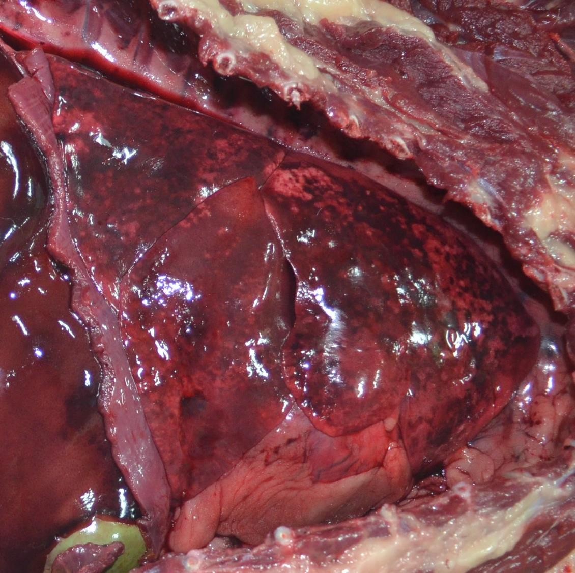
The animal was obese, and the body was in moderate autolysis. The thoracic cavity contained approximately 15-20 ml of red-tinged fluid and the parietal pleura lining the intercostal spaces had coalescing dark red areas (hemorrhage). The lungs had bilateral multifocal to coalescing dark red to black, variably sized foci widespread throughout the pleural surface (Figure 1). The caudal aspect of the left cranial lung lobe had a focally extensive gray and firm area that sunk in formalin (consolidation). The trachea was filled with abundant, red-tinged froth and the mucosa was diffusely reddened. The tracheobronchial lymph nodes were enlarged approximately 2X and diffusely dark red, with no corticomedullary distinction.
Laboratory Results:
Bacteriology (aerobic culture), Lung:
Heavy growth of Streptococcus equi subsp. zooepidemicus and Escherichia coli.
Microscopic Description:
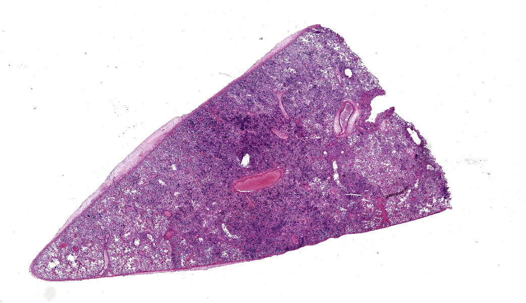
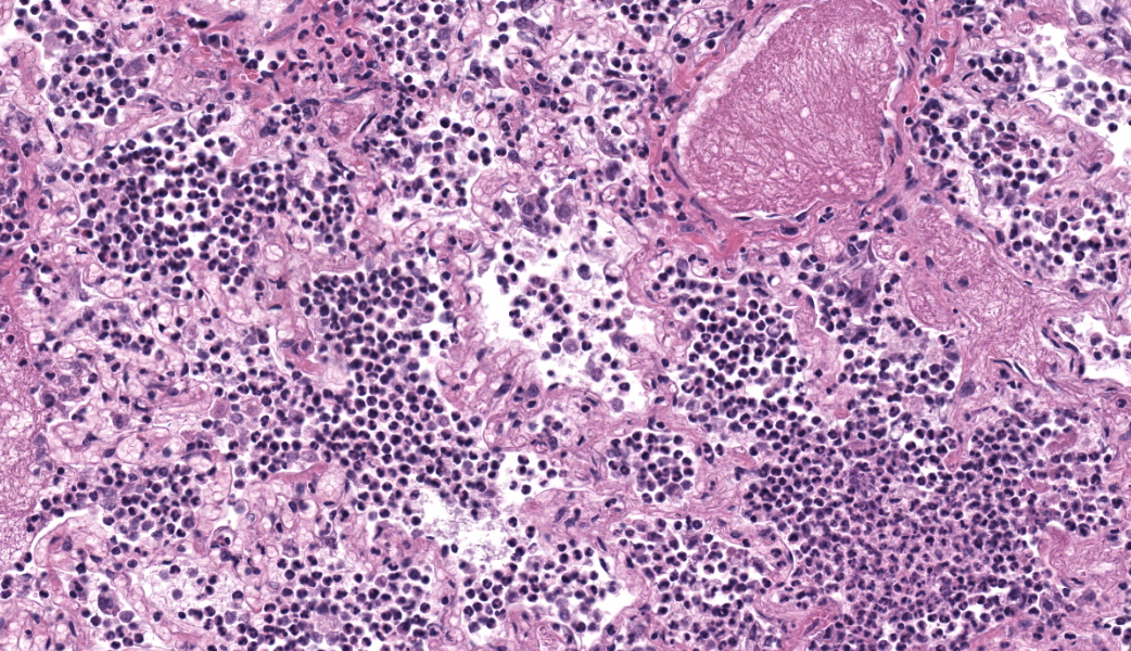
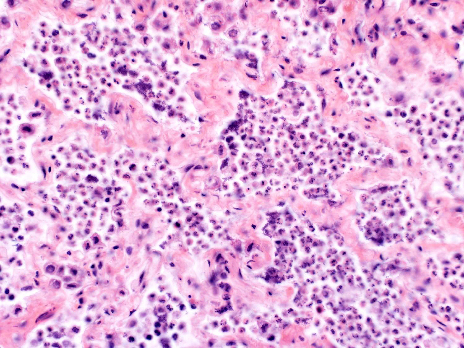
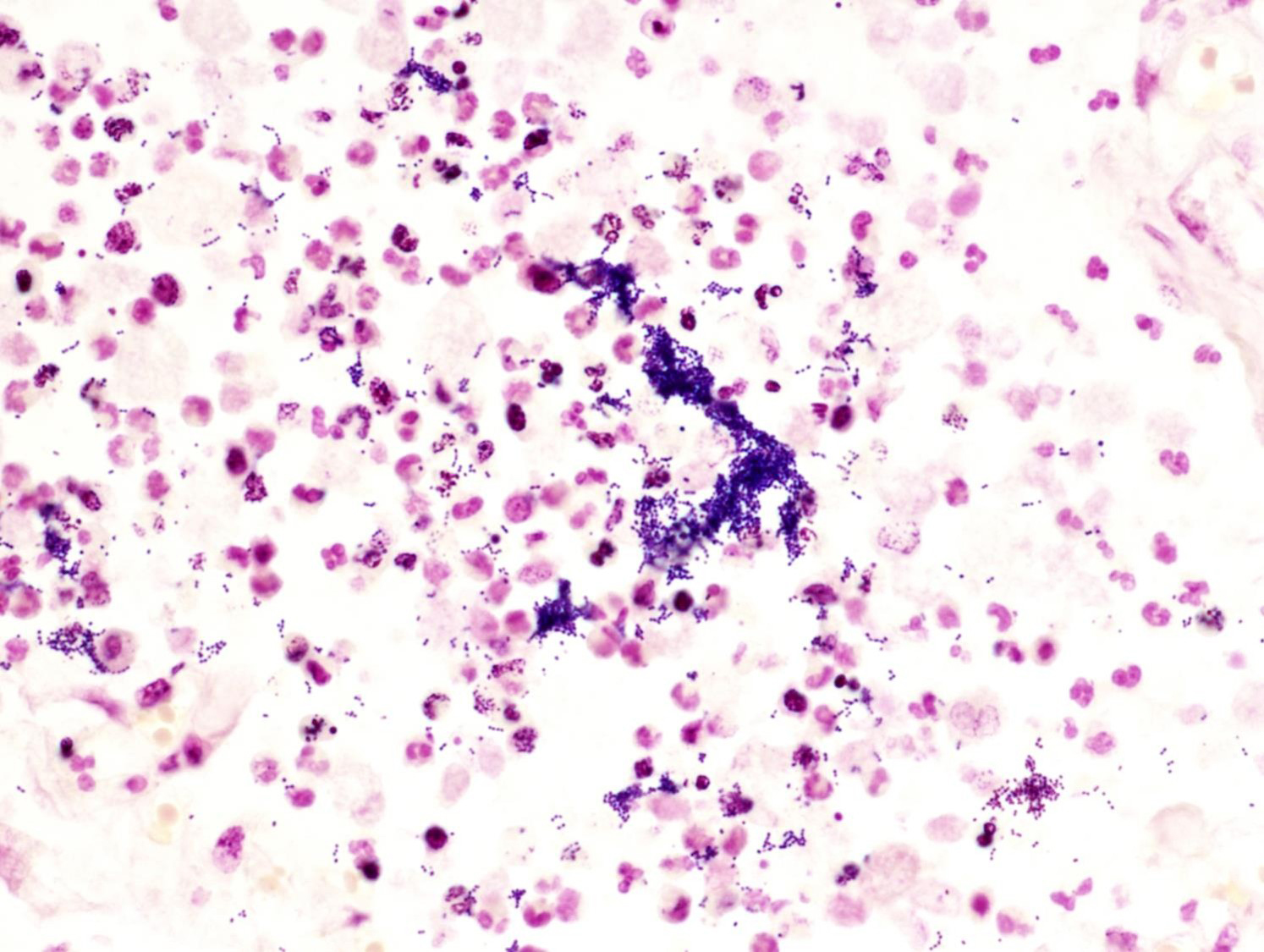
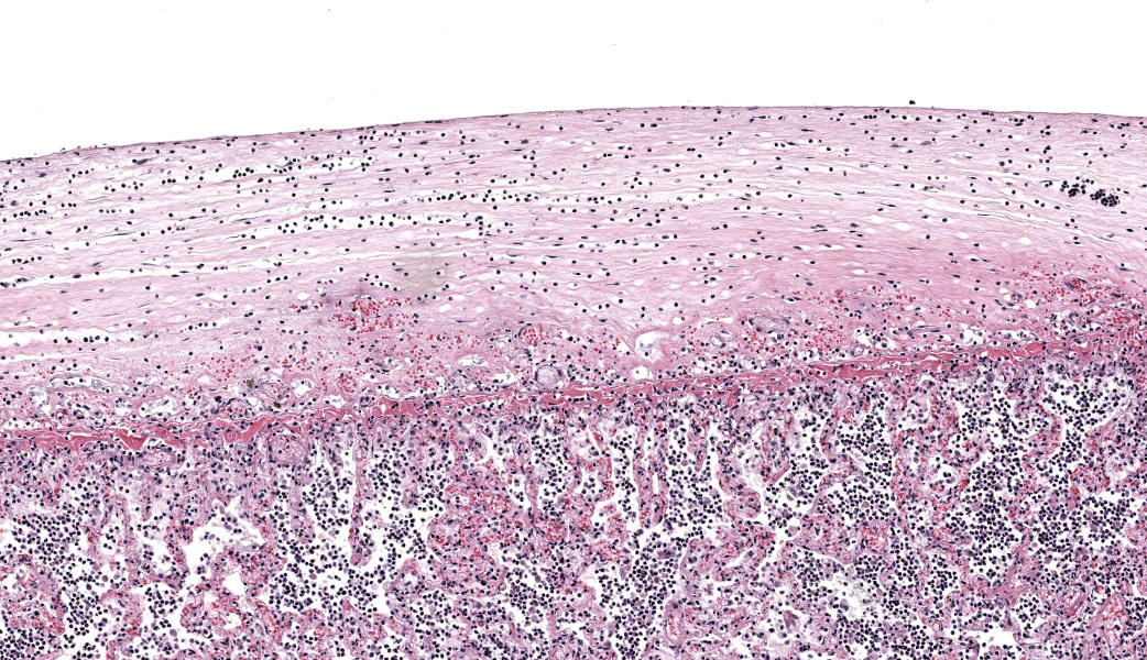
Lung: The alveolar spaces are diffusely flooded by numerous viable and degenerate neutrophils and macrophages, with occasional fibrin exudation and edema fluid. Within alveoli, there are rare colonies of cocci, occasionally organized in short chains; bacteria are also observed within macrophages and adhered to the membrane of inflammatory cells. Multifocal bronchioles are filled with cellular debris and neutrophils. The tunica adventitia and the tunica submucosa of multifocal blood vessels and bronchi are expanded by moderate edema. Occasionally dilated lymphatic vessels contain intraluminal fibrin and few neutrophils. Rare small caliber blood vessels are occluded by fibrin thrombi. The pleura is segmentally expanded by abundant fibrin exudate, edema, and moderate numbers of neutrophils.
Contributor's Morphologic Diagnoses:
Lung: Bronchopneumonia, fibrinosuppurative and hemorrhagic, multifocal to coalescing, marked, acute, with intralesional coccoid bacteria and locally extensive fibrinous pleuritis
Contributor's Comment:
Bacterial pneumonia caused by opportunistic agents is fairly common in dogs. Involved bacteria include generally Bordetella bronchiseptica, Staphylococcus spp., Pasteurella multocida, E. coli, Klebsiella pneumoniae, Pseudomonas aeruginosa, Acinetobacter spp., and Streptococcus spp.4 In a review of 393 cases of streptococcal infections in dogs, several streptococcal species (?-hemolytic Streptococcus spp., ?-hemolytic Streptococcus spp., S. canis, S. dysgalactiae subsp. equisimilis, and S. equi subsp. zooepidemicus) were associated with pneumonia.12 spp., S. canis, S. dysgalactiae subsp. equisimilis, and S. equi
Streptococcus equi subsp. zooepidemicus (S. zooepidemicus) is an opportunistic zoonotic bacterial pathogen capable of causing disease in several animal species.1,2,5,10,15 These are gram-positive, Lancefield group C, ?-hemolytic cocci commonly isolated from the upper respiratory and lower genital tract of horses9 that can affect different organ systems and express variable clinical presentations, including pneumonia, endometritis, placentitis, meningitis or septicemia.5,8,14,19 In alpacas and llamas, this bacterium is known to cause sepsis, also called "alpaca fever".6 In one reported case, cocci were demonstrated within enteric lymphatic vessels of a neonate, suggesting a possible lymphatic dissemination of this agent.6S. zooepidemicus has also been shown to be transmitted to humans from different animal species such as guinea pigs, horses and dogs, causing severe pneumonia, bacteremia, septic arthritis, and meningitis.1,10,13,19S. zooepidemicus can also cause pharyngitis, with the potential to trigger acute post-streptococcal glomerulonephritis (APSGN) in humans.17 In swine, the first documented outbreak of S. zooepidemicus in the United States occurred during September and October 2019, with high mortality rates ranging from 10-50% in groups of pigs over the period of 8-10 days at a buying station in Ohio and at an abattoir in Tennessee. Previous outbreaks in pigs were also reported in China, resulting in significant economic losses.5
S. zooepidemicushas been associated with severe respiratory disease in dogs for a number of years.16 However, more recently, its importance has been underscored by fatal outbreaks in kenneled dog populations in several countries.3,14,18 Hence, the bacterium has been recognized as one of the components of the multifactorial Canine Infectious Respiratory Disease (CIRD, Kennel Cough), which includes most commonly the bacterium Bordetella bronchiseptica and viruses such as Canine Herpes Virus (CHV) and Canine Parainfluenza Virus (CPIV).14,16 In the present case, it is believed that the dog, which was likely immune-suppressed due to routine treatment with anti-histamines and corticosteroids for allergies, was exposed to a large burden of the bacterium during a visit to the veterinary clinic. No indications of other pathogens related to active disease were observed. While in this case there was heavy growth of both E. coli and S. zooepidemicus from lung samples, only intralesional gram-positive coccoid bacteria frequently organized in chains a Gram stain of affected lung sections revealed. Therefore, the E. coli isolate was interpreted as a contaminant.
The disease caused by S. zooepidemicus in dogs frequently has a brisk clinical course and high morbidity, and deaths have been reported to occur as soon as 2 days after exposure.14 Clinically, the dogs present with severe acute respiratory distress associated with severe, acute fibrinosuppurative, necrotizing and/or hemorrhagic pneumonia, as well as hemothorax.14,16 The pathogenesis of S. zooepidemicus infection in dogs is incompletely understood;15,16 however, environmental stress, extenuating physical exercise and overcrowding can be determinant aspects in the spread and development of this disease by triggering the release of stress hormones such as cortisol and norepinephrine and consequently reducing re- sistance to microbial invasion.11,18
The involvement high morbidity, and deaths have been reported to occur as soon as 2 days after exposure.14 Clinically, the dogs present with severe acute respiratory distress associated with severe, acute fibrinosuppurative, necrotizing and/or hemorrhagic pneumonia, as well as hemothorax.14,16 The pathogenesis of S. zooepidemicus infection in dogs is incompletely understood;15,16 however, environmental stress, extenuating physical exercise and overcrowding can be determinant aspects in the spread and development of this disease by triggering the release of stress hormones such as cortisol and norepinephrine and consequently reducing resistance to microbial invasion.11,18 The involvement of stress hormones can also indirectly explain the large numbers of extracellular cocci observed in acutely infected dogs, indicating rapid proliferation and evasion of clearance mechanisms.18
Gross examination of S. zooepidemicus infected dogs reveals features similar to the ones observed in this case, such as mottled dark to bright red lungs with variably sized areas of consolidation or alveolar collapse, multifocal areas of hemorrhage in tracheobronchial lymph nodes, and hemothorax.14,18 Histological features consist of extensive alveolar flooding by neutrophils and macrophages with occasional fibrin (fibrinosuppurative pneumonia), with gram-positive cocci arranged in short chains and occasional clusters within alveoli; these findings are also similar to the ones observed in the present case.2,16
Episodes of pneumonia caused by S. zooepidemicus in dogs often lead to acute clinical disease with a high mortality rate despite intensive treatment, as even early antimicrobial therapy may not successfully mitigate the inflammatory events that lead to hypotension and multiple organ dysfunction.15 This can be particularly devastating in sheltered dog populations and, therefore, prevention of spread through adequate management practices is desirable. Survival rates are more likely to improve after follow-up measures, such as designation of intake rows, depopulation programs, and establishment of quarantine facilities are in effect, and these measures can also help prevent further significant respiratory disease outbreaks.3,7
Contributing Institution:
Louisiana Animal Disease Diagnostic Laboratory (LADDL), School of Veterinary Medicine, Louisiana State University (LSU) http://www.vetmed.lsu.edu/laddl/
JPC Morphologic Diagnosis:
Lung: Bronchopneumonia, fibrinosuppurative and necrotizing, subacute, diffuse, marked, with multifocal pleuritis and intra-alveolar cocci and bacilli.
JPC Comment:
The contributor has provided an excellent review of streptococcocal infections and specifically, Streptococcus equi subsp. zooepidemicus (S. zooepidemicus). The widespread nature of streptococcal diseases in many vertebrate species exemplifies how "big things may arrive in small packages." The small size of streptococci belies the many virulence factors that they possess and their ability to cause severe and life-threatening disease.
The major virulence factors of Streptococcus zooepidemicus allow it to escape a variety of the body's attempts at neutralizing this pathogen. Antiphagocytic factors include hyaluronic acid capsule which is identical to that seen in the body, providing a form of molecular mimicry and camouflage.20 The M-like protein, SzP, helps to prevent opsonization, which significantly reduces phagocytosis by macrophages. An additional M-like protein, SzM, binds plasminogen and allows streptococci to degrade fibrin (a common product of their infection), promoting additional bacterial spread. 20 Streptokinase acts in a similar fashion, cleaving host plasminogen into active plasmin, dissolving thrombi and allowing the bacteria to spread. IgG binding proteins bind to the Fc region of immunoglobulins, allowing S to evade the body's humoral response. 20 Hydrolytic enzymes include C5a peptidases, which destroy this fragment of complement, impairing the recruitment of neutrophils to the site of infection. 20S. zooepidemicus secretes a range of 11 pyrogenic exotoxins which act as superantigens, non-specifically activating and result in a massive release of immune mediators ("cytokine storm") in affected individuals.15, 20 Lipoteichoic acid (once referred to as the "endotoxin" of streptococci), a component of the wall of these cocci, acts in a similar fashion, and while its release requires the destruction of the bacterium, acts synergistically, with the combination resulting in pyrexia, systemic inflammation, and vascular permeability and is a major driver of "scarlet fever" in humans, as well as necrotizing fasciitis and toxic shock syndrome, among many other syndromes.20, 21 Finally, streptolysins are potent pore-forming proteins which may resulting in damage to epithelial linings of many tissues, allow dissemination through underlying vasculature, as well as outright resulting in the destruction of inflammatory cells, as well as enhancing production of IL-1, TNF, and IL-6.15, 20
References:
- Abbott Y, Acke E, Khan S, et al. Zoonotic transmission of Streptococcus equi subsp. zooepidemicus from a dog to a handler. J Med Microbiol. 2010;59: 120-123.
- Blum S, Elad D, Zukin N, et al. Outbreak of Streptococcus equi subsp. zooepidemicus infections in cats. Vet Microbiol. 2010;144: 236-239.
- Byun JW, Yoon SS, Woo GH, Jung BY, Joo YS. An outbreak of fatal hemorrhagic pneumonia caused by Streptococcus equi subsp. zooepidemicus in shelter dogs. J Vet Sci. 2009;10: 269-271.
- Caswell JL, Williams KJ. Respiratory System. In: MAXIE MG, ed. Jubb, Kennedy, and Palmer's Pathology of Domestic Animals. Volume 2, 6th ed. St. Louis, MO: Elsevier; 2016:577,578.
- Chen X, Resende-De-Macedo N, Sitthicharoenchai P, et al. Genetic characterization of Streptococcus equi subspecies zooepidemicus associated with high swine mortality in United States. bioRxiv. 2019: 2019.2012.2012.874644.
- Corpa JM, Carvallo F, Anderson ML, Nyaoke AC, Moore JD, Uzal FA. Streptococcus equi subspecies zooepidemicus septicemia in alpacas: three cases and review of the literature. J Vet Diagn Invest. 2018;30: 598-602.
- Crossland N, Kawabata A, Fowlkes N, Kim K, Timoney J, Del Piero F: An outbreak of Streptococcus equi zooepidemicus bronchopneumonia in a Louisiana kennel: Then and now. In: 2014 American College of Veterinary Pathologists Annual Meeting. Atlanta, GA, 2014.
- Eyre DW, Kenkre JS, Bowler IC, McBride SJ. Streptococcus equi subspecies zooepidemicus meningitis--a case report and review of the literature. Eur J Clin Microbiol Infect Dis. 2010;29: 1459-1463.
- Fulde M, Valentin-Weigand P. Epidemiology and pathogenicity of zoonotic streptococci. Curr Top Microbiol Immunol. 2013;368: 49-81.
- Gruszynski K, Young A, Levine SJ, et al. Streptococcus equi subsp. zooepidemicus infections associated with guinea pigs. Emerg Infect Dis. 2015;21: 156-158.
- Jaeger G, Skogmo HK, Kolbjørnsen O, Larsen HJS, Bergsjø B, Sørum H. Haemorrhagic pneumonia in sled dogs caused by Streptococcus equi subsp. zooepidemicus - one fatality and two full recoveries: a case report. Acta Vet Scand. 2013;55: 67-67.
- Lamm CG, Ferguson AC, Lehenbauer TW, Love BC. Streptococcal infection in dogs: a retrospective study of 393 cases. Vet Pathol. 2010;47: 387-395.
- Pelkonen S, Lindahl SB, Suomala P, et al. Transmission of Streptococcus equi subspecies zooepidemicus infection from horses to humans. Emerg Infect Dis. 2013;19: 1041-1048.
- Pesavento PA, Hurley KF, Bannasch MJ, Artiushin S, Timoney JF. A clonal outbreak of acute fatal hemorrhagic pneumonia in intensively housed (shelter) dogs caused by Streptococcus equi subsp. zooepidemicus. Vet Pathol. 2008;45: 51-53.
- Priestnall S, Erles K. Streptococcus zooepidemicus: An emerging canine pathogen. Vet J. 2011;188: 142-148.
- Priestnall SL, Erles K, Brooks HW, et al. Characterization of pneumonia due to Streptococcus equi subsp. zooepidemicus in dogs. Clin Vaccine Immunol. 2010;17: 1790-1796.
- Torres R, Santos TZ, Bernardes AFL, Soares PA, Soares ACC, Dias RS. Outbreak of glomerulonephritis caused by Streptococcus zooepidemicus SzPHV5 type in Monte Santo de Minas, Minas Gerais, Brazil. J Clin Microbiol. 2018;56.
- Velineni S, Timoney JF, Russell K, et al. Clones of Streptococcus zooepidemicus from outbreaks of hemorrhagic canine pneumonia and associated immune responses. Clin Vaccine Immunol. 2014;21: 1246-1252.
- Zahlanie Y, Almatrafi M, Filkins L, Hsiang MS. Possible canine source of Streptococcus equi subspecies zooepidemicus causing meningitis in an infant. ID Cases. 2019;17: e00568.
- Girlando V, De Angelis L, D'Egidio G, et al. From Infection to Autoimmunity: pyogenesas a Model Pathogen. Microorganisms. 2025 Jun 16;13(6):1398.
- Alper S, Warg LA, De Arras L, et. al. Novel Innate Immune Genes Regulating the Macrophage Response to Gram Positive Bacteria. Genetics. 2016 Sep;204(1):327-36. doi: 10.1534/genetics.115.185314. Epub 2016 Jun 29. PMID: 27356610; PMCID: PMC5012397.