Joint Pathology Center
Veterinary Pathology Services
Wednesday Slide Conference
2018-2019
Conference 11
5 December, 2018
CASE III: E2337 (JPC 4090973)
Signalment: 13-year old female domestic European shorthair cat (Felis catus)
History: The affected skin displayed a plaque-like lesion with irregular edges.
Gross Pathology: The tissue sample submitted for histopathological examination had an extension of 1,2 cm consisting of skin and subcutis.
Laboratory results: None given.
Microscopic Description:
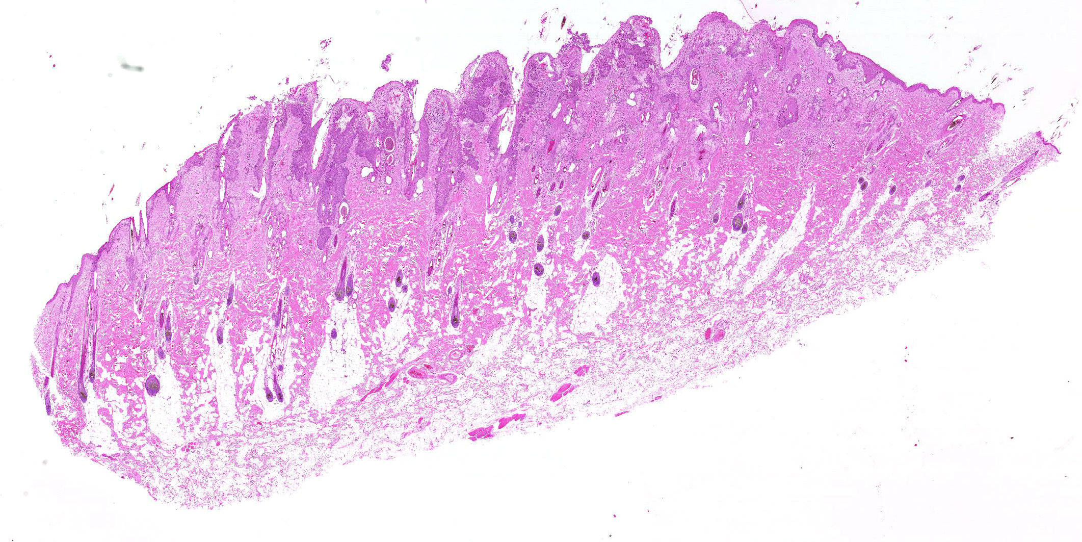
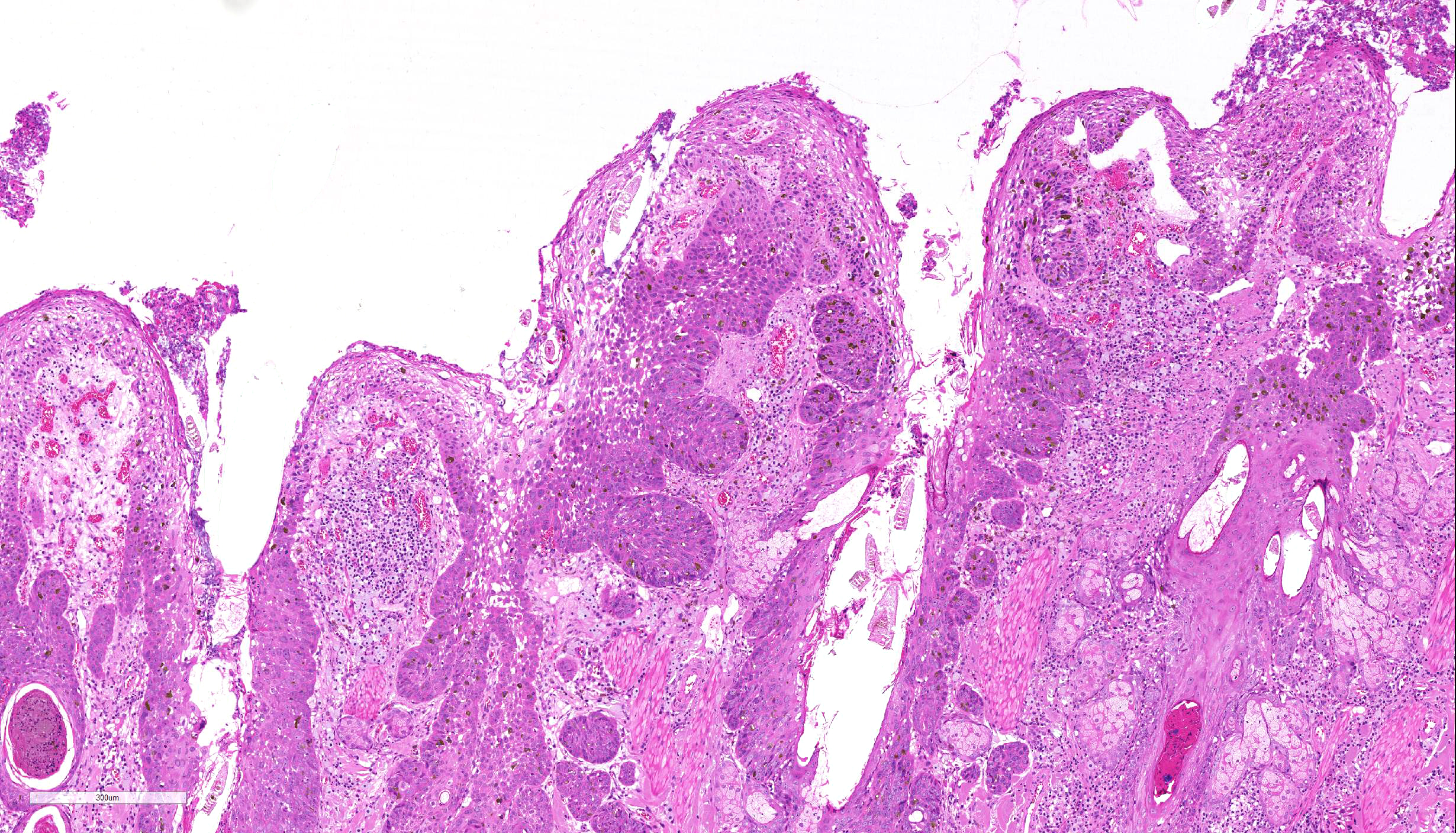
Haired skin: There is an area of irregular epidermal hyperplasia with formation of rete ridges, also affecting the hair follicle infundibulum; part of the lesion is covered by a serocellular crust. There is a mild hyperkeratosis and mild to severe hyperpigmentation. In most areas, the basement membrane is intact with neoplastic cells confined to the epidermis with dysplasia of all layers with loss of the polarity of the cell nuclei and loss of the normal stratification of the keratinocytes. Some keratinocytes are small with hyperchromatic nuclei, others are rather large with vesicular nuclei.
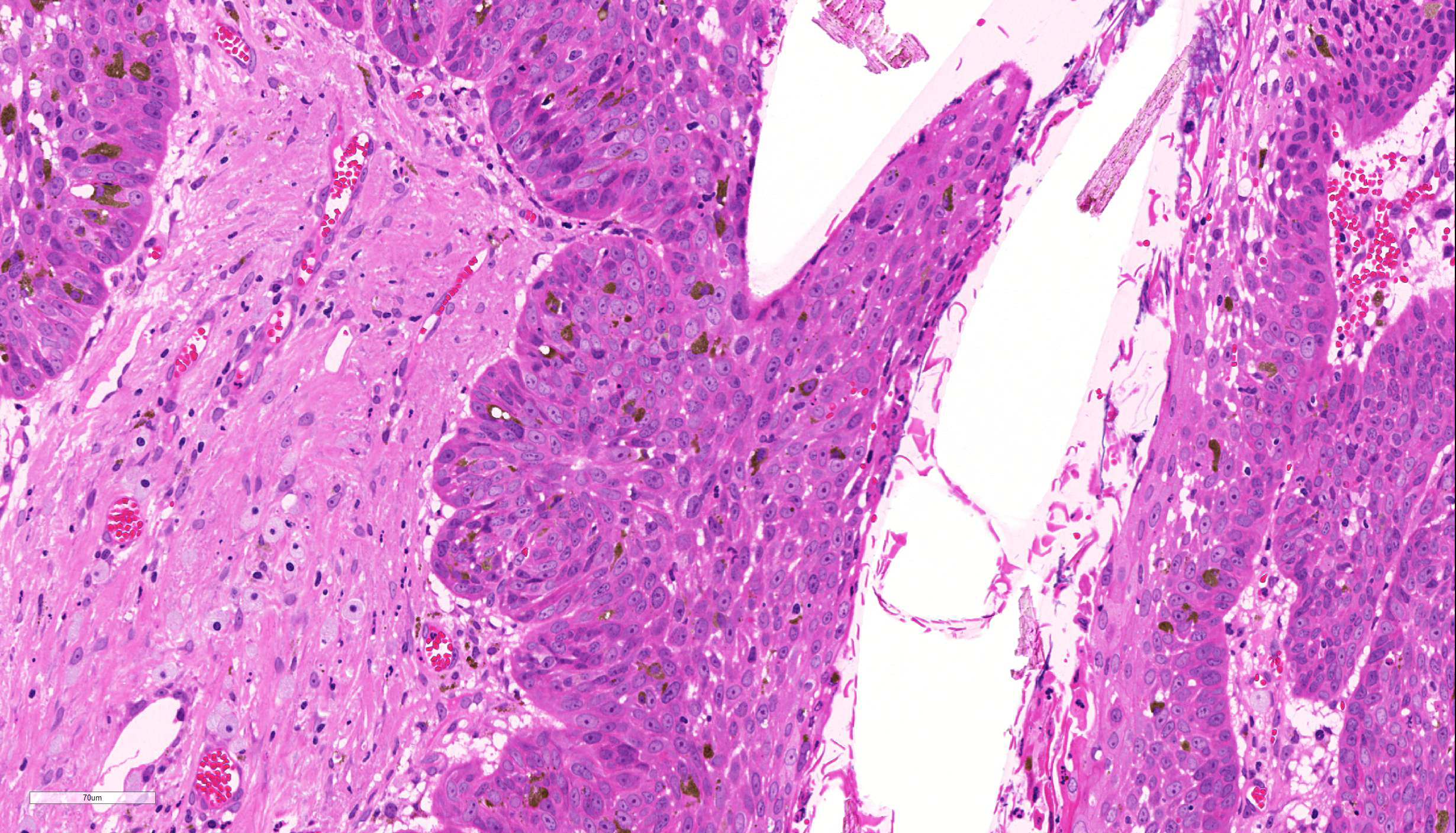
Groups of cells are dorsoventrally protracted and bent in one direction, exhibiting a "wind-blown” appearance. Nuclei are large, round to oval, centrally placed, and vesicular with finely stippled chromatin and one to two prominent round magenta nucleoli. There is a mild to moderate anisocytosis and anisokaryosis. The number of mitotic figures range from 0 to 4 per high power field. Multifocally, dark basophilic round structures are present within the neoplastic cells, interpreted as apoptotic bodies. In the dermis a mild, perivascular infiltration of neutrophils, lymphocytes and macrophages is present and few mast cells are observed. Multifocally there is moderate dermal fibrosis.
Contributor’s Morphologic Diagnoses:
Epidermal hyperplasia and dysplasia, focally extensive, severe (Bowenoid in situ carcinoma – BISC)
Contributor’s Comment: The morphologic findings are compatible with a Bowenoid in situ carcinoma (BISC) or Bowen-like disease, an uncommon premalignant lesion in middle-aged to old cats, that has been rarely reported in dogs.5,6
Grossly, irregular, slightly elevated to heavily-crusted plaques and verrucous or papillary lesions up to 5.0 cm in diameter are found on haired, pigmented or non-pigmented skin at any site of the body.3,5,6 Usually, multiple lesions occur, but also solitary lesions are seen infrequently.4
Lesions are reported to be chronic, not painful, and only mild pruritus is noted.3 There is neither a causal correlation to sunlight exposure nor a breed or sex predilection.5,6
Several studies suspect that papillomavirus infection may be associated with feline BISC in situ.3,7,8 In two studies, papillomavirus-antigen was detected in 11% and 48% of BISC in situ, respectively, using immunohistochemistry.7,8
Contributing Institution:
Institute of Veterinary Pathology at the Centre for Clinical Veterinary Medicine LMU, Munich
Veterinaerstr. 13; 80539 Munich Germany
JPC Diagnosis: Haired skin: Bowenoid squamous cell carcinoma in situ.
JPC Comment: John Templeton Bowen, MD was a quiet professor of dermatology who spent his days at Harvard Medical School dodging patients, preferring to read slides. In 1912, he published a paper on a particular form of precancerous chronic dermatosis. As his proposed name of “dyskeratosis lenticularis et discoides” didn’t appear to catch on widely, Dr. Jean Darier (who already had Darier’s disease named after him, so he wasn’t really in the game anymore) suggested in his rather aptly named 1914 textbook on dermatology, A Textbook on Dermatology, that this condition simply be called Bowen’s disease.1 Bowenoid papulosis, a proliferative lesion of the genitals resulting from infection by human papillomavirus 16 or 18, was named in Bowen’s honor in 1977, 37 years after his death, when he couldn’t refuse.
In humans, Bowen’s disease is the eponym commonly applied to cutaneous squamous cell carcinoma in situ. It appears as a sharply marginated erythematous scaly or verrucous, often pigmented plaque. It may be appear in sun-exposed areas of skin, or other areas where it may be associated with HPV infection or exposure to inorganic arsenic.9
In the cat, Bowenoid in situ carcinoma (BISC) presents as one or multiple thickened areas of the epidermis, which may present as a thick plate of keratin due to its propensity for overlying orthokeratotic or parakeratotic hyperkeratosis.4 This keratinization may extend into follicles which may become dilated and plugged with keratin debris. Over time, BISCs may progress to a nodular mass which bulges into the underlying dermis.4
The histologic features of the risks are characteristic. Poorly lesions display basal cell crowding and a loss of nuclear polarity and stratification. Cells within the stratum spinosum and even more superficial levels are enlarged with prominent nuclei and nucleoli are more consistent with cells of the basal layer. Mitoses may be present at any level in the dermis. Alternatively, as seen in this case discs may demonstrate a more basaloid pattern, with proliferation of epithelial cells with minimal cytoplasm and large often hyperchromatic nuclei. Melanization of these of the epithelial cells as well as macrophages in the underlying dermis is often seen in these lesions.4
Feline papilloma virus has been repeatedly isolated from lesions in this type, and cytopathic effects, such as koilocyte formation, associated with papillomavirus infection may be seen in early lesions. Similar changes are rarely seen in advanced lesions.4
Cats that develop one BISC are likely to develop additional lesions over time. Histologic differentials include invasive squamous cell carcinoma and actinic keratosis. The differentiation between a BISC and a SCC is made by careful examination to insure that all cells are confined within the basement membrane. Differentiation between actinic keratosis and a BISC may be more difficult unless papilloma virus-induced cell changes are present. As opposed to the BISC, actinic keratosis demonstrates random loss of nuclear polarity of the basal cells only, a poorly defined interface between normal and affected epidermis, and most importantly a lack of thickening and dysplasia of the follicular infundibulum.4
The association between feline papilloma virus infection in the cat in the development of BISC is well-known and consistent with an emerging body of knowledge associating papillomavirus infection and malignant transformation of epithelial neoplasms in cats and many other species. In the cat, Felis catus papilloma virus 2 and 3 (FcPV-2 and -3) have been isolated from BISC lesions. Other lesions associated with feline papillomavirus infection including feline viral plaques (FcPV-1 and -2) and FcPV-2 been detected not only in BISCs but in up to 50% of invasive SCC lesions in this species. Feline sarcoid is a rare neoplasms of the skin of the nose limbs, or digits of young to middle-aged cats; however, the papillomavirus that has been isolated from these lesions has extensive similarity in the DNA to bovine papillomavirus 1 and 2 and may represent a novel bovine papilloma virus.
The moderator discussed the requirements and methods for differentiation between papillomavirus-induced lesion and similar lesions that might be induced by actinic damage. The moderator suggested that the cellular proliferation extending deep into hair follicles as well as the bulbous outgrowth of neoplastic cells perpendicularly outward from the affected follicles is more suggestive of viral induction than actinic induction.
References:
- Arora H, Arora S, Sha V, Nouri K. John Templeton Bown. JAMA Derm; 2015; 191(13):1329.
- Baer KE, Helton K. Multicentric squamous cell carcinomas in situ resembling Bowen`s disease in cats. Vet Pathol.1993; 30: 535-543.
- Favrot C, Welle M, Heimann M, Godson DL, Guscetti F. Clinical, histologic and immunohistochemical analyses of feline squamous cell carcinoma in situ. Vet Pathol. 2009; 46:25-33.
- Goldschmidt MH. Munday JS, Scruggs JL, Klopfleisch R. Kiupel M. In Surgical Pathology of Tumors of Domestic Animals Vol. 1: Epithelial tumors of the skin. 3rd ed., Gurnee, IL: C.L. Davis and S.W. Thompson Foundation; 2018: 47-51.
- Gross TL, Ihrke PJ, Walder EJ, Affolter VK. Skin diseases of the dog and cat: clinical and histopathological diagnosis, 2nd ed. Oxford, UK: Blackwell science; 2005: 148-151, 575-584.
- Mauldin EA, Peters-Kennedy J. Integumentary System. In: Maxie MG, Hrsg. Jubb, Kennedy, and Palmer's Pathology of Domestic Animals. 6. Aufl. Edinburgh: Elsevier; 2016; 706-714.
- Munday JS, Kiupel M. Papillomavirus-associated cutaneous neoplasia in mammals.Vet Pathol. 2010; 47: 254-264.
- Nicholls PK, Stanley MA. The immunology of animal papillomaviruses. Veterinary Immunology and Immunopathology 2000; 73: 101–27.
- Patterson JW, Wick MR. Epidermal tumors. In AFIP Atlas of Tumor Pathology, Fourth Series. Non-melanocytic lesions of the skin. ARP Press, Bethesda MD. 2006: pp. 32-35.


