Signalment:
36-year-old male chimpanzee, (
Pan troglodytes).The chimpanzee was found dead. There was no history of prior illness, other than a single episode of vomiting several days prior to death.
Gross Description:
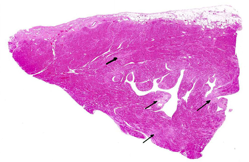
The intact heart is moderately, diffusely enlarged and weighs 497.3 grams. The heart:BW ratio is 0.68 (normal 0.54 - 0.60). There are multifocal streaks of pale, firm fibrosis, predominantly within the left ventricular free wall, interventricular septum, and apex of the heart. The right ventricle appears mildly dilated. Additionally, there is a focal discrete, white, firm 2 cm diameter multicystic nodule within the superficial to mid cortex of the right kidney.
Histopathologic Description:
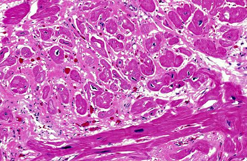
There is moderate to severe, multifocal to coalescing myocardial fiber degeneration, characterized by myocardiocyte pallor and loss of cross striations. There is multifocal, moderate cardiac myofiber disorganization and separation and replacement of myofibers by pale pink, predominantly mature fibrous connective tissue (fibrosis) and adipose tissue. There is significant fibrosis at the base of the mitral valve. The cardiac myocardial fiber nuclei are frequently moderately enlarged, basophilic, and bizarre. Minimal, light brown intracytoplasmic granular pigment (lipofuscin) is present in scattered myocytes. Rare, minimal to mild atherosclerotic changes are seen in random coronary arterioles, characterized by few mural macrophages, foamy cells and eosinophilic matrix material. There is multifocal mild lymphocytic and lesser histiocytic inflammation within the areas of fibrosis or surrounding degenerate myofibers and within the epicardial connective tissue.
Morphologic Diagnosis:
Heart: Moderate to severe, multifocal to coalescing, chronic myocardial degeneration and fibrosis with mild multifocal chronic myocarditis.
Condition:
Cardiomyopathy of great apes
Contributor Comment:
Cardiomyopathy was first reported in the chimpanzee in 1984, and the initial case report describes a 26-year-old male chimpanzee with history of heart murmur progressing to weight loss, heart failure, and coma over a 10 month period.(2) Gross lesions in this case included dilated cardiomyopathy, pericardial effusion, cerebral infarct, and hepatomegaly, and cardiac histopathology showed myocardial fibrosis and coronary atherosclerosis.(2) Subsequently, interstitial myocardial fibrosis has emerged as a major cause of heart disease and sudden death in chimpanzees, and in most cases there are no associated lesions of atherosclerosis.(3) Sudden death is thought to be secondary to conduction abnormalities and arrhythmias. A 2009 study of 87 adult chimpanzees found a 68% prevalence of heart disease and a 52% prevalence of idiopathic cardiomyopathy, with heart disease the primary cause of death.(5) In another study, interstitial myocardial fibrosis was identified as the most common cause of sudden death in chimpanzees, and was present in 92% cases of sudden cardiac death and 81% cases of all sudden death.(5) In humans, myocardial fibrosis occurs secondary to systemic hypertension, myocarditis, cardiomyopathy, or as response to myocardial injury, and can be a replacement (scarring) response to injury such as myocardial infarction or reactive and triggered by external stimuli such as pressure or volume overload.(5) In great apes, myocardial fibrosis with atrophy and hypertrophy of cardiac myocytes occurs with minimal or no inflammation and often no apparent cause or associated disease.(5) Potential biomarkers for myocardial fibrosis in chimpanzees have been investigated, and brain natriuretic protein and cardiac troponin I were elevated in cases of cardiovascular disease in one report.(1) In this study, neither a lipid panel including cholesterol, LDL, and triglycerides nor hsCRP, one of the best biomarkers for indicating ischemic disease in man, were useful in the diagnosis of heart disease in chimpanzees.(1)
JPC Diagnosis:
Heart: Fibrosis, multifocal, moderate, with myofiber degeneration, atrophy and loss.
Conference Comment:
The contributor provides an excellent overview of cardiomyopathy in great apes; the microscopic lesions in this case are consistent with this well-documented condition. Interestingly, myocyte degeneration and fibrosis appear most severe in the subendocardial and subepicardial tissue, while sparing the rest of the myocardium; the clinical significance of this finding is unknown. Additionally, moderate numbers of Anitschkow cells, also known as caterpillar cells due to their wavy nuclei, are present within affected areas of myocardium. These are thought to be macrophages or attempts at myofiber regeneration and are found in the myocardium in certain disease states.(4) Other striking features include widespread cardiomyocyte hypertrophy, characterized by fiber thickening and a change in nuclear morphology from spindle/cigar shaped to a box car like (rectangular) silhouette (see
WSC 2013-2014, case conference 17, case 2), as well as frequently enlarged, bizarre cardiomyocyte nuclei. Conference participants speculated that these nuclear changes may represent abortive attempts at regeneration.Â
References:
1. Ely JJ, Zavaskis T, Lammey ML, et al. Association of brain-type natriuretic protein and cardiac troponin I with incipient cardiovascular disease in chimpanzees (Pan troglodytes). Comp Med. 2011;61:163-169.
2. Hansen JF, Alford PL, Keeling ME. Diffuse myocardial fibrosis and congestive heart failure in an adult male chimpanzee. Vet Path. 1984;21:529-531.
3. Lammey ML, Baskin GB, Gigliotto AP, et al. Interstitial myocardial fibrosis in a captive chimpanzee (Pan troglodytes) population. Comp Med. 2008;58:389-394.
4. Maxie MG, Robinson WF. Cardiovascular system. In: Maxie MG, ed. Jubb, Kennedy and Palmers Pathology of Domestic Animals. Vol 3. 5th ed. Philadelphia, PA: Elsevier; 2007:35.
5. Seiler BM, Dick EJ, Guardado-Mendoza R, et al. Spontaneous heart disease in the adult chimpanzee (Pan troglodytes). J Med Primatol. 2009;38:51-58.

