Wednesday Slide Conference, 2025-2026, Conference 5, Case 3
Signalment:
11-years-old, male, mixed breed canine, Canis familiaris.History:
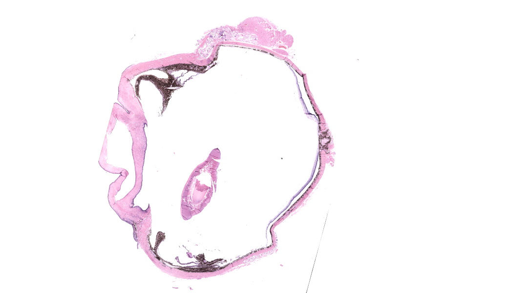
“Enucleated right eye”. No other history was provided.Gross Pathology:
An entire formalin-fixed eye was submitted for histopathology.Laboratory Results:
N/AMicroscopic Description:
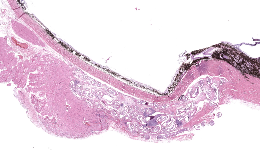
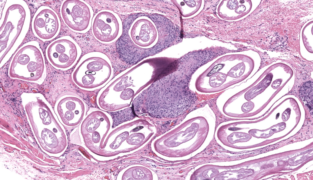
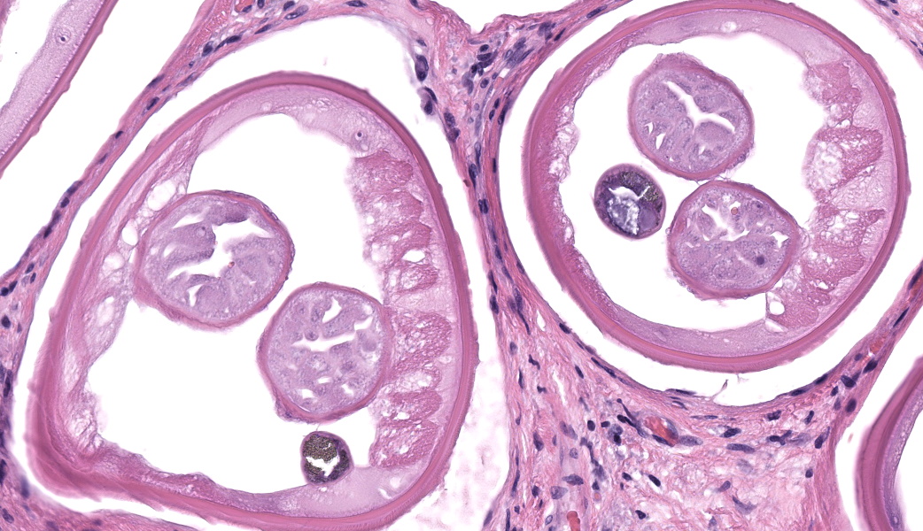
Enucleated right eye: Expanding and infiltrating the sclera, and occasionally extending to and dissecting the periocular skeletal muscle, are multiple cross and oblique sections of nematode parasites. Nematodes are characterized by a thick cuticle with annular ridges seen as raised areas in oblique sections, coelomyarian/polymyarian musculature that is atrophied and multifocally replaced by hypodermal tissue, a very small intestinal cross section, and reproductive organs. Mild hemorrhage, fibrin, and edema are present in affected sclera, and occasional nematode cross sections are surrounded by epithelioid macrophages. The cornea is hypercellular with low numbers of neutrophils and areas of neovascularization.Contributor's Morphologic Diagnoses:
Granulomatous scleritis and myositis with intralesional Onchocerca lupiContributor's Comment:
Onchocerca lupi is a parasitic, filarid nematode that is the causative agent of ocular onchocerciasis in domestic dogs.1 Cases have also been described in cats, one case was reported in a wolf, and coyotes in the southwestern United States have been reported to be probable reservoirs.1,2,3 Canine infections with Onchocerca lupi have been reported in Europe, North America, and in Asia.1,3,4,5Onchocerca lupi is also zoonotic, and human cases of Onchocerca lupi have been reported in the United States, Europe, North Africa, and in the Middle.4,6,7 Outside of the United States, reported human cases are mostly in adults, manifesting as localized ocular disease, however, human cases in the southwestern United States have been predominately seen in children, including at least 4 neuroinvasive cases.7,8The life cycle of Onchocerca lupi has not been completely described, but it is thought to be similar to that of other filarid nematodes. Canids are described as definitive hosts.4 Adult male and female nematodes develop within canine periocular tissues, and, after mating, females release microfilariae, which travel to the subcutaneous tissues, especially of the head, and the inter-scapular and lumbar areas.4 Rare cases of intraocular onchocerciasis, and of extensive aberrant migration of onchocerciasis have been reported in domestic dogs.9,10Wolbachia spp. have been described in adult Onchocerca lupi, and this bacteria is necessary for the reproduction and survival of other Onchocerca species, as well as of Dirofilaria immitis , another filarid nematode.8 Presumably, the intermediate host becomes infected with microfilaria while feeding on the definitive host. The exact intermediate host is unknown, but is thought to be a species of black fly (Simulium spp), as is the case in other Onchocerca species, and Onchocerca lupi DNA has been isolated from Simulium tribulatum in California, USA.11Onchocerca lupi DNA has also been found within Ceratopogonidae sp. (biting midges) in Northern Arizona and New Mexico, USA.12 Presumably, the larvae develop into infective L3s within the intermediate host, which then infects the definitive host during feeding.4
Clinically, canine cases of Onchocerca lupi may present with single or multiple, variably sized nodules on the bulbar conjunctiva, conjunctival hyperemia, episcleral injection, chemosis, periocular swelling, retinal detachment, and corneal edema.13,14 Typical gross lesions include nodules affecting the conjunctiva, episcleral tissue, and periorbital soft tissues.13 Some cases do not have reported clinical signs directly attributable to Onchocerciasis, and are diagnosed as an incidental lesion on biopsy secondary to other presenting causes like neoplasia.13 Bilateral involvement is described.14
Onchocerca lupi is typically diagnosed via histopathology, and to our knowledge no commercially available polymerase chain reaction (PCR) tests is available, although PCR assays have been described in the peer reviewed literature.7,14 Typical histologic lesions include nematodes with very small intestinal cross sections, atrophied coelomyarian/polymyarian musculature, and annular cuticular ridges that can be seen in longitudinal or oblique sections. Microfilaria may be seen within female worms. Variable amounts of inflammation ranging from mild to severe, and including macrophages, lymphocytes, and eosinophils may be seen, as can fibroplasia. We have seen pyogranulomatous inflammation as well in our laboratory. Currently, a commercially available serology test is not available, although serologic tests are in development.15
Contributing Institution:
NMDA Veterinary Diagnostic Services, 1101 Camino de Salud NE, Alberquerquw, NM 87102, https://nmdeptag.nmsu.edu/labs/veterinary-diagnostic-services.htmlJPC Diagnoses:
- Globe and periocular tissue: Scleritis and orbital rhabdomyositis, granulomatous, chronic, multifocal, moderate, with adult filarids.
- Retina, ganglion cell layer: Degeneration and atrophy, chronic, diffuse, severe.
- Globe, lens: Cataract.
- Globe, cornea: Keratitis, neutrophilic and histiocytic, chronic, multifocal, moderate.
JPC Comment:
This third case stimulated excellent discourse on ocular pathology and the associated changes seen in this eye. While the nematodes in the periocular tissues were striking, the other ocular lesions were a point of discussion focus for conference participants to ensure residents in attendance understood the importance of not getting “pigeon-holed” onto one lesion before examining the tissues in their entirety. This eye had multiple other lesions, some of which were subtle, and these caught the attention of the more experienced participants in the room. The keratitis in this eye served as a springboard to discuss the other changes, and conference participants suspect the keratitis was secondary to melanosis and inflammation causing occlusion of the drainage angle in this case, resulting in secondary glaucoma (as evidenced by degeneration of inner retinal cells and retinal ganglion cell loss) and subsequent buphthalmia. One of the more common sequelae to buphthalmia is an exposure keratitis due to inability of the eyelids to completely close over the enlarged eye. Coupled with possible irritation from the nematodes, participants felt that the keratitis and precluding factors were important features to discuss in this case.Conference participants unanimously agreed that the nematodes in section were consistent with Onchocerca lupi. For the sake of educational discussion, differentials for ocular nematodes in a dog were: Toxacara canis (ascarid), Onchocerca lupi (spirurid), Angiostrongylus vasorum (metastrongylid), Dirofilaria immitis (spirurid), Ancylostoma caninum (strongylid), Trichinella spiralis (aphasmid), and Thelazia spp. (spirurids). Of these, Thelazia spp. are the true ocular parasite and tend to reside in the conjunctival sacs and lacrimal ducts of infected animals. Onchocerca spp. in other species were briefly discussed, and honorable mentions included O. cervicalis, which sets up shop in the nuchal ligament of horses, O. gutturosa and O. lienalis, both of which invade the nuchal and gastrosplenic ligaments of cattle, and O. volvulus, the causative agent of “river blindness” in humans.
References:
- Labelle AL, Maddox CW, Daniels JB et al. Canine ocular onchocercosis in the United States is associated with Onchocerca lupi. Vet Parasitol. 2013;193(1-3):297-301.
- Tudor P, Iona?cu I, Mateescu CI, Bezerra-Santos MA et al. Feline ocular onchocercosis by Onchocerca lupi: Phylogenetic insights and implication for veterinary health. Acta Trop. 2023; 237:106723.
- Roe CC, Yaglom H, Howard A, et al. Coyotes as Reservoirs for Onchocerca lupi, United States, 2015-2018. Emerg Infect Dis. 2020 Dec;26(12):2989-2993.
- Rojas A, Morales-Calvo F, Salant H, Otranto D, Baneth G. Zoonotic Ocular Onchocercosis by Onchocerca lupi.Yale J Biol Med. 2021;94(2):331-34
- McGarry JW, Carrozza R, Bradley C, Latrofa MS, Makepeace BL, Otranto D. Onchocerca lupi in imported dogs in the UK: implications for animal and public health. BMC Vet Res. 2022 Feb;18(1):66.
- Otranto D, Sakru N, Testini G, Gürlü VP, Yakar K, Lia RP, Dantas-Torres F, Bain O. Case report: First evidence of human zoonotic infection by Onchocerca lupi (Spirurida, Onchocercidae). Am J Trop Med Hyg. 2011;84(1):55-8.
- Bowers Wu D, Ko B, Lopez Hernandez G, et al. Neuroinvasive Onchocerca lupiInfection in a Ten-Year-Old Girl. Case Rep Infect Dis. 2022; 2022:9773058
- Cantey PT, Weeks J, Edwards M, et al. The Emergence of Zoonotic Onchocerca lupi Infection in the United States--A Case-Series. Clin Infect Dis. 2016 15;62(6):778-83
- Komnenou AT, Thomas AL, Papadopoulos E, Koutinas AF. Intraocular localization of Onchocerca lupi adult worm in a dog with anterior uveitis: A case report. Vet Ophthalmol. 2016 ;19(3):245-249.
- Wallitsch K, Jaffey JA, Ferguson S, et al. Extensive Aberrant Migration of Onchocerca lupi in a Dog. Top Companion Anim Med. 2022;49:
- Hassan HK, Bolcen S, Kubofcik J, et al. Isolation of Onchocerca lupi in Dogs and Black Flies, California, USA. Emerg Infect Dis. 2015;21(5):789-96.
- Roe CC, Holiday O, Upshaw-Bia K, et al. Biting midges (Diptera: Ceratopogonidae) as putative vectors of zoonotic Onchocerca lupi(Nematoda: Onchocercidae) in northern Arizona and New Mexico, southwestern United States. Front Vet Sci. 2023 15;10:1167070.
- Dubielzig RR, Ketring KL, McLellan GJ, Albert DM. Veterinary Ocular Pathology: A comparative review. Saunders Elsevier; 2010.
- McLean NJ, Newkirk K, Adema CM. Canine ocular onchocerciasis: a retrospective review of the diagnosis, treatment, and outcome of 16 cases in New Mexico (2011-2015). Vet Ophthalmol. 2017;20(4):349-356.
- Latrofa MS, Louzada-Flores VN, Maia C, et al. Development of an indirect ELISA for the serodiagnosis of canine infection by Onchocerca lupi. Sci Rep. 2024;14(1):3348