Wednesday Slide Conference, 2025-2026, Conference 2, Case 4
Signalment:
7 year old male neutered Domestic Shorthair Cat.History:
A red plaque from the right upper conjunctiva was submitted for biopsy.Gross Pathology:
One irregular piece of pale tan, soft to firm tissue with no natural borders was received.Laboratory Results:
N/AMicroscopic Description:
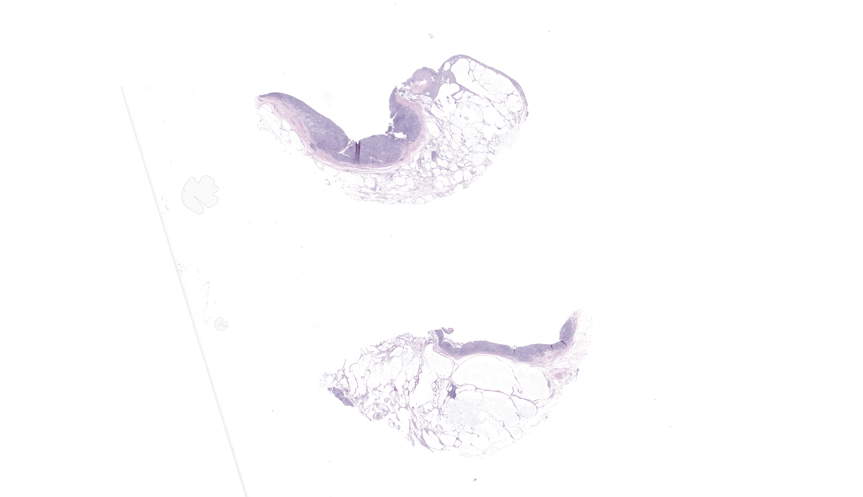
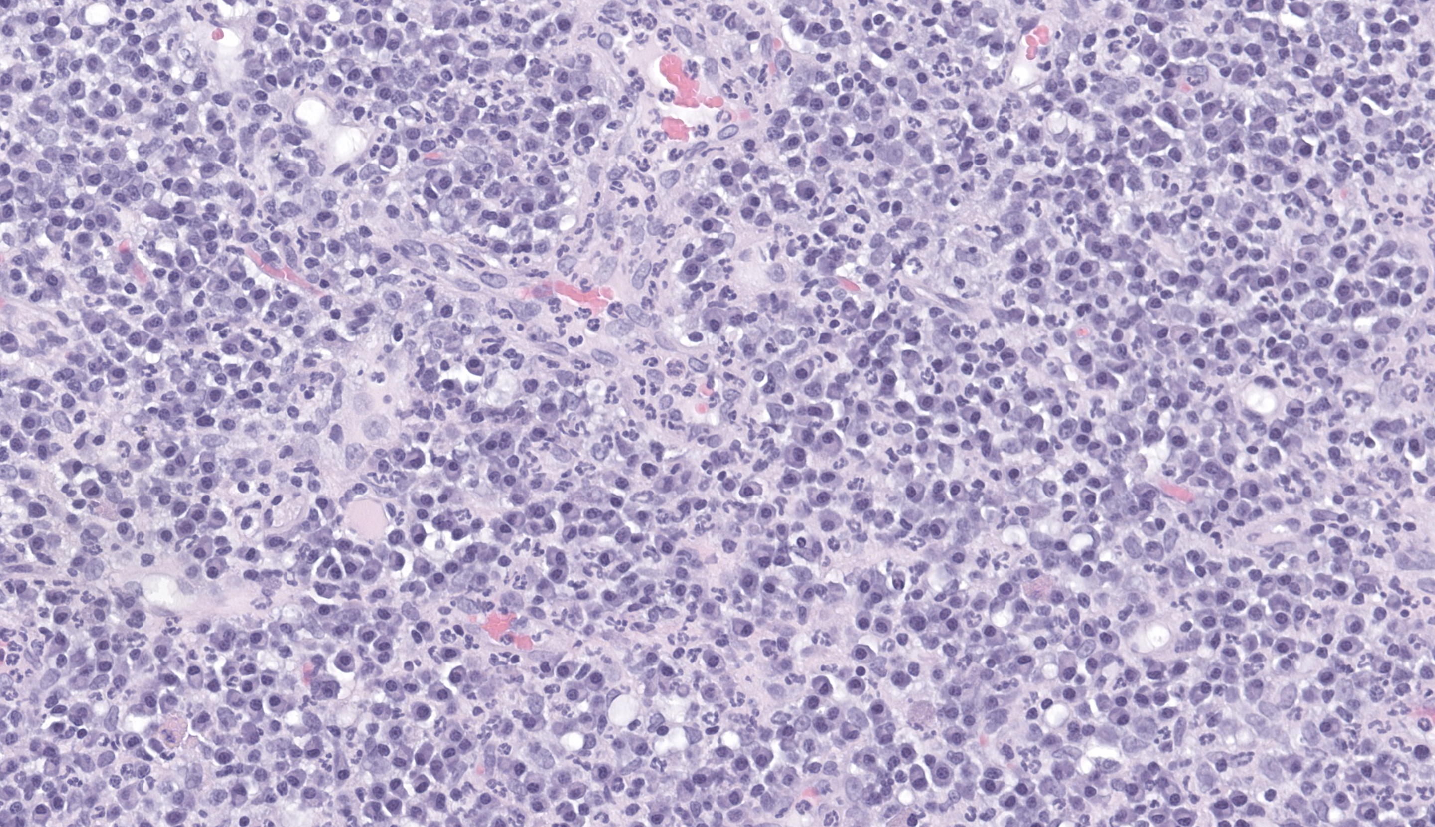
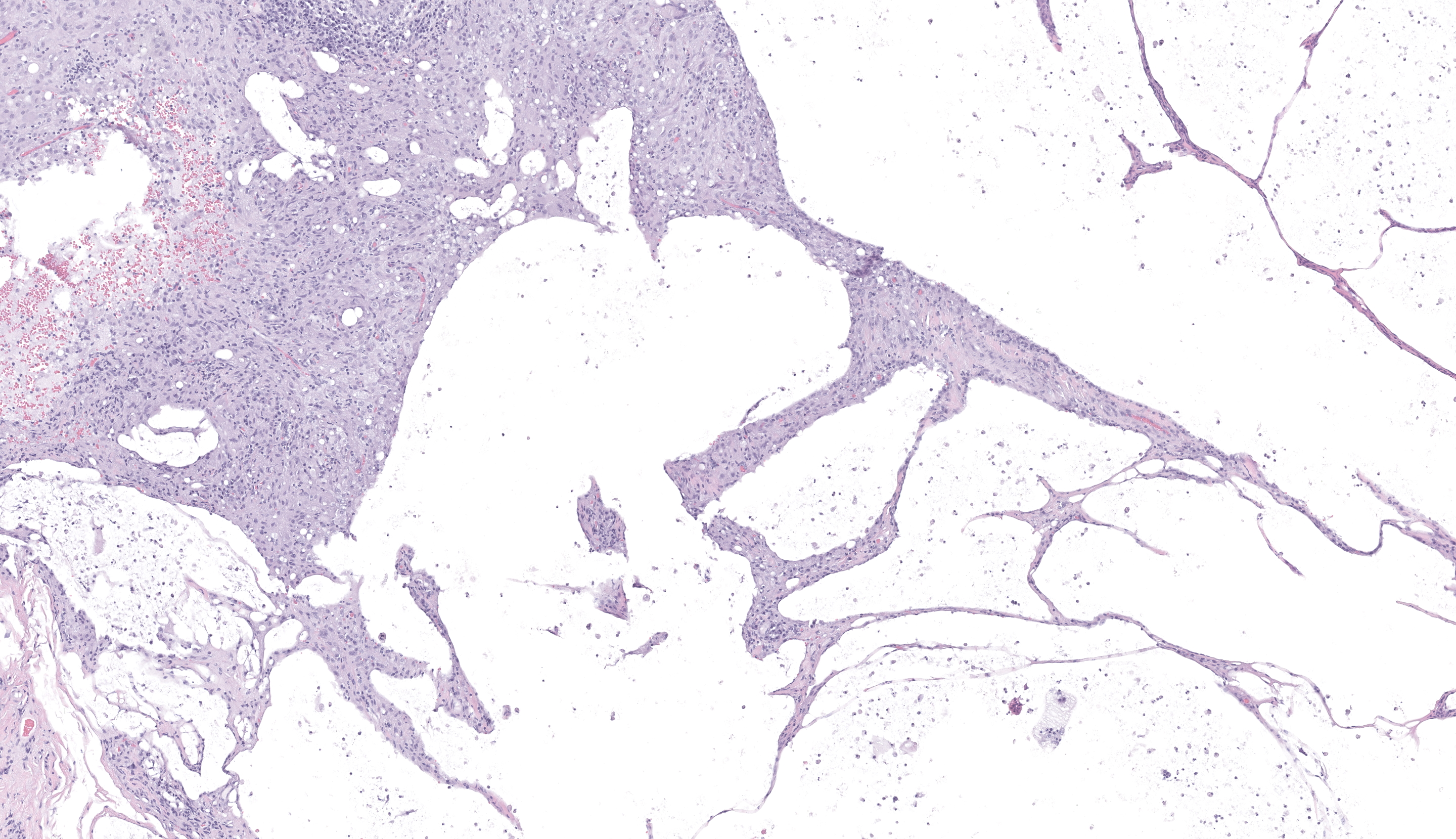
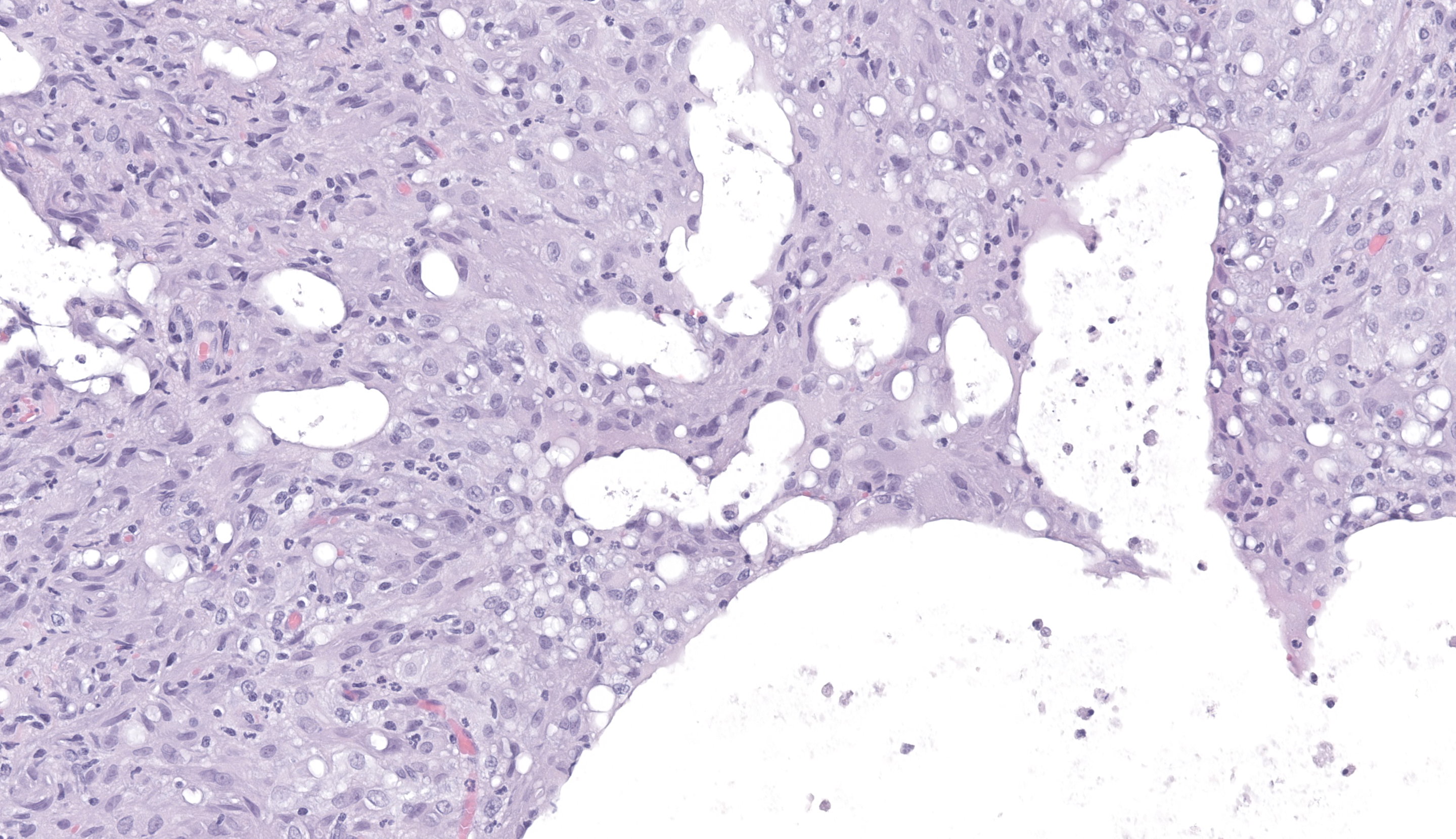
Skin (right upper palpebral conjunctiva). One bisected tissue is examined. The substantia propria is markedly expanded by coalescing, variably sized clear spaces (presumptive lipid), occasionally containing low numbers of macrophages, and surrounded by a small amount of fibrosis, few macrophages, and rare multinucleated giant cells. Larger aggregates of macrophages are present superficially. The superficial substantia propria is moderately expanded by low numbers of lymphocytes and few plasma cells.Contributor's Morphologic Diagnoses:
Skin (right upper palpebral conjunctiva): Marked chronic lipogranulomatous conjunctivitisContributor's Comment:
These findings are consistent with the uncommon condition known as Feline Lipogranulomatous Conjunctivitis. This condition results in nodular lesions, most commonly affecting the palpebral conjunctiva, and it occurs secondary to leakage of lipid-rich material into the tissue. Cats with sparse periocular pigment (such as white or orange cats) may be predisposed. One source reports that nearly a quarter of cats with this condition also had concurrent periocular neoplasia; there was no evidence of neoplasia in these sections. Complete excision is expected to be curative for this benign lesion.Contributing Institution:
University of Tennessee College of Veterinary Medicine, Department of Biomedical and Diagnostic Sciences https://vetmed.tennessee.edu/academics/biomedical-and-diagnostic-sciences/JPC Diagnoses:
Conjunctiva: Conjunctivitis, lipogranulomatous and lymphoplasmacytic, chronic, diffuse, severe.JPC Comment:
The last conference case of the day represented a unique feline entity that lent itself to a difficult tissue ID for conference participants. Many thanks to our contributor for this unique case! Conference participants waffled on anatomic location initially but eventually settled on either conjunctiva or mucocutaneous junction of some kind. Dr. Williams steered participants in the right direction to conjunctiva and drew special attention to the large lakes of clear space within the subconjunctival tissues. He turned those lakes into yet another lesson about paying special attention to what is NOT present. He stated that, when there is clear space, it should make the pathologist consider, “Is this air (emphysema), clear fluid, or fat?” In this case, those clear spaces were what was left of large lakes of lipid, characteristic in this condition, following tissue processing.Feline lipogranulomatous conjunctivitis includes characteristic non-ulcerated, smooth, white, conjunctival nodules within the palpebral conjunctiva. These are almost always adjacent to the eyelid margin and usually involve the meibomian glands.1,3 These nodules often cluster, are usually bilateral, and may be present on both upper and lower eyelids.1,3 However, lesions are usually more severe on the upper lid than the lower. This condition doesn’t generally respond well to treatment and surgery is usually recommended if there is irritation. Luckily, these cats typically have a good prognosis post-op. Feline lipogranulomatous conjunctivitis is similar to chalazia in dogs, except that chalazia are usually the result of Meibomian gland neoplasia and do not tend to cluster.3 Differential diagnoses for conjunctival inflammation with nodule formation in cats should include meibomian gland infections (i.e. marginal blepharitis or styes), eosinophilic keratoconjunctivitis, feline herpesvirus 1 (FHV-1), Chlamydia felis, feline calicivirus (FCV), and Mycoplasma spp. Conjunctival neoplasia should also be considered in these cases, as approximately 25% of cats with lipogranulomatous conjunctivitis have had concurrent neoplasms, usually squamous cell carcinomas.2,3
Inflammation within the Meibomian glands is usually minimal, but abnormal squamous epithelium may be seen within ducts. Usually, there is fibrosis surrounding the gland.3
The pathogenesis is currently thought to involve actinic (UV or solar) radiation-induced damage to the Meibomian glands, resulting in blockage of normal glandular secretion and leakage of sebaceous material into the surrounding tissues, which incites a granulomatous inflammatory response.
It is thought that the UV radiation leads to DNA damage and oxidative stress, followed by cytokine release and immune activation with subsequent damage to the gland. This might help explain why cats with low levels of periocular pigment are more susceptible to the condition, as well as why a quarter of these cats also have concurrent UV-related neoplasms.
On microscopic examination, the nodules are characterized by variably sized lipid lakes within the submucosal connective tissue. These lakes are usually surrounded by macrophages, multinucleated giant cells, and low numbers of plasma cells and lymphocytes.3 Macrophages frequently will have phagocytosed lipid droplets and/or cholesterol crystals.
References:
- Dubielzig, RR, Ketring, K, McLellen, GJ, Albert, DM. Veterinary Ocular Pathology. Elsevier; 2010. pp. 174, 179.
- Foster TM, Newbold GM, Miller EJ, Jeong YJ, Premanandan C, Husbands BD. Periocular fibrosarcoma with lipogranulomatous conjunctivitis in a cat. Vet Ophthalmol. 2025;28(3):646-650.
- Read, RA, Lucas, J. Lipogranulomatous conjunctivitis: clinical findings from 21 eyes in 13 cats. Veterinary Ophthalmology. 2001;4:93-98.