Wednesday Slide Conference, Conference 21, Case 2
Signalment:
An adult female Sprague Dawley rat (Rattus norvegicus)
History:
Patient presented with a multilobulated, firm facial mass on the left maxilla. Fine needle aspirate not consistent with abscessation or inflammation.
Gross Pathology:
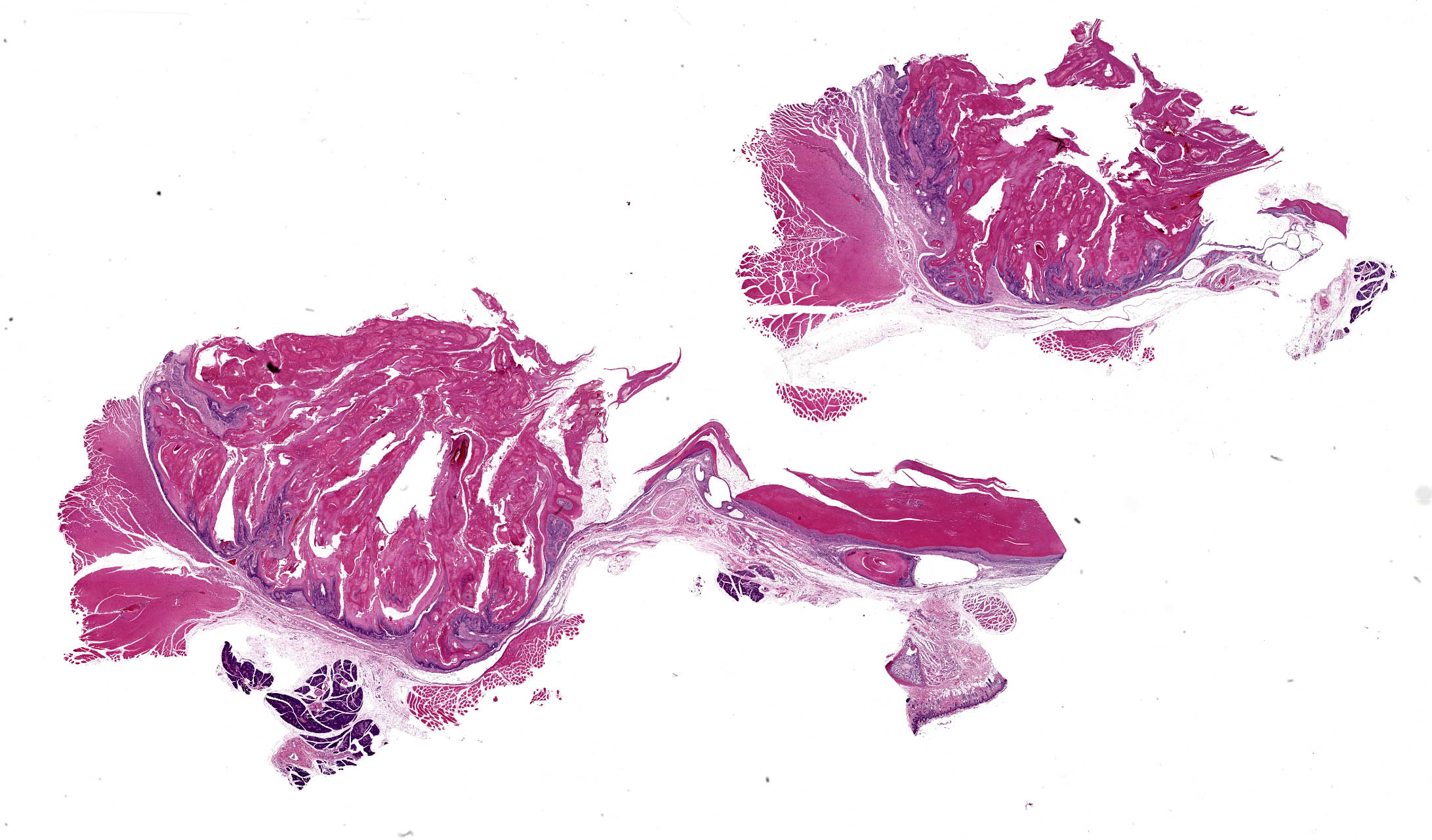
The subcutis at the base of the left pinna is expanded by a firm, bilobed mass. The upper lobe is spherical and measures 2 cm along the widest axis; the lower lobe is spherical and measures 1 cm along the widest axis. On cut surface, the mass is well circumscribed, has a fibrous capsule, and centrally contains copious amounts of friable, tan to pink material. The central material often displays a laminar appearance.
Microscopic Description:
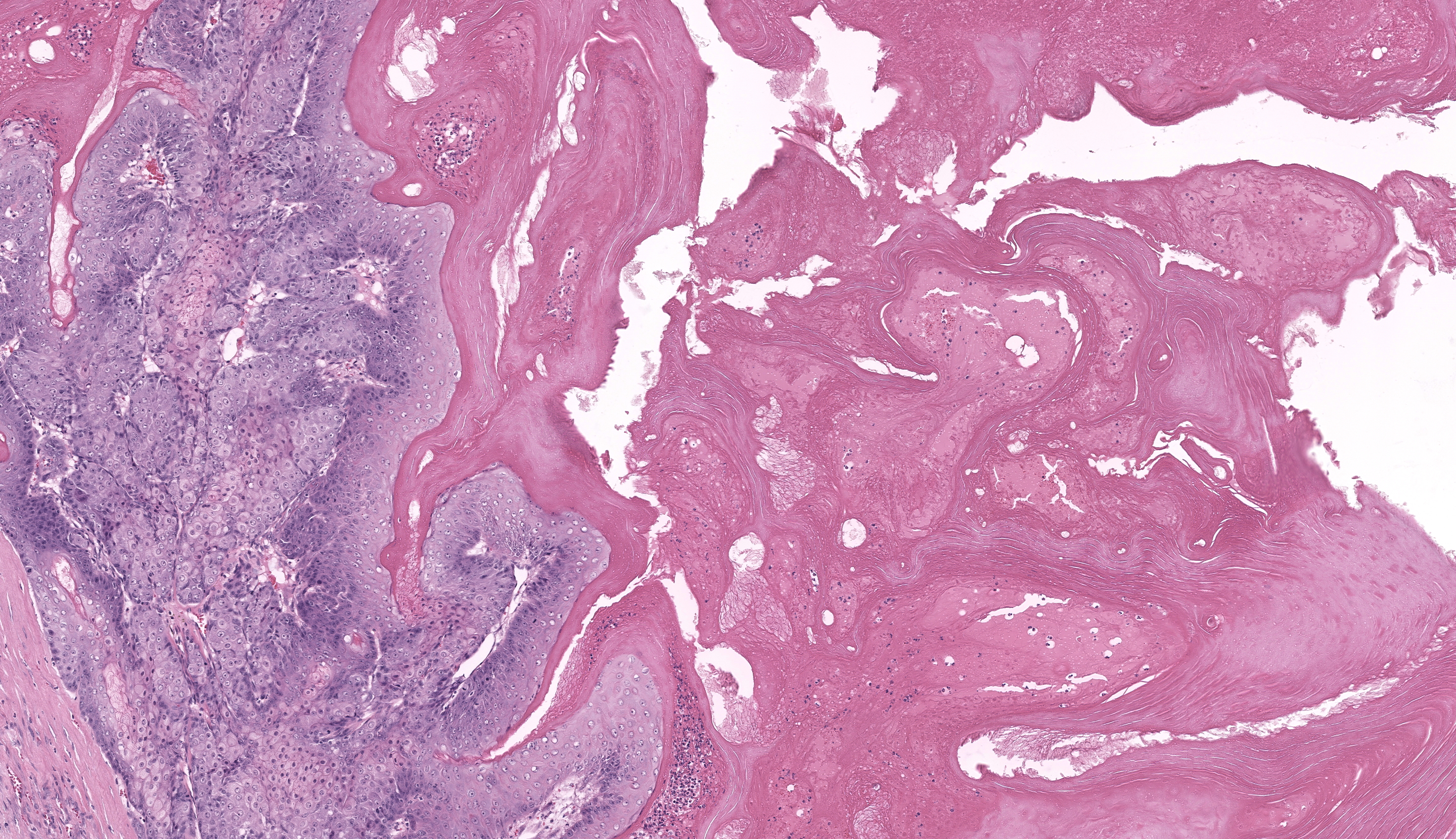
Haired skin: Examined are two sections of haired skin containing the grossly reported mass, skeletal muscle, and dermal adnexa. The mass is an expansile, well demarcated, and variable cellular neoplasm composed of well differentiated squamous epithelium arranged in trabeculae, islands, and papillary projections supported by pre-existing fibrovascular stroma. The neoplastic cells often exhibit sebaceous differentiation, with the mass centrally displaying squamous differentiation leading to abrupt keratinization organized into lamellated keratin and keratin pearls. The neoplastic cells have distinct cell borders, moderate to abundant finely vacuolated, eosinophilic cytoplasm, and round to ovoid nuclei containing finely stippled chromatin with one to two prominent nucleoli. Anisokaryosis and anisokaryosis are mild. Nine mitotic figures are observed in 10 HPF (2.37mm2). Few aggregates of lymphocytes, plasma cells, eosinophils, and neutrophils infiltrate the dermis adjacent to the mass. Apocrine glands adjacent to the mass are moderately dilated and contain scant, pale eosinophilic, acellular material.
Contributor’s Morphologic Diagnosis:
Zymbal’s gland: Adenoma
Contributor’s Comment:
Histologic evaluation of the mass is consistent with a Zymbal’s gland tumor due to cellular morphology (specifically sebaceous differentiation of neoplastic cells) and anatomic location. An adenoma is diagnosed based on the lack of invasion into the underlying stroma. Zymbal’s gland adenomas arise from Zymbal’s holocrine glands at the base of the external ear.1,2 These tumors often exhibit vacuolated cytoplasm, well differentiated squamous epithelium, and robust keratinization which are features in this case as well.1,2
Differentiation between an adenoma and a carcinoma of the Zymbal’s gland can be achieved based on histomorphology and growth patterns.1 Diagnostic features associated with adenomas include well-differentiated neoplastic cells, lobulated structure, a mixture of both basaloid and mature sebaceous cells, cystic areas, and lack of nuclear atypia.2 Diagnostic features associated with carcinomas include ulceration, irregular acini, papillary projections into cystic cavities, frequent mitoses, and increased cellular pleomorphism.2 Differential diagnoses for an adenoma include glandular hyperplasia or a well-differentiated carcinoma while those for a carcinoma include an adenoma (for well-differentiated carcinomas) or squamous papilloma.2
Zymbal’s glands are mostly observed and described in rats, however the structure is also present in mice.2 Zymbal’s gland typically measures approximately 3 to 5 mm in diameter, and is composed of three to four lobules.2 While spontaneous tumors are uncommon, Zymbal’s gland tumors are one of the most common tumor types in chemical associated carcinogenesis studies in laboratory rodents.3,4,5 Specifically, Zymbal’s gland tumors have been shown to be inducible by mutagenic compound 2-amino-3methylimidazo[4,5-5]quinoline among other chemical mutagens.5
Contributing Institution:
North Carolina State University College of Veterinary Medicine Department of Population Health and Pathobiology
https://cvm.ncsu.edu/php/
JPC Diagnosis:
Zymbal’s gland: Adenoma
JPC Comment:
This second case is a WSC classic with elements such as the parotid salivary gland, auricular cartilage, and cheek muscle aiding in tissue identification. Conference participants discussed differential diagnoses including papilloma and keratoacanthoma due to the abundant keratin in section. As the contributor notes, sebaceous differentiation of neoplastic cells is a key feature to distinguish a Zymbal’s gland tumor. Likewise, serial sections of this neoplasm might appear widely different (more or less sebaceous cells present) given the heterogenous distribution of these cells. We agree with the contributor that this neoplasm is an adenoma given the lack of invasion and bland cellular features. We attribute the presence of mixed inflammatory cells and edema to the inflammatory stimulus of abundant keratin. Dr. Radaelli emphasized that Zymbal’s gland tumors resemble clitoral and preputial gland neoplasms given the cell of origin, though the supporting architecture is different.2
References:
- Percy DH, Barthold SW, eds. Pathology of Laboratory Rodents and Rabbits. 4th ed. Blackwell Publishing; 2016.
- Rudmann D, Cardiff R, Chouinard L, et al. Proliferative and Nonproliferative Lesions of the Rat and Mouse Mammary, Zymbal’s, Preputial, and Clitoral Glands. Toxicologic Pathology. 2012;40(6_suppl):7S-39S.
- National Toxicology Program. Toxicology and Carcinogenesis Studies of 8-Methoxypsoralen (CAS No. 298-81-7) in F344/N Rats (Gavage Studies). Natl Toxicol Program Tech Rep Ser. 1989 Jul;359:1-130.
- Pucheu-Haston CM, Brandão J, Jones KL, et al. Zymbal gland (auditory sebaceous gland) carcinoma presenting as otitis externa in a pet rat (rattus norvegicus). Journal of Exotic Pet Medicine. 2016; 25(2):133–138.
- Makino H, Ishizaka Y, Tsujimoto A, et al. Rat p53 gene mutations in primary Zymbal gland tumors induced by 2-amino-3-methylimidazo[4,5-f]quinoline, a food mutagen. Proc Natl Acad Sci U S A. 1992 Jun 1;89(11):4850-4.