CASE II:
Signalment
One-month-old, male, European Shorthair cat (Felis catus)
History
The animal was handed over to a local animal shelter with multiple open, oozing skin wounds on several parts of the body. Treatment with amoxicillin showed no improvement. PCRs on feline herpes- and caliciviruses were negative. As the disease progressed, cutaneous nodules with purulent exudation began to occur. The cat died spontaneously and at the same time littermates started to show similar skin lesions.
Gross Pathology
At necropsy, multiple, extensive, erosive to predominantly ulcerative, partly proliferative skin lesions on the frontal, ventral, and lateral surfaces of the trunk and limbs as well as the inner surfaces of the nostrils and auricles were present. The superficial cervical and popliteal lymph nodes were moderately enlarged. The lung parenchyma showed multifocal firm white-gray foci.
Laboratory Results
A PCR on affected skin and lung tissue samples for Orthopoxvirus was positive. Immunohistochemical staining of lung tissue and skin for Orthopoxvirus antigen (mouse monoclonal ?Orthopox 5B1, German Armed Forces, Munich) was positive.
Microscopic Description
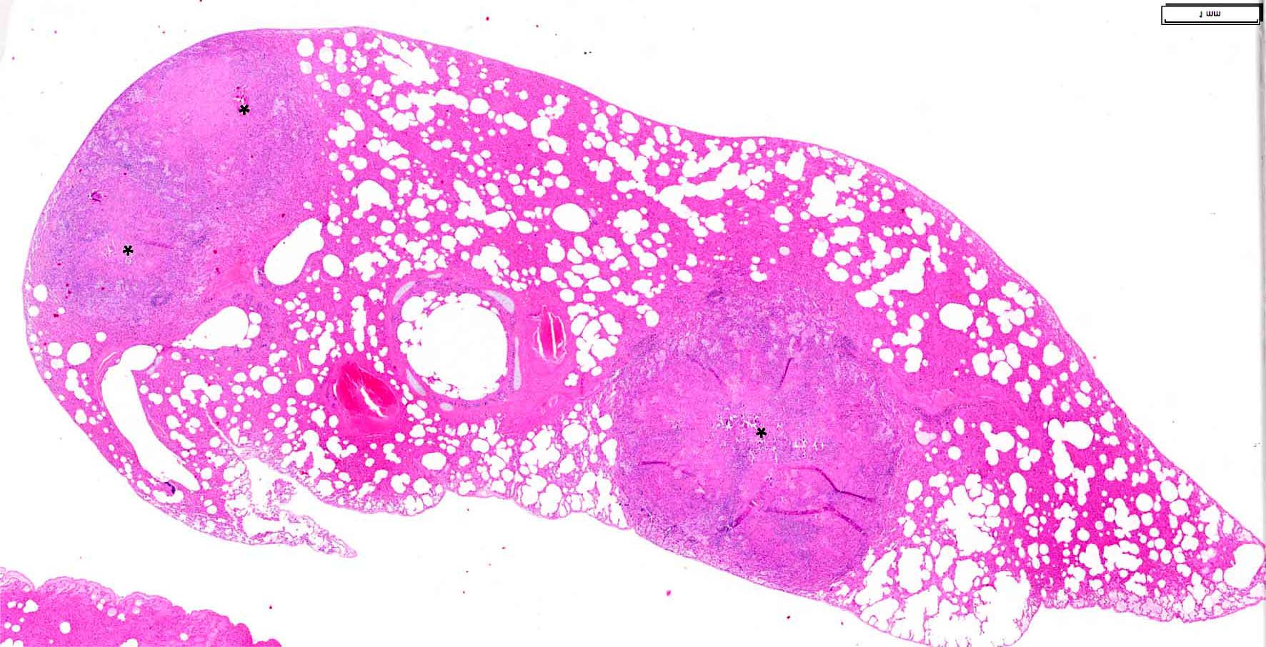
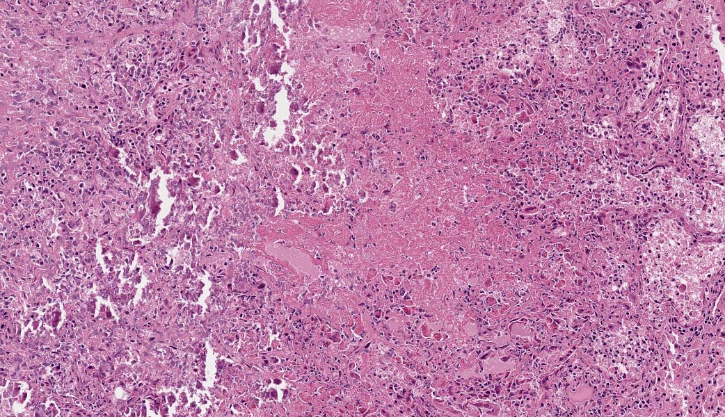
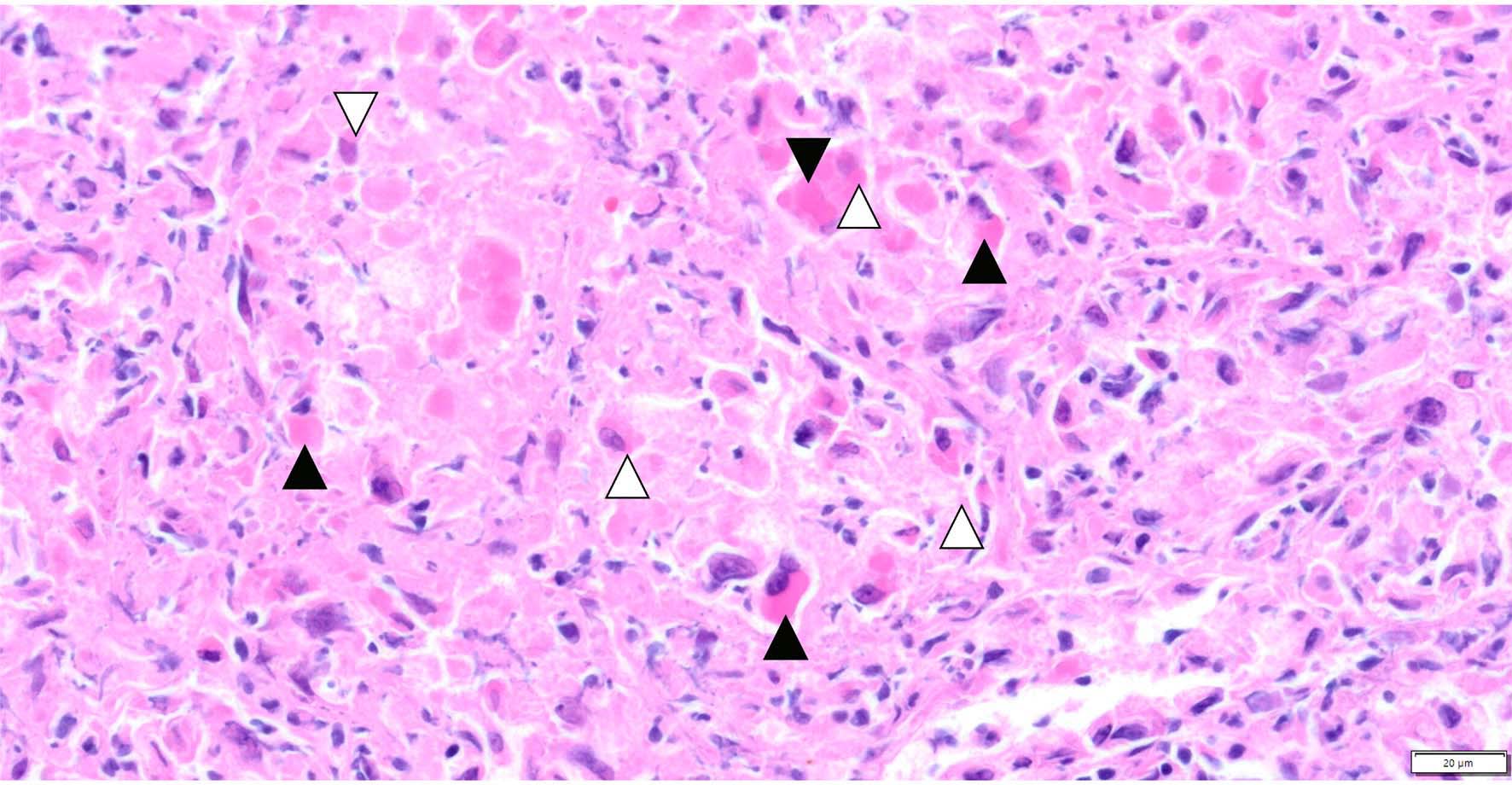
In the lung there are multifocal to coalescing consolidated areas with loss of the organotypical architecture. These areas measure up to 2.8 mm in diameter and are composed of karyorrhectic, -lytic and &emdash;pyknotic cells and homogeneous eosinophilic cellular debris (lytic necrosis). Necrotic foci contain moderate to large numbers of predominantly degenerated neutrophils, fewer macrophages, lymphocytes, plasma cells and occasional multinucleated giant cells. Central areas of the necrotic foci multifocally show mild mineralization of cellular debris (Fig. 2-2). Numerous macrophages and degenerated cells within the affected areas contain up to 10 &miro;m large, cytoplasmic, bright eosinophilic viral inclusion bodies (A-type inclusions, Downie bodies, Fig. 2-3). Occasionally small eosinophilic, perinuclear inclusion bodies were visible (B-type inclusions, Guarnieris bodies, Fig. 2-3). Adjacent alveolar spaces contain moderate numbers of neutrophils and macrophages admixed with fibrin and homogeneous proteinaceous fluid. Bronchiolar lumina are filled multifocally with moderate amounts of cellular debris and degenerated immune cells. The epithelium of bronchi, bronchioles, and terminal bronchioles show varying degrees of necrosis and squamous metaplasia.
Contributor's Morphologic Diagnoses:
Lung, pneumonia, necrosuppurative, lymphohistiocytic and plasmacytic, multifocal to coalescing, severe, subacute with numerous eosinophilic, cytoplasmic viral inclusion bodies, feline.
Contributor's Comment:
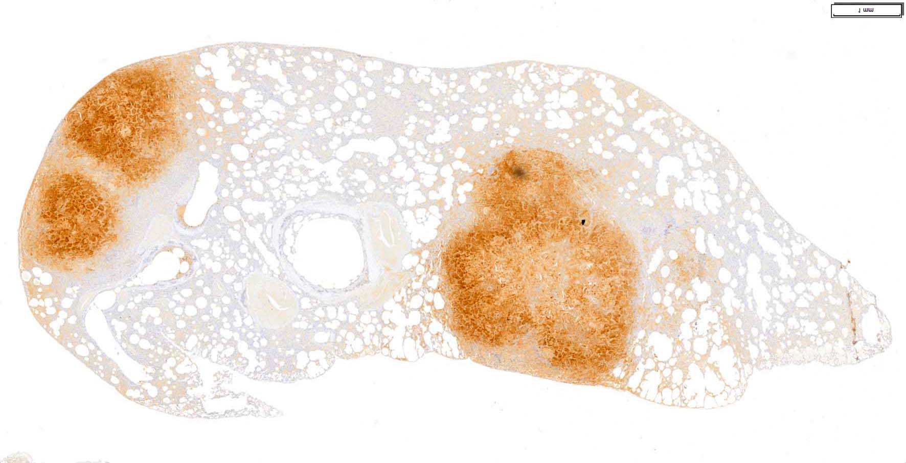
Orthopoxvirus (OPV) infections in domestic cats manifest as cutaneous and generalized forms of disease.1-5,12,13 Gross lesions, histopathological and immunohistochemical (Fig. 2-4 and 2-5) as well as molecular findings are consistent with a generalized OPV infection as described previously by Schulze et al. (2007).13 As in the case presented here the affected animal exhibited generalized skin lesions and severe viral pneumonia.13
OPV infections of domestic cats, first described in 1978, are mainly caused by members of the genus Cowpox virus (OPXV) and are usually restricted to the skin. 2,10,13 Initial infection is thought to occur during hunting or subsequent feeding on rodent reservoir species such as rats, bank voles and wood mice.10,13,14, Affected animals develop a primary skin lesion usually in exposed areas such as head, neck, forelimbs, or paws10 Typical lesions develop from pink or red colored macules to papules and fluid filled vesicles that further develop to pustules. The typical pox virus lesion is thereafter formed upon eruption of the pustule by a raised hemorrhagic ulcer.1,7 The epitheliotropic virus replicates in infected cells initiating leukocyte infiltration and epidermal hyperplasia resulting in lesion formation.1 Secondary cutaneous lesions develop during the viremic phase 1-3 weeks post infection, characterized by multiple ulcerating, rarely vesicular papules. Amelioration of lesions, usually occurring after 2-3 weeks, is thereafter characterized by thick gray crusts. Most animals recover within 5 weeks.10
Unlike other DNA viruses, OPVs replicate in the cytoplasm of infected cells. For several OPVs, two types of inclusion bodies can be differentiated.9 B-type inclusions, also called "Guarnieris bodies," represent juxtanuclear viral factories responsible for poxvirus DNA replication, intermediate and late gene transcription, protein synthesis, and assembly of virions. All poxviruses form B-type inclusions and manifest as small, round to oval, slightly basophilic perinuclear inclusions.6,9,8,11 A-type inclusions, also known as "Downie bodies," are only formed by certain strains of the species Cowpox virus, Ectromelia virus, Fowlpox virus, Canarypox virus, Raccoonpox virus, and Volepox virus. They are made up of large quantities of mature virions embedded in a cytoplasmic matrix of A-type inclusion protein. They are thought to increase virion stability after cell lysis.6,8 Histologically, they are characterized by one or multiple large, acidophilic cytoplasmic inclusions.11
Generalized infection with pneumonia represents a rare disease manifestation in domestic felids and is mainly seen in immunocompromised individuals or in association with systemic corticosteroid treatment.4,10,12,13 Previous reports include underlying infections with feline leukaemia virus, feline immunodeficiency virus (FIV), feline panleukopenia virus and feline herpesvirus.2,10 While OPV-induced pneumonia in juvenile cats seems mostly to occur in combination with widespread skin lesions, infection in older domestic cats or wild felids can occur in absence of typical secondary skin lesions.2,4,10,13
Although human infections are overall rare, multiple transmission events from cats or rats to their handlers have been reported.5,13-15 Human infection is characterized by typical cutaneous lesions like those described above. As in cats, lesions are mainly restricted to the hands and face of infected individuals, but generalized, even fatal, disease can develop in immunocompromised persons.5,14
Contributing Institution
Department of Pathology, University of Veterinary Medicine, Hannover Buenteweg 17, 30559 Hannover, Germany. http://www.tiho-hannover.de/kliniken-institute/institute/institut-fuer-pathologie/
JPC Diagnoses
Lung: Pneumonia, necrotizing, subacute, multifocal, marked with syncytia and numerous intracytoplasmic viral inclusions.
JPC Comment
The contributor provides an excellent, in-depth review of cowpox infection in felids, the species that, in the last decade, has become the most prominent source of zoonotic infections, particularly in Europe, where this infection is considered endemic. Rodents, rather than cattle, are the reservoir host for this poorly named virus and are the obvious source of feline infections (and likely many infections in zoo animals and few infections in cattle and humans). Cowpox infection has also been documented in large felids, anteaters, mongooses, rhinoceroses, alpacas, okapis, elephants, and cotton-top tamarins.16 Coxpox isolates from these various species have been grouped into numerous genotypes which vary significantly in their pathogenicity.16
As mentioned by the contributor, orthopoxviruses include cowpox, variola virus (the causative agent of smallpox), and vaccinia virus, which has been used for immunization since the 19th century, when Edmund Jenner first used it to vaccinate against smallpox. The actual identity of the virus used by Jenner (or perhaps his mentor, Dr. James Fuster, another bit of historical controversy) is unclear, a several possibilities exist for the true identify of the pocks on the hands of milkmaid Sarah Nelms, as there is no way that Jenner could definitively identify the causative agent. At that time, the pocks could also result from a range of other diseases, to include bovine papular stomatitis, pseudo-cowpox or para-vaccinia ("milker's nodules)", herpes mammillitis and papillomatosis, not to mention variola virus itself (the causative agent of smallpox.)17 It is likely that Jenner believed that he was actually using the agent of horsepox, a disease that was occasionally transferred from horses to cattle by farm workers. Interestingly, horsepox is a disease that does not exist today (the last natural case was seen in 1976 in Mongolia), but over the centuries, it likely evolved into what we call "vaccinia virus" today.17
While not "extinct" like horsepox, clinical cowpox in cattle is now extremely rare, but occurs sporadically in enzootic areas (Europe and Russia). Cowpox virus produces lesions on the teats and the contiguous parts of the udder of cows, and is spread through herds by the process of milking.16
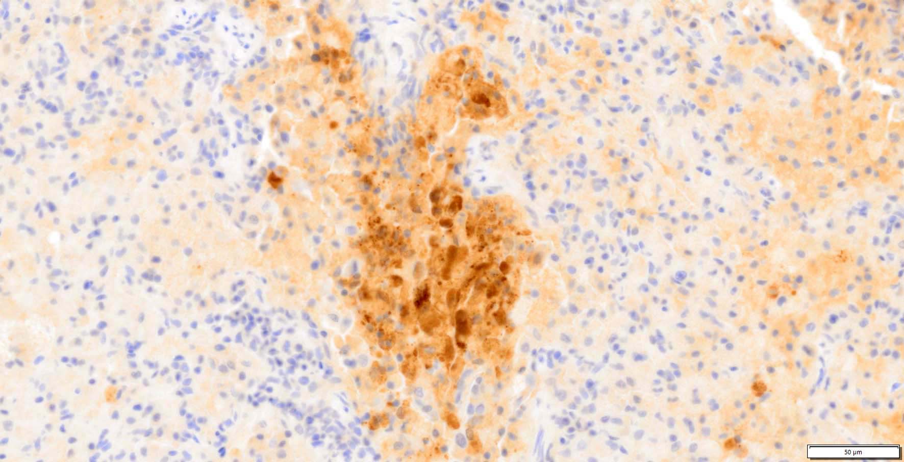
Vaccinia virus, in addition to being the orthopoxvirus most closely related to cowpox, is also the most closely studied orthopoxvirus. Because orthopoxvirus structural proteins are highly conserved, immunization with VACV provides cross-protection against variola and other orthopoxviruses, such as monkeypox. (This close antigenic relationship is crudely demonstrated in Fig. 2-6, in which an anti-vaccinia immunomarker was applied to the section in this case). But the utility of vaccinia virus extends well beyond its antigenic similarity and its role for hundreds of years as a vaccination proxy for smallpox. As significant disease is rarely seen with vaccinia inoculation (life-threatening infections are limited to approximately 1-2 individuals per million), it is also widely used in recombinant vaccines in humans carrying immunizing antigens of influenza, hepatitis B, rabies, herpes virus, HIV, among others. It has also been used to develop an oral vaccine against rabies for wildlife.
A few comments on the morphologic diagnosis in this case are likely appropriate. Based solely on histologic evaluation of the slide, it is very difficult to classify this particular pneumonia. The pathogenesis of the cowpox lesion is typical of an interstitial pneumonia, however, the multinodular appearance of the neoplasm in the submitted sections and the lack of diffuse interstitial thickening within the excellent sampling of this lung made diagnosis of an interstitial pneumonia difficult. In addition, the largest nodule in these sections surrounds a large airway. Because of the difficulties that classification presents in this particular H&E section, JPC staff has chosen discretion over valor and simply classified this case as "pneumonia".
Another interesting feature of this disease which made it into the morphologic diagnosis, is the presence of virally infected syncytia. This is an uncommon feature of most poxviral infections, but is well documented in cowpox. A previous case of cutaneous cowpox in a cat (WSC 2018, Conf 11, Case 2) also demonstrates syncytial cell formation.
References
- Bruneau RC, Tazi L, Rothenburg S. Cowpox Viruses: A zoo full of viral diversity and lurking threats. Biomolecules. 2023;13.
- Caswell JL, Williams KJ. Respiratory System. In: Maxie MG, ed. Jubb, Kennedy & Palmer's Pathology of Domestic Animals: Volume 2 (6th Edition). W.B. Saunders; 2016:465-591.
- Herder V, Wohlsein P, Grunwald D, et al. Poxvirus infection in a cat with presumptive human transmission. Vet Dermatol. 2011;22:220-224.
- Hinrichs U, van de Poel H, van den Ingh TS. Necrotizing pneumonia in a cat caused by an orthopox virus. J Comp Pathol. 1999;121:191-196.
- Hobi S, Mueller RS, Hill M, et al. Neurogenic inflammation and colliquative lymphadenitis with persistent orthopox virus DNA detection in a human case of cowpox virus infection transmitted by a domestic cat. Br J Dermatol. 2015;173:535-539.
- Howard AR, Moss B. Formation of orthopoxvirus cytoplasmic A-type inclusion bodies and embedding of virions are dynamic processes requiring microtubules. J Virol. 2012;86:5905-5914.
- James WD, Berger TG, Elston DM, Odom RB. Andrews' diseases of the skin : clinical dermatology, 10th ed. Philadelphia: Saunders Elsevier; 2006.
- Kastenmayer RJ, Maruri-Avidal L, Americo JL, Earl PL, Weisberg AS, Moss B. Elimination of A-type inclusion formation enhances cowpox virus replication in mice: implications for orthopoxvirus evolution. Virol. 2014;452-453:59-66.
- Leite JA, da Fonseca FG, de Souza Trindade G, et al. A-type inclusion bodies: a factor influencing cowpox virus lesion pathogenesis. Arch Virol. 2011;156:617-628.
- Mauldin EA, Peters-Kennedy J. Integumentary System. In: Maxie MG, ed. Jubb, Kennedy & Palmer's Pathology of Domestic Animals: Vol 1 (6th Ed.). W. B. Saunders; 2016:509-736.
- Pfeffer M, Meyer H. Poxvirus diagnostics. In: Mercer AA, Schmidt A, Weber O, eds. Poxviruses. Basel: Birkhäuser Basel; 2007:355-373.
- Schöniger S, Chan DL, Hollinshead M, et al. Cowpox virus pneumonia in a domestic cat in Great Britain. Vet Rec. 2007;160:522-523.
- Schulze C, Alex M, Schirrmeier H, et al. Generalized fatal cowpox virus infection in a cat with transmission to a human contact case. Zoonoses Publ Health. 2007;54: 31-37.
- Stolz W, Götz A, Thomas P, et al. Characteristic but unfamiliar--the cowpox infection, transmitted by a domestic cat. Dermatol. 1996;193:140-143.
- Wolfs TF, Wagenaar JA, Niesters HG, et al. Rat-to-human transmission of Cowpox infection. Emerg Infect Dis. 2002;8:1495-1496.
- In: Fenner's Veterinary Virology. Academic Press, 2016, pp. 157-174.
- Huygelen C. Jenner's koepokentstof in het licht van de moderne vaccinologie [Jenner's cowpox vaccine in light of current vaccinology]. Verh K Acad Geneeskd Belg. 1996;58(5):479-536; discussion 537-8. Dutch. PMID: 9027132.
- Jacobs BL, Langland JO, Kibler KV, Denzler KL, White SD, Holechek SA, Wong S, Huynh T, Baskin CR. Vaccinia virus vaccines: past, present and future. Antiviral Res. 2009 Oct;84(1):1-13. doi: 10.1016/j.antiviral.2009.06.006. Epub 2009 Jun 26. PMID: 19563829; PMCID: PMC2742674.
- Vanderplasschen A, Pastoret PP. The uses of poxviruses as vectors. Curr Gene Ther. 2003 Dec;3(6):583-95. doi: 10.2174/1566523034578168. PMID: 14683453.