CASE IV:
Signalment:
Four-month-old female castrated Berkshire pig
History:
Eighteen fair pigs were purchased from multiple sources. All pigs are showing respiratory signs and coughing. Twelve have responded to antibiotic treatment, two are still sick, and four are dead. Three of the dead pigs stopped eating and died over 10 days despite antibiotic treatment. This animal was wobbly, ataxic, and dragging her legs several days prior to being found dead.
Gross Pathology:
The eye is grossly unremarkable. Other pertinent gross findings include pale yellow to orange fluid in the pericardial sac and abdominal cavity that contains tan, friable, stringy material that attaches to the adjacent organs (heart or serosal organs respectively). The right coxofemoral joint contains tan, mucoid joint fluid and flocculant white strands, and similar material is present in the tarsal and metatarsal joints bilaterally.
Laboratory Results:
Mycoplasma culture: Growth of Mycoplasma hyorhinis
Respiratory culture: Moderate Streptococcus suis
No growth of Salmonella species
PRRS virus PCR: Positive
Microscopic Description:
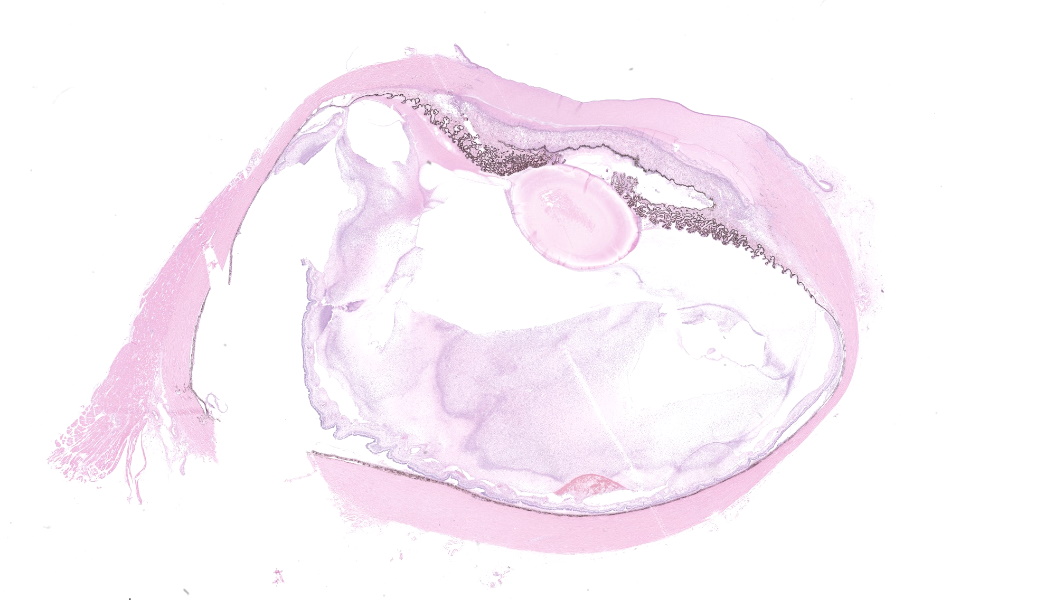
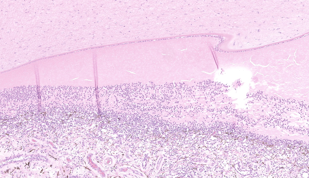
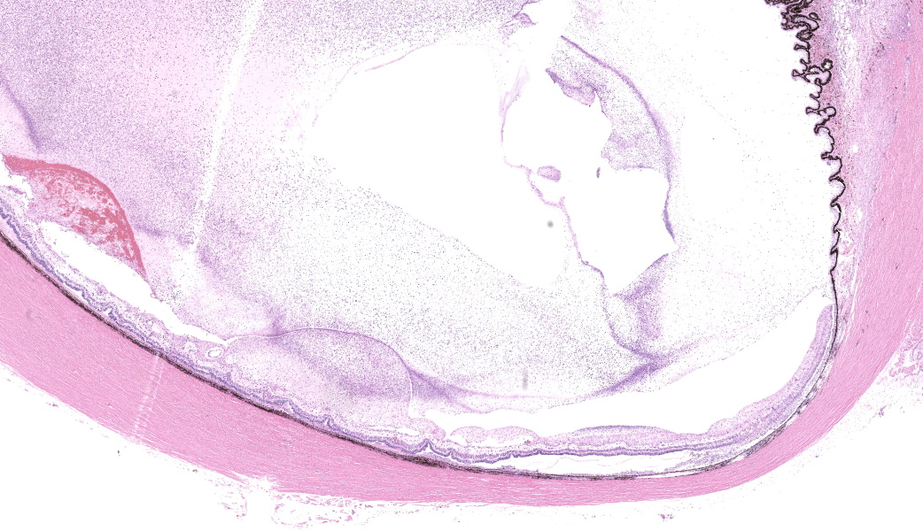
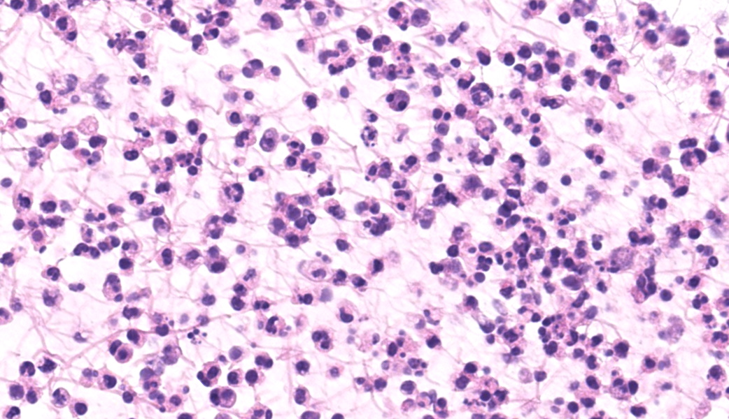
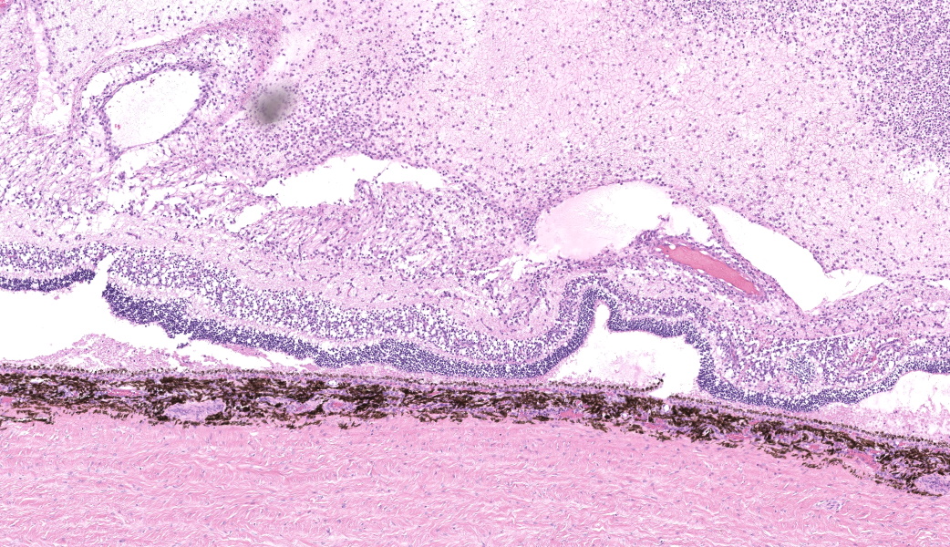
The vitreal space of the eye is filled with proteinaceous eosinophilic fluid, fibrin, and large numbers of neutrophils and foamy macrophages that contain dozens of small, round basophilic organisms that are gram-positive on a Gram stain. The inflammatory population extends into the ciliary body and retina. The retina is detached and multifocally obscured by eosinophilic amorphous material (necrosis), thin wispy eosinophilic material (fibrin), neutrophils, histiocytes, and lymphocytes. Fragments of retina and retinal vessels are frequently separated from the intact retina and surrounded by necrosis and similar inflammatory cells. The inflammatory population extends into the subretinal space, the sclera, and the conjunctiva.
Contributor's Morphologic Diagnoses:
Eye:
Severe fibrinosuppurative endophthalmitis and moderate lymphohistiocytic panuveitis
Severe diffuse retinal necrosis
Contributor's Comment:
Polyserositis is a condition affecting multiple organ systems of young pigs, typically from age 4-8 weeks. Polyserositis is a condition of economic concern, as it is associated with high morbidity and high mortality. There are several etiologic agents that can cause polyserositis, including Glaesserella parasuis (formally Haemophilus parasuis and the agent responsible for Glässer's disease), Streptococcus suis, and Mycoplasma hyorhinis.1 In all cases, the mechanisms of action are vasculitis and acute inflammation, which leads to leakage of the vessels into third spaces.1-3 Although this review focuses on Streptococcus suis, many points are true for the other common bacterial causes as well.
It is important to note that most healthy pigs carry Streptococcus suis.3 Disease occurs when the bacterium is introduced to immunosuppressed animals or in instances of stress such as overcrowding and poor ventilation. Transmission is through aerosolized mucus droplets that enter the body via the oral or respiratory tract.1
A presumptive diagnosis of polyserositis is often based on clinical signs, animal age, and gross findings. Necropsy can be highly suggestive of the condition. Tissue bacterial cultures are required to identify the exact etiologic agent and are often used to determine antibiotic susceptibility.3
Gross findings are often very distinct for this disease and include fibrinous peritonitis, pleuritis, and/or pericarditis as well as edema fluid in one or more cavities. The surfaces of the joints and meninges can also be affected. Clinically, these findings may result in trouble breathing, coughing, inadequate cardiac function, high fever, and lameness or neurological diseases.4 These bacteria can be found in conjunction with viruses such as Porcine Respiratory and Reproductive Syndrome virus (PRRSV) or porcine circovirus-2 (PCV-2), which can cause further mucociliary apparatus dysfunction.5 While the animal in this case was positive for PRRSV, the vaccination status of this animal was unknown. If vaccinated recently, the positive PCR result could be a result of recent vaccination rather than a true contributor to disease.
Histologic lesions may be reflective of the gross findings; there is often chronic multiorgan inflammation and fibrin. Vasculitis may or may not be present depending on the chronicity and severity of the disease. Bacterial organisms may or may not be seen. In this case, Gram positive round to elongated bacteria were identified, consistent with Streptococcus suis. 6
Disseminated septicemia is common in polyserositis but spread to the eyes is uncommon. There is one case report of disseminated Streptococcus suis to the eye of a man following the consumption of contaminated raw pork.7 This slide represents an interesting case of a common disease in an uncommon location.
Contributing Institution:
National Institutes of Health CBSTP
37 Convent Dr.
Bldg. 37, Rm. 2002
Bethesda, MD 20892
https://nih-cbstp.nci.nih.gov/
JPC Diagnoses:
Eye: Endophthalmitis, fibrinosuppurative, subacute, diffuse, severe, with vasculitis, Fiddesian synechia, retinal detachment and necrosis, and intra- and extracellular cocci.
JPC Comment:
The contributor provides a concise description of polyserositis in swine. This condition is typically associated with three particular agents in swine: Streptococcus suis, Glasserella (formerly Hemophilus) parasuis, and Mycoplasma hyopneumoniae. The disease known as polyserositis is often associated with systemic sepsis and abundant deposition of fibrin within the potential spaces
, which typically include the pleura, peritoneum (which may extend along the vaginal tunics in boars), pericardial space, meninges, and joints. They are also each a cause of pneumonia in this species, which may not involve the potential spaces.
Their involvement in ocular disease in swine is less well documented. Of the three, Mycoplasma hyorhinis is the agent most well associated with ocular disease in pigs. First identified from conjunctival isolates in swine in 1976, outbreaks of conjunctivitis in three unrelated swine farms in the Midwest were documented by Resende et al. in 2019.9 Affected pigs demonstrated initial lymphofollicular hyperplasia of the palpebral and bulbar conjunctiva, which progressed to diffuse lymphoplasmacytic inflammation. In the following year, isolates of this agent, which had initially been thought to be M.hyorhinis, were identified through MALDI-TOF as a unique Mycoplasma species.10
Mycoplasma spp. also result in conjunctivitis in several other species. M. synoviae and Mycoplasma gallisepticum are well known as causes of conjunctivitis in turkeys, and M. gallisepticum causes a similar lymphoplasmacytic conjunctivitis in finches and other songbirds. M. bovoculi and M. bovis may result in conjunctivitis in cattle, and M. conjunctivae has been reported to cause conjunctivitis and keratoconjunctivitis in small ruminants. M. felis may result in disease in cats, and M. genitalium has been reported to cause conjunctivitis in human patients with concurrent urethritis.
With regards to a more widespread evaluation of ocular pathology in pigs, the subject has rarely appeared in the Wednesday slide conference, with the sole previous submission of a pig eye occurring in 1982, in a pig with pseudo-rabies-induced conjunctivitis (the connection between alpha herpes virus infection and keratoconjunctivitis is well known in numerous species).
Eye disease is not commonly reported in swine, but is certainly important in any production species, as pigs with ocular disease will likely have a decreased feed-to-gain ratio due to discomfort, decreased ability to find/compete for feed, and other reasons. While husbandry issues (ammonia) are likely the most common cause of ocular disease in pigs (salt toxicity may also result in blindness), a wide range of other infectious agents are reported.1 The systemic effects of a range of viral disease may affect the eye, to include pseudorabies, African swine fever, classical swine fever, porcine cytomegalovirus, and influenza A.1 Bacterial agents affecting the eye (beyond those discussed above) include Chlamydia sp. Staphylococcus hyicus, and the effects of atrophic rhinitis.1 Riboflavin deficiency may result in cataracts, and finally a range of toxins may result neurologic disease resulting in blindness (arsenic, lead, mercury).
One helpful clue as to etiology for the observant pathologist (and mentioned in the contributor's morphologic description) are the multiple foci of polymerized fibrin in the retina. While this is suggestive of vasculitis in itself (and supportive of a diagnosis of S. suis infection in this case), close inspection of retinal vessels will demonstrate more definitive signs of vasculitis (hypereosinophilia of vessels wall, necrosis and cellular debris within the vessel wall itself, and perivascular hemorrhage and fibrin polymerization.) Like many pathogenic streptococci, S. suis possesses lipoteichoic acid on its surface, which, along with facilitating adhesion to host tissues and biofilm formation, is a potent stimulator of the immune system, resulting in cellular damage and tissue inflammation similar to that seen in the lipopolysaccharide (endotoxn) of gram-negative bacilli. The effects of lipoteichoic acid is extremely damaging to blood vessels, and vascular damage similar to than seen with traditional endotoxemia may be seen in streptococcal infections as well.
References:
- Zimmerman JJ, ed. Diseases of Swine. 10th ed. Wiley-Blackwell; 2012.
- Glässer Disease in Pigs - Generalized Conditions. Merck Veterinary Manual. Accessed June 27, 2025. https://www.merckvetmanual.com/generalized-conditions/glasser-disease/glässer-disease-in-pigs
- Streptococcus suis Infection in Pigs - Generalized Conditions. Merck Veterinary Manual. Accessed June 27, 2025. https://www.merckvetmanual.com/generalized-conditions/streptococcal-infections-in-pigs/streptococcus-suis-infection-in-pigs
- Staats JJ, Feder I, Okwumabua O, Chengappa MM. Streptococcus Suis: Past and Present. Veterinary Research Communications. 1997;21(6):381-407. doi:10.1023/A:1005870317757
- Obradovic MR, Segura M, Segales J, Gottschalk M. Review of the speculative role of co-infections in Streptococcus suis-associated diseases in pigs. Vet Res. 2021;52(1):49. doi:10.1186/s13567-021-00918-w
- Reams RY, Glickman LT, Harrington DD, Thacker HL, Bowersock TL. Streptococclcs Suis Infection in Swine: A Retrospective Study of 256 Cases. Part II. Clinical Signs, Gross and Microscopic Lesions, and Coexisting Microorganisms. J VET Diagn Invest. 1994;6(3):326-334. doi:10.1177/104063879400600308
- Li Z, Xu M, Hua X. Endogenous endophthalmitis caused by Streptococcus suis infection: a case report. BMC Ophthalmol. 2022;22(1):165. doi:10.1186/s12886-022-02389-9.
- Friis NF. A serologic variant of Mycoplasma hyorhinis recovered from the conjunctiva of swine. Acta Vet Scand. 1976;17(3):343-53.
- Resende TP, Pieters M, Vannucci FA. Swine conjunctivitis outbreaks associated with Mycoplasma hyorhinis. J Vet Diagn Invest. 2019 Sep;31(5):766-769.
- Hennig-Pauka I, Sudendey C, Kleinschmidt S, Ruppitsch W, Loncaric I, Spergser J. Swine Conjunctivitis Associated with a Novel Mycoplasma Species Closely Related to Mycoplasma hyorhinis. Pathogens. 2020 Dec 25;10(1):13. doi: 10.3390/pathogens10010013. PMID: 33375690; PMCID: PMC7824142.