Signalment:
Gross Description:
Histopathologic Description:
Eye (not on the slide): ulceration of the cornea; small foci of infiltrating lymphocytes and plasma cells at the limbus.
Morphologic Diagnosis:
Lab Results:
Condition:
Contributor Comment:
Rabbits can be easily infected experimentally and act as an animal model for human herpes simplex infection.(7) Trigeminally innervated areas, as for example the cornea, serve as the portal of entry for the human herpes simplex virus.(7) Furthermore, nasal infection is known to lead to focal lesions in the brain.(8)
JPC Diagnosis:
Conference Comment:
HSV can also result in fatal disease in nonhuman primates. In Old World primates, the course of HSV infection is comparable to humans with localization to the mucocutaneous tissues and relatively mild disease. However, New World primates (NWP) are considered highly susceptible and infection often leads to severe systemic disease and death with a rapid clinical course in many cases. In some NWPs such as owl monkeys and marmosets, they also develop ulceration of the oral mucous membranes but it is accompanied by hemorrhage and necrosis in the cerebral cortex as well as many other organs. In most of the reported cases in non-human primates, close contact with an infected human was found to be the source of infection. The most characteristic lesions in HSV infection in NWPs are oral ulcerations and lesions at the mucocutaneous junction, which cannot be grossly differentiated from lesions caused by herpesvirus T (Herpes tamarinds) infection. Molecular methods are needed to differentiate infections caused by the two viruses due to similarity of lesions.(4)
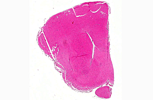
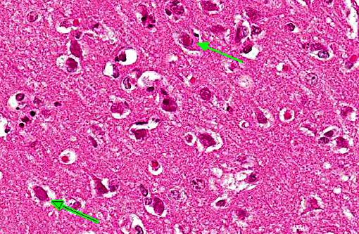
The conference description included multifocal, random areas of neuronal necrosis within the superficial gray matter accompanied by gliosis, satellitosis and low numbers of infiltrating heterophils. Perivascular cuffing by mononuclear cells is present multifocally within the gray matter and a similar mononuclear infiltrate as well as edema expand the meninges. The lack of neuronal degenerative changes such as swelling was noted by some participants; however, in some viral infections, neuronal necrosis (even in acute infection) may be the defining lesion.
References:
1. Grest P, Albicker P, Hoelzle L, Wild P, Pospischil A. Herpes simplex encephalitis in a domestic rabbit (Oryctolagus cuniculus). J Comp Pathol. 2002; 126: 308-11.
2. Hinze HC. New Member of the Herpesvirus Group Isolated from Wild Cottontail Rabbits. Infect Immun. 1971; 3: 350-354.
3. Nesburn AB. Isolation and characterization of a herpes-like virus from New Zealand albino rabbit kidney cell cultures: a probable reisolation of virus 3 of Rivers. J Virol. 1969; 3: 59-69.
4. Matos R, Russell D, Van Alstine W, Miller A. Spontaneous fatal Human herpesvirus 1 encephalitis in two domestic rabbits (Oryctolagus cuniculus). J Vet Diag Invest. 2014; 26(5):689-694.
5. M+â-ñtz-Rensing K, Jentsch KD, Rensing S, Langenhuyzen S, et al. Fatal Herpes simplex infection in a group of common marmosets (Callithrix jacchus). Vet Pathol. 2003; 40:405-411.
6. Onderka DK, Papp-Vid G and Perry AW: Fatal herpesvirus infection in commercial rabbits. Can Vet J. 1992; 33:539-543.
7. Paeivaerinta MA, Roeyttae M, Hukkanen V, Marttila RJ. Nervous system inflammatory lesions and viral nucleic acids in rabbits with herpes simplex virus encephalitis-induced rotational behaviour. Acta Neuropathol. 1994; 87: 259-68.
8. Stroop WG and Schaefer DC. Production of encephalitis restricted to the temporal lobes by experimental reactivation of herpes simplex virus. J Infect Dis. 1986; 153: 721-731.
9. Weissenboeck H, Hainfellner A, Berger J, Kasper I and Budka H. Naturally occurring herpes simplex encephalitis in a domestic rabbit (Oryctolagus cuniculus). Vet Pathol. 1997; 34: 44-7.


