CASE II:
Signalment:
12-year-old, male neutered, Quarter horse, Equus ferus caballus
History:
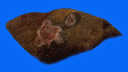
An ulcerated skin lesion, 4 x 3 cm in size, was submitted in toto with surrounding haired skin for surgical biopsy from the horse. The horse recovered uneventfully from surgery.
Gross Pathology:
The horse had an ulcerated skin lesion of the left cervical region.
Laboratory Results:
A formalin-fixed paraffin-embedded tissue block of the skin lesion was submitted for further speciation of the fungus. Panfungal polymerase chain reaction identified Curvularia spicifera with 100% DNA homology with reference strains for this species.
Microscopic Description:
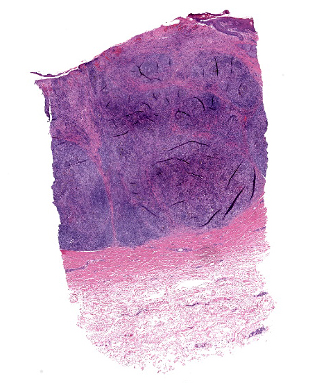
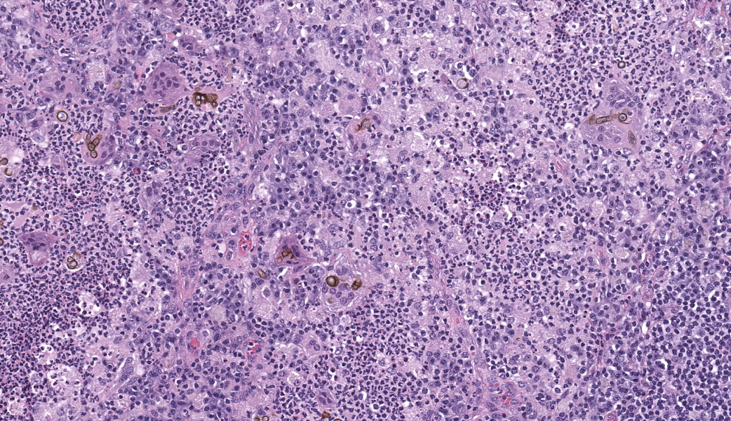
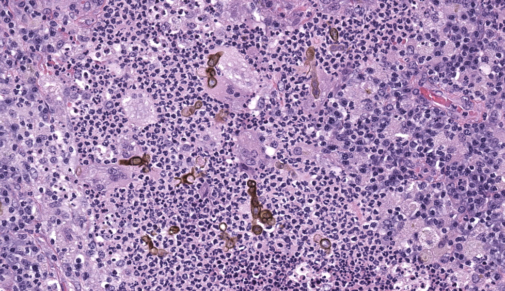
Haired skin: Sections of multifocally ulcerated haired equine skin and subcutis were examined. In the dermis and superficial subcutis, multi nodular and coalescent foci of pyogranulomatous inflammation were associated with pigmented fungi that were usually at the center of the pyogranulomas. The organisms were primarily arranged in pseudohyphae of spherical yeast-like cells that were 8-10 µm diameter and protruded from slightly larger (10-15 µm diameter) ovoid sclerotia. Germinating buds from the terminal yeasts of the short chains of pseudohyphae were consistent with germ tubes and these were 3-4 µm diameter. All stages of the organisms were translucent and uniformly pigmented pale tan, with the melanin pigment confined to retractile fungal cell walls. Degenerate neutrophils were concentrated in the central regions surrounding fungal elements. Langhan's giant cells, admixed with the neutrophilic debris, often contained various fungal elements as well. Smaller numbers of macrophages, neutrophils, lymphocytes and a few additional Langhan's giant cells were located in the tissue and debris surrounding the pyogranulomas and fungi. The ulcers were covered with serocellular debris and degenerate neutrophils. The flanking epidermis was hyperplastic. Within the intact epidermis, deep rete ridges contained a mildly elevated mitotic index of the stratum basale. Hypergranulosis with keratinohyaline granules of the cells of the middle epidermal strata was noted. Laminar hyperkeratosis of the stratum corneum was apparent as well in the remaining epidermis. Within the submitted tissue sections, inflammation and fungi did not extend to margins of the surgical biopsy tissue.
Contributor's Morphologic Diagnoses:
Haired skin: dermatitis, ulcerative and pyogranulomatous, multinodular and coalescent, chronic, moderate, with pigmented fungal elements, Quarter horse, (Equus ferus caballus).
Contributor's Comment:
Many species of pigmented (dematiaceous) fungi are known as plant pathogens and a number of them as opportunistic pathogens in human and animal diseases.1,2 Pigmented fungi can be identified on a microscopic level in tissues, however identification to a species level requires molecular techniques.3 Human cases of systemic phaeohyphomycosis are reportedly more common as a result of immune suppression during cancer therapy, renal dialysis and steroid therapy.4 Superficial phaeohyphomycosis in veterinary species, as with the equine patient in this case, is far more common than systemic disease and immune suppression is not likely involved in the pathogenesis.5 In one reference, the presence of the fungal granulomas primarily on the head and neck of a series of horses suggests a traumatic event as a common cause of infection.5 Chromomycosis is a more inclusive term for phaeohyphomycosis and chromoblastomycosis and may indicate either superficial infection or systemic disease.6
The description of dematiaceous fungi as primary plant pathogens and occasional opportunistic pathogens of the skin of horses would support the ubiquity of this group of organisms in association with plants and subsequent injuries from affected plant awns and branches.2
Dogs, cats, cattle, horses, and goats, as well as humans are affected by cutaneous dematiaceous fungal species.4,6,7 The identification of pigmented fungal agents to a species level is complicated by the large numbers of pigmented fungi that can infect animals and the histomorphological similarities between species. Molecular diagnostics are needed for speciation when speciation is necessary.3
Curvularia species are in the Ascomycota and sexual reproduction involves production of ascospores in the sexual stages. The asexual stages are the survival stages that evolve to escape harsh environmental conditions.1,2,8 Recent molecular studies have refined speciation of the genus Curvularia.2 The asexual stage of most Curvularia species was known as Bipolaris until molecular characterization.1
A few virulence factors have been described for the pigmented fungi. Melanin is the most universal virulence factor for pigmented fungi9,10. As with melanin in animals, it acts as a protection from environmental stressors such as ultraviolet light and host cell oxidative and immune mechanisms. The chemical structures of various melanin molecules varies, however, in fungi, melanin acts as a structural polymer that is linked to chitin in the cell wall.11 As with the Curvularia spicifera in the horse in this report, the pale tan fungal melanin is confined on a microscopic level to the cell wall.
In many species of fungi, the dormant stages are also considered virulence attributes. In Curvularia species and many other diverse groups of fungi, sclerotia form in adverse conditions to protect the organisms from drying, ultraviolet light, tissue cytokines and other noxious influences.12. Numerous germ tubes and mycelia were noted in the tissue of the horse in this case, and many originated from the pigmented sclerotia.
Mycoviruses are mostly RNA viruses that act as virulence factors for some endophytic fungi, notably Curvularia protuberata, an endophytic fungus of panic grass (Dichanthelium lanuginosum). The mycovirus, Curvularia thermotolerance virus (CThTV) confers thermotolerance to panic grass when present as an endogenous virus of C. protuberata, thus allowing the grass to grow at elevated temperatures.13 To the author's knowledge, there are no mycoviruses associated with enhanced pathogenicity of chromomycotic agents when infecting animals, however.
Systemic chromomycoses, though rare in animals, most often involve fungal infections of the brain and spinal cord that are invariably fatal.14 Occasional respiratory manifestations and generalized spread also occur. Most of these infections metastasize from environmental contamination in immune-suppressed humans and animals. Phaeohyphomycoses are considered emerging diseases among humans with chronic immune suppression. Systemic animal chromomycoses are also considered more prevalent with compromise of the immune system. Cutaneous infections in animals are more common and not linked to any depression in immune capabilities.5
Contributing Institution:
New Mexico Department of Agriculture Veterinary Diagnostic Services 1101 Camino de Salud NE Albuquerque, NM 87102 New Mexico Department of Agriculture
JPC Morpholoic Diagnoses:
Haired skin: Dermatitis, pyogranulomatous, chronic, multifocal to coalescing, severe, with pigmented fungal hyphae.
JPC Comment:
The contributor provides an excellent writeup on the breadth of pigmented fungal infections in animal species. These agents have been well represented in the Wednesday Slide Conference over the years, with 14 cases dating back to 1987. In these cases, horses and cats have been overrepresented, with cutaneous and nervous system infections predominating (which is quite representative of the species and system predilection for these agents when infecting mammals). In cats, solitary cutaneous lesions on the face and paws predominate, while multiple lesions on various parts of the body are most often seen in horses. As mentioned by the contributor, most infections in domestic species, as opposed to humans, are seen in immunocompetent individuals.15
There are a number of additional "virulence factors" described for these pathogens, which assist in protection both in the environment and following infection of animal tissues. The presence of melanin is easily the most well-researched in pathogenic fungi, with Cryptococcus neoformans and the various dematiaceous yeasts being the most renowned employers of this compound. In the environment, this compound protects the fungi from the stresses of ultraviolet and electromagnetic radiation, promotes thermotolerance, and assists in protection from unicellular predators, like amoeba.16 During infection of animal tissue, melanization helps shield C. neoformans and dematiaceous fungi from engulfment and killing via the oxidative burst by host macrophages.16 In addition, melanin likely promotes neurotropism in these fungi16 - melanin is a very common product of cells derived the neural tube, and its presence likely results in a form of protective mimicry for these fungi when infecting the nervous system.
Additional factors that likely contribute to their pathogenicity both in the environment and within animal tissue include the presence of carotene (also protective from a variant of oxidative stresses), formation of thick cell walls, presence of yeast-like phases (easily the most environmentally sound of the various stages of fungal evolution), adhesion, hydrophobicity, and production of siderophores (a virulence factor best known for its employment by many forms of pathogenic bacteria.5
Finally, the confusing terminology associated with dematiaceous fungal infections probably bears some review. A recent review article by Rodrigues et al.17 provides an excellent resource for the terminology of all fungal infections in animal tissues, including those of the pigmented fungi. The term "phaeohyphomycosis" refers to a subset of pigmented fungal infections (including this case), in which the infection is caused by yeast-like cells and hyphae. "Chromoblastomycosis" is also caused by pigmented fungi, predominantly in the form of extremely thick-walled yeast-like cells referred to as "sclerotic bodies" or "Medlar cells". The term "chromomycosis" was historically used to refer to both conditions but has largely fallen out of favor.17 Budding mycologists may also be confused as to the terms "pseudohyphae" and "hyphae", especially in infections characterized by a yeast form. The prototypical pathogenic yeast infection that produces both pseudohyphae and hyphae, Candia albicans, best provides this lesson in morphology, with pseudohyphae having constrictions at the sites of septation, and true hyphae having straight walls over the septations. Agents associated with phaehyphomycosis typically have pigmented hyphae, with no pseudohyphae.17
This case also reinforced for all participants the pitfalls of trying to speciate fungi based solely on morphology. In this par-ticular case, participants identified definite constrictions at the sites of septation (see image 2-4), while reports of Curvularia suggest that they only form hyphae, not pseudohyphae. For this reason, plans for definitive speciation (if desired) must include PCR.
References:
- Manamgoda DS, Rossman AY, Castlebury LA, Chukeatirote E, Hyde KD. A taxonomic and phylogenetic re-appraisal of the genus Curvularia (Pleosporaceae): human and plant pathogens. Phytotaxa. 2015; 212:175-198.
- Marin-Felix Y, Hernandex-Restrepo M, Crous PW. Multi-locus phylogeny of the genus Curvularia and description of ten new species. Mycological Progress. 2020; 19:559-588.
- Meason-Smith C, et al. Panfungal polymerase chain reaction for identification of fungal pathogens in formalin-fixed animal tissues. Veterinary Pathology. 2017; 54:640-648.
- Revankar SG, Sutton DA. Melanized fungi in human disease. Clin Microbiol Rev. 2010; 23:884-928.
- Valentine BA, Taylor GH, Stone JK, Halse RR. Equine cutaneous fungal granuloma: a study of 44 lesions from 34 horses. Vet Dermatol. 2006; 17:266-272.
- Gross TL, Ihrke PJ, Walder EJ, Affolter VK. Skin diseases of the dog and cat. 2nd Cutaneous infections of other opportunistic fungi. pp301-303. Blackwell Publishing. 2005.
- Sevedmousavi S, Guillot J, de Hoog GS. Phaeohyphomycoses, emerging opportunistic diseases in animals. Clin Microbiol Rev. 2013; 26:19-35.
- Madrid H, da Cunha KC, Gene J, Dijksterhuis J, Cano J, Sutton DA, Guarro J, Crous PW. Novel Curvularia species from clinical specimens. Persoonia. 2014; 33:48-60.
- Rizner TL, Wheeler MH. Melanin biosynthesis in the fungus Curvularia lunata (teleomorph: Cochliobolus lunatus). Can J Microbiol. 2003; 49:110-119.
- Smith DFQ, Casadevall A. The role of melanin in fungal pathogenesis for animal hosts. Curr Top Microbiol Immunol. 2019; 422: 1-30.
- Solano F. Melanins: skin pigments and much more ? types, structural models, biological functions, and formation routes. New Journal of Science. 2014; Article ID 498276.
- Singh UP, Singh SK, Koya Sugawara, Srivastava JS, Sarma BK, Prithiviraj B. Studies on sclerotium formation in Curvularia Mycobiology. 2001; 29:154-159.
- Chebgke Liu, Cleckler B, Morsey M. Development of an expression vector to overexpress or downregulate genes in Curvularia protuberata. J Fungi (Basel). 2018; 4:54.
- Arcobello JT, Revankar SG. Phaeohyphomycosis. Semin Respir Crit Care Med. 2020; 41:131-140.
- Velazquez-Jimenez Y, Hernandez-Castro R, Romero-Romero R. Feline phaeohyphomycotic cerebellitis caused by Cladosporium cladosporioides-complex: case report and review of literature. J Comp Path. 2019:170:78-85.
- Baker RP, Chrissian C, Stark RE, Casadevall A. Cryptococcus neoformans melanization incorporates multiple catecholamines to produce polytypic melanin. J Biol Chem. 2022 Jan;298(1):101519. doi: 10.1016/j.jbc.2021.101519. Epub 2021 Dec 20. PMID: 34942148; PMCID: PMC8760516.
- Rodrigues Hoffmann A, Ramos MG, Walker RT, Stranahan LW. Hyphae, pseudohyphae, yeasts, spherules, spores, and more: A review on the morphology and pathology of fungal and oomycete infections in the skin of domestic animals.Vet. Pathol.2023;60(6):812-828