CASE III:
Signalment:
Adult female horse (Equus caballus) 9 years old
History:
A 9-year-old Belgian warm blood mare presented initially with a history of reduced appetite, less energy than usual, and no longer responding when the owners called her. The veterinarian examined her and referred her to the veterinary clinic with a suspected endotoxemia. During the day, she is in the paddock with another pony, and at night, she is kept in the stable. Vaccinations for influenza, tetanus, and EHV-1, as well as deworming, are up to date.
General examination at the veterinary clinic: the animal is lethargic, shows negative menace reflexes, and positive pupillary reflexes. Ultrasound revealed a clearly enlarged left kidney. The walls of the small intestines are thickened. The urinary bladder is large and contains urine with sediment. Blood test results: T /F 8.5, Anemic, Total protein 96, Albumin 28, Triglycerides 2.3, NH3 6. The following day, there was a rapid deterioration in clinical symptoms. The animal was standing with a wide stance, followed later by lateral recumbency. A spinal cord puncture was caried out. Results: cerebrospinal fluid: suspicion of infection with Halicephalobus spp. The animal was euthanized.
Gross Pathology:
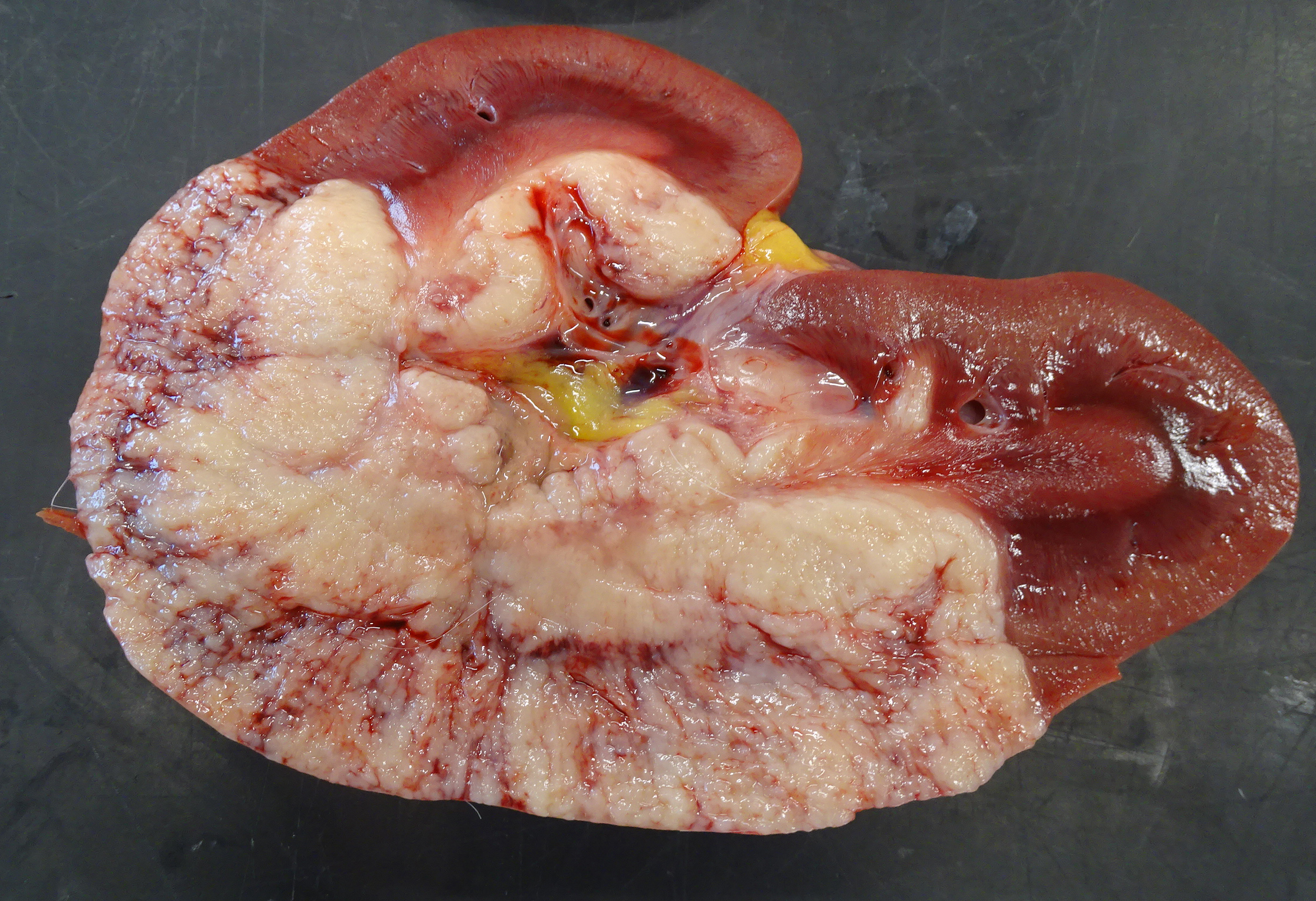
The carcass was in fair body condition and good postmortem condition. Lungs showed pulmonary congestion and edema with frothy fluid in the trachea. Liver, heart: no gross abnormalities. The left kidney was enlarged. A raised, white to beige, well-demarcated and multilobulated mass was extending through the entire depth of the kidney and obliterating approximately 70% of the renal parenchyma. In the right kidney, there was a focal nodule, (about 2 x 2 cm), with similar appearance to the lesion in the left kidney. Left renal lymph node showed a nodule (about 1 x 0.5 cm), with the same gross appearance as the lesion in the left kidney. Brain and spinal cord were dissected after fixation: no gross abnormalities. Samples of the kidneys, left renal lymph node, brain and spinal cord were taken for histopathological examination.
Microscopic Description:
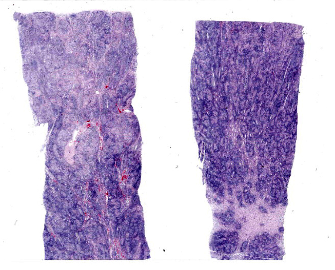
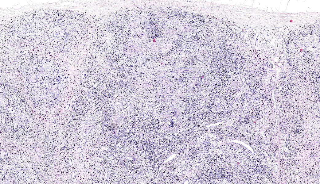
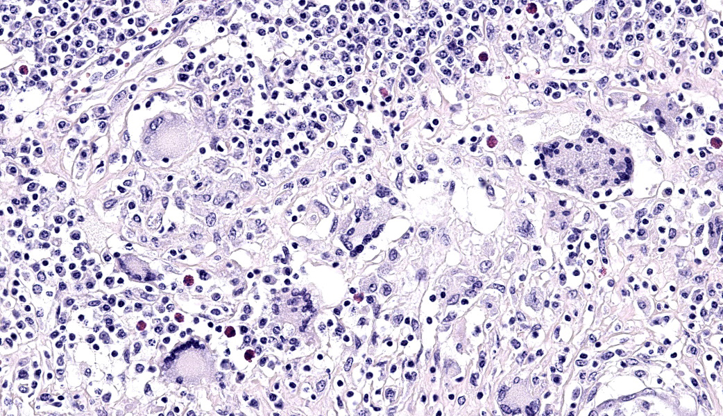
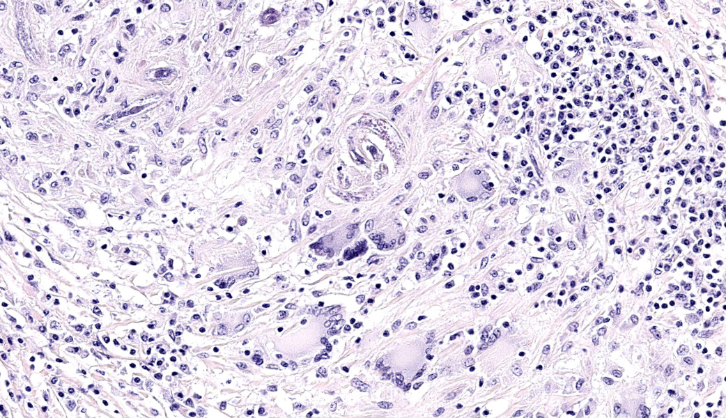
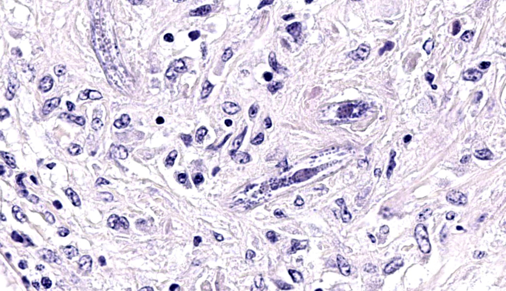
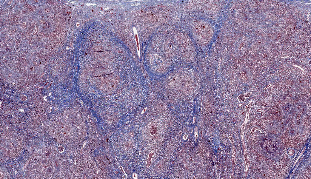
Left and right kidney: obliterating approximately 80% of the renal parenchyma within the section are multifocal to coalescing, variably sized, moderately to densely cellular aggregates of inflammatory cells composed predominately of large number of macrophages, giant cells, and lymphocytes and some plasma cells, with a lesser amount of eosinophils, separated by some fibrous tissue. Within these areas, numerous tangential and cross sections through metazoa (adults and larvae) are present. The adult nematodes are approximately 15-20 microns in diameter, and have a thin cuticle, platymyarian-meromyarian musculature, intestinal tract, and characteristic rhabditiform esophagus with a corpus, isthmus and terminal bulb. Multifocally, there are areas where a centrally located, strongly basophilic, granular material has accumulated (dystrophic mineralization).
In addition to nephritis, microscopic examination of the brain and left renal lymph node from this horse identified both meningoencephalitis and lymphadenitis with intralesional rhabditoid nematodes that were consistent with Halicephalobus gingivalis.
Contributor's Morphologic Diagnoses:
Severe chronic granulomatous and eosinophilic nephritis with intralesional Halicephalobus gingivalis.
Contributor's Comment:
Halicephalobus gingivalis, a free living, saprophytic nematode of the order Rhabditidia, family Panagrolaimidae. They are found in water, soil, decaying organic matter, and has been known to parasitize various species including humans, horses, a zebra and a donkey.
The life cycle and pathogenesis is still poorly understood. Infection can occur through wounds in oral and nasal mucosal membranes or in skin. Mucosal invasion results in local inflammation, with dissemination throughout, the body possibly via haematogenous or lymphatic spread. The most commonly reported sites of infection in horses are the brain, kidneys, lymph nodes, oral and nasal mucosa, and adrenal glands. H. gingivalis is characterized by its distinct morphology. It has a diameter of approximately 15-25µm, a smooth cuticle and
platymarian-meromyarian musculature. It shows a rhabditiform oesophagus with a corpus, isthmus and bulb. The gastrointestinal tract is lined by uninucleate cuboidal cells. It has a didelphic reproductive tract with dorsiflexed ovaries. Within well-developed lesions, tangential and cross sections of adults as well as larva are easily visualized, with intermixed 10-l5µm diameter, ovoid, embryonated eggs.
Aberrant halicephalobiasis manifests as granulomatous lesions and often carries a high mortality rate in both humans and horses.
Both males and females nematodes exist in the environment, but only female nematodes have been observed within microscopic lesions, suggesting that within a host, reproduction occurs asexually via parthenogenesis.
Contributing Institution:
Department of Veterinary Pathology, Faculty of Veterinary Medicine, Ghent University, Merelbeke, Belgium https://www.ugent.be/di/di0S/nl
JPC Diagnoses:
Kidney: Nephritis, granulomatous, chronic, diffuse, severe, with rhabditid adults, larvae, and eggs.
JPC Comment:
Halicephalobus gingivalis is also a frequent visitor to the Wednesday Slide Conference, with sixteen instances since 1990, in which it has appeared most frequently in its current nomenclature (Halicephalobus gingivalis), but also in previous scientific names including Halicephalobus deletrix and Micronema deletrix.
The contributor provides a concise review of Halicephalobus gingivalis infection in the horse, and more basic information may be gained from review of previous iterations in the WSC.
While the majority of publications involve H. gingivalis infection in the horse, H. gingivalis appears to be a rare parasite of humans as well with 8 cases reported since 2015.3,4 It has a worldwide distribution but with reports in humans in Europe, North America, Africa, and Australia. In each case, definitive diagnosis of infection was made at autopsy. In each case, the clinical manifestation was meningoencephalitis with several cases demonstrative meningomyelitis as well.3 Rarely, nematodes have been found in the lungs and liver as well. Histologic lesions include perivascular acute and chronic granulomatous inflammation, similar to that seen in the horse.3
In addition to horses and humans, fatal meningoencephalitis has also been reported in a 7-week-old bovine Holstein calf from a dairy operation. While the source of the infection was not identified, the authors suggested either the umbilicus or milk.5
Finally, a few recent reports of H. gingivalis infection in horses bear review. Due to the relative rarity of reported cases, many report document the presence of the nematodes in one or two organs. Some early reports discuss swelling of the jaws as a result of oral migration from the infiltrating nematodes from the gingiva (hence the name H. gingivalis) into the underlying maxilla and mandible, resulting in osteomyelitis. A 2024 report chronicled a bone lesion resulting in a comminuted fracture of the tibia6. (A characteristic lesion, likely similar to that seen in this case, was present in the left kidney as well.)6 The pathogenesis of the tibial lesion was not identified, although contamination of a wound in the region could not be excluded.
Differing from the typical report of H. gingivalis infection in one or two organs (if one is the brain, the rapidly progressive and devastating effects of neurologic involvement in a horse often results in euthanasia early on in disease progression), a 2025 report7 describes a "disseminated" infection with H. gingivalis. In this case, the agent was found in the heart, kidneys, oral cavity, tongue, left eye, lungs, CNS, adrenal glands, liver, lymph nodes and the spleen (which has not been previously reported). The cause of the prolonged survival (~8 weeks) of this animal following initial diagnosis (following enucleation of an infected eye) may have been facilitated by the administration of corticosteroids and ivermectin, allowing for additional distribution of the parasites via vascular channels before neurologic signs necessitated the animal's euthanasia.7
Finally, transplacental transmission and abortion due to H. gingivalis was identified in a Connemara mare, who, despite having successful deliveries in previous pregnancies, aborted in two consecutive pregnancies. In both cases, larva of Halicephalobus gingivalis were identified within placental tissue. The authors of this study concluded that not all H. gingivalis infections may result in disseminated or life-threatening infections and that infection may be widespread than previously thought.8 A single previous case of verminous mastitis and transmammary H. gingivalis delivery to a foal (who subsequently died) has been reported.9
References:
- Calewaert A et al. Neurological dysfunction and fatal encephalitis due to Halicephalobus gingivalis in two horses in Belgium. Equine Veteripary Education.2024 https: //doi.org/10.1111 /eve.14068
- Gardiner CH, Poynton SL. Morphologic characteristics of rhabditoids in tissue section. In: An Atlas of Metazoan Parasites in Animal Tissues. Washington DC, AFIP Press, pp. 14-16.
- Onyiche TE et al. Parasitic and zoonotic meningoencephalitis in humans and equids: Current knowledge and the role of Halicephalobus gingivalis. Parasite Epidemiol Control. 2018;3(1): 36-42.