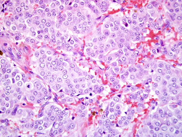
Signalment:
Gross Description:
Histopathologic Description:
Morphologic Diagnosis:
Lab Results:
Condition:
Contributor Comment:
JPC Diagnosis:
Conference Comment:
C-cell neoplasms exhibit positive cytoplasmic immunoreactivity for calcitonin and are negative for thyroglobulin. The sensitivity for thyroglobulin for thyroid carcinomas is 90.5% alone, but if it is combined with TTF-1, that sensitivity increases to 95.2%.16 TTF-1, thryoid transcription factor, is expressed in the thyroid, brain, and lung during early embryogenesis, and the thyroid cells and bronchioloalveolar epithelial cells following birth. In the lung, it activates surfactant proteins and Clara cell secretory protein gene promoters.1 In the thyroid gland, it activates many factors including thyroglobulin, thyroperoxidase, thyrotropin receptor, and thyroid peroxidase.5,6,8,10,18 When positive, TTF-1 is diffusely located within the nucleus and never in the cytoplasm. In one study, approximately 50% of the C-cell neoplasms also stained positive for TTF-1, therefore it is not suitable to use as a single marker.16
C-cell tumors in bulls, often occur concurrently with bilateral pheochromocytomas and pituitary adenomas.4 Multiple endocrine tumors are thought to arise due to a simultaneous neoplastic mutation of multiple endocrine cell populations of neural crest origin in the same individual.3,23 In humans, multiple endocrinen neoplasia type 2 (MEN-2) occurs in an autosomal dominant pattern, and is classified into three clinical manifestations.17 MEN-2A is characterized by the presence of a medullary thyroid carcinoma in addition to a pheochromocytoma and multiple tumors of the parathyroid gland.7 MEN-2B consists of a medullary thyroid carcinoma, a pheochromocytoma, ganglioneuromatosis, and marfanoid habitus.21 FMTC syndrome, is the third form of MEN-2 and is defined as the development of a medullary thyroid carcinoma and a low incidence of other clinical manifestations of either MEN-2A or MEN-2B.7
References:
2. Capen CC and Block H E; Calcitonin secreting ultimobranchial neoplasms of the thyroid gland in bulls: An animal model for medullary thyroid carcinoma in man. Am J Pathol. 1974; 74:371-380
3. Capen CC: Tumors of the endocrine glands. In: Tumors in Domestic Animals, ed. Meuten DJ, 4th ed., pp. 657-664. Blackwell Publishing, Ames, IA, 2002
4. Carver JR, Kapatkin A, Patnaik AK: A comparison of medullary thyroid carcinoma and thyroid adenocarcinoma in dogs: A retrospective study of 38 cases. Vet Surg 24:315-319, 1995
5. Civitareale D, Castelli MP, Falasca P, Saiardi A: thyroid transcription factor 1 activates the promoter of the thyrotropin receptor gene. Mol Endocrinol 7:1589-1595, 1993
6. Civitareale D, Lonigro R, Sinclair AJ, Di Lauro R: A thyroid-specific nuclear protein essential for tissue-specific expression of the thyroglobulin promoter. EMBO J 8:2537-2542, 1989
7. Donis-Keller H, Dou S, Chi D, Carlson KM, Toshima K, Lairmore TC, Howe JR, Moley JF, Goodfellow P, Wells SA: Mutations in the RET proto-oncogene are associated with MEN 2A and FMTC. Hum Mol Genet 2:851-856, 1993
8. Endo T, Kaneshige M, Nakazato M, Ohmori M, Harii N, Onaya T: Thyroid transcription factor-1 activates the promoter activity of rat thyroid Na+/I- symporter. Mol Endocrinol 11:1747-1755, 1997
9. Hillage CJ, Sanecki RK, Theodorakis MC: Thyroid carcinoma in a horse. J Amer Vet Med Assoc 181:711-714, 1982
10. Kikkawa F, Gonz+�-�lez FJ, Kimura S: Characterization of a thyroid-specific enhancer located 5.5 kilobase pairs up-stream of the human thyroid peroxidase gene. Mol Cell Biol 10:6216-6224, 1990
11. Krook L., Lutwak L, McEntee K: Dietary calcium and osteopetrosis in the bull: Syndrome of calcitonin excess? Am J Clin Nutr 22:115-118, 1969
12. Leav I, Schiller AL, Rijiiberk A, Kegg MB, der Kinderen PJ: Adenomas and carcinomas of the canine and feline thyroid. Am J Pathol 83:61-64, 1976
13. McEntee K, Hall CE, Dunn HO: The relationship of calcium intake to the development of vertebral osteophytosis and ultimobranchial tumors in bulls. In: Proceedings of the Eight Technical Conference on Artifical Insemination Reproduction, pp. 45-47, 1980
14. Melvin KW, Miller HH, Tashjean AH Jr: Early diagnosis of medullary carcinoma of the thyroid gland by means of calcitonin assay. N Engl J Med 285:1115-1120, 1971
15. Peterson ME, Randolph JF, Zaki FA, Heath H: Multiple endocrine neoplasia in a dog. J Amer Vet Med Assoc 180:1476-1478, 1982
16. Ramos-Vara JA, Miller MA, Johnson GC, Pace LW: Immunohistochemical detection of thyroid transcription factor-1, thyroglobulin, and calcitonin in canine normal, hyperplastic, and neoplastic thyroid gland. Vet Pathol 39:480-487, 2002
17. Raue F, Frank-Raue K: Multiple endocrine neoplasia type 2: 2007 update. Horm Res 68:101-104, 2007
18. Saji M, Shong M, Napolitano G, Palmer LA, Taniguchi S-I, Ohmori M, Ohta M, Suzuki K, Kirshner SL, Giuliani C, Singer DS, Kohn LD: Regulation of major histocompatibility complex class I gene expression in thyroid cells. J Biol Chem 272:20096-20107, 1997
19. Spoonenberg DP, VcEntee K: Phoechromocytoma and ultimobranchial (C Cell) neoplasms in the bull: Evidence of autosomal dominant inheritance in the Guernsey breed. Vet Pathol 20:369-400, 1983
20. Thomas RG: Vertebral body osteophytes in bulls. Pathol Vet 6:1-46, 1969
21. Vasen HF, van der Feltz M, Raue F, Nieuwenhuyzen Kruseman A, Koppeschaar HPF, Pieters G, Seif FJ, Blum WF, Lips CJM: the natural course of multiple endocrine neoplasia type 2b. Arch Intern Med 152:1250-1252, 1992
22. Wadsworth PF, Lewis DJ, Jones DM: Medullary carcinoma of the thyroid in a Mouflon (Ovis musimon). J Comp Pathol 91:313-316, 1981
23. Weichert RF III: The neural ectodermal origin of the peptide-secreting endocrine gland: a unifying concept for the etiology of multiple endocrine adenomatosis and the inappropriate secretion of peptide hormones by nonendocrine tumors. Amer J Med 49:232-241, 1970
