Wednesday Slide Conference, 2025-2026, Conference 5, Case 4
Signalment:
2.5-year-old spayed female domestic shorthair cat (Felis catus)History:
Dental procedure performed 6 weeks prior with multiple extractions, including right maxillary arcade. Right eye discomfort was noted by the owner one day post-surgery. Medical management (full details not available) was attempted by the primary veterinarian, but the patient was refractory to treatment. The cat was referred to an ophthalmologist who documented severe anterior uveitis, iris bombe, and lens capsule rupture via ultrasound. Enucleation was performed due to suspicion of septic endophthalmitis secondary to dentistry-related ocular trauma.Gross Pathology:
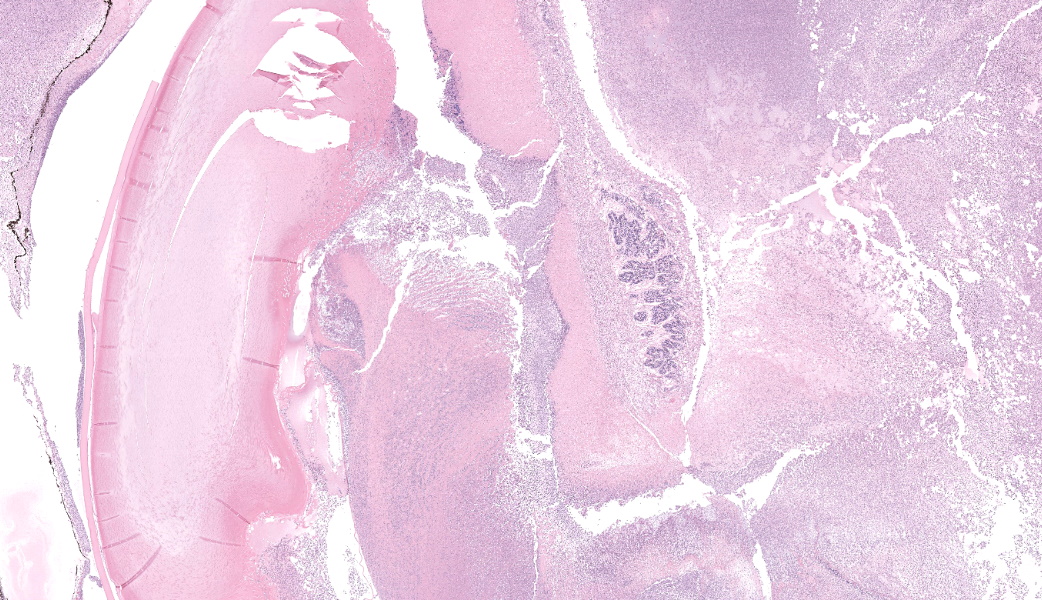
Submitted fixed in 10% neutral buffered formalin for evaluation is an enucleated eye (diameter: 31mm; cornea diameter: 18mm; axial length: 22mm) with a less than 1mm segment of optic nerve. On section of the eye, the ocular chambers contain abundant amounts of thick off-white purulent material.Laboratory Results:
N/AMicroscopic Description:
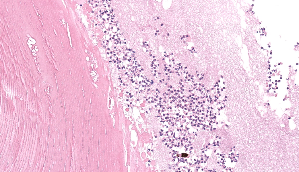
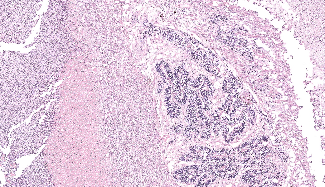
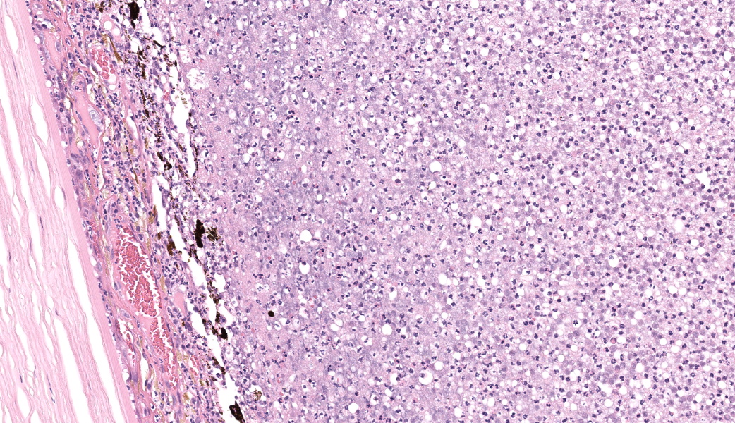
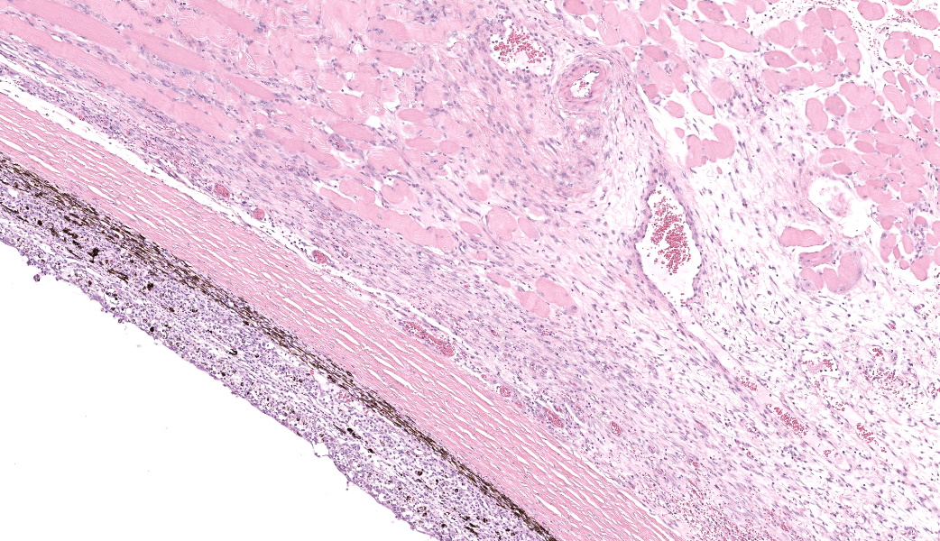
Filling all ocular chambers and coating the surfaces of intraocular structures are vast accumulations of exudate consisting of myriad degenerate and intact neutrophils, many macrophages, abundant amounts of fibrinous to proteinic material, abundant necrotic cellular debris, occasional pools of extravasated erythrocytes, and multifocal colonies of mixed bacteria (rods and coccobacilli). Centrally incorporated into the inflammatory exudates is a ruptured lens which lacks a capsule in many areas and has highly undulating free capsule margins regionally. The lenticular stroma is variably vacuolated with the following features: streaks of pallor, Morgagnian globules, many infiltrating leukocytes (mostly neutrophils), and occasional pockets of similar bacteria. The retina is diffusely detached and largely inapparent apart from remnant segments of atrophied and degenerate retina enmeshed within the exudate. Suppurative to pyogranulomatous inflammatory infiltrates multifocally extend into the iris, ciliary body, choroid, optic nerve which is significantly gliotic with rarefied neuropil, and optic nerve meninges. The iris is displaced anteriorly and multifocally abuts the posterior aspect of the cornea. The anterior chamber is severely narrow, and the drainage angle is collapsed and inapparent. The corneal stroma is moderately to markedly oedematous and contains small to moderate numbers of scattered neutrophils. The anterior corneal epithelium appears attenuated in areas. The sclera is variably thinned with multifocal often perivascular infiltrates of lymphocytes and plasma cells with variable numbers of admixed neutrophils and occasional macrophages. There are increased numbers of perilimbal pigmented cells. A thin to moderately thick layer of oedematous and inflamed granulation tissue regionally lines the mid and posterior scleral margins outside the globe and extends into a bundle of periocular skeletal muscle. In addition to infiltrates of the aforementioned inflammatory cells, the granulation tissue also contains many golden-brown pigmented macrophages (siderophages) which are concentrated at the level of the ciliary body. There is abundant haemorrhage in the retrobulbar loose connective tissue.Contributor's Morphologic Diagnoses:
Endophthalmitis, diffuse, suppurative to pyogranulomatous, severe, with lens rupture, retinal detachment and degeneration/atrophy with intraocular exudation and intralesional mixed bacteria, drainage angle collapse, keratitis, corneal oedema, scleritis and regional periscleral fibrosisContributor's Comment:
Ophthalmic complications associated with scleral penetrating injuries are infrequently reported in dogs and cats with a recent history of dental procedures. Iatrogenic globe trauma as a complication of routine dentistry may occur secondary to slippage of dental instruments during maxillary tooth extraction or inadvertent needle perforation of the globe when placing infra-orbital and maxillary nerve blocks. Endophthalmitis is a common sequela of dental-associated globe penetration due to implantation of bacteria and/or lens rupture. Medical management is often poor and most cases of post-dentistry endophthalmitis lead to palliative and diagnostic enucleation.Compared with other species, cats may be at increased risk of traumatic globe injury during exodontic procedures due to a large globe size that nearly fills the orbit, lack of bony orbital floor, and close proximity of maxillary tooth roots (especially PM4 and M1) to the ventral orbit. Bone resorption associated with periodontal and endodontic disease may further compromise the narrow rim of alveolar bone surrounding the roots of the maxillary premolars and molars, increasing vulnerability to iatrogenic ocular trauma. Older cats may be at increased risk of penetrating ocular trauma during dental procedures compared with younger cats due to age-associated atrophy of orbital fat pads and closer globe position to the oral cavity.
Clinical findings in animals following orbital penetration during a dental procedure include vision loss, epiphora, mucopurulent ocular discharge, blepharospasm, exophthalmos, buphthalmos, glaucoma, hyphema, hypopyon, miosis, iris bombe, cataract, and orbital cellulitis. Gross examination of the globe commonly demonstrates opacification of the aqueous and vitreous humor and may reveal a site of scleral penetration. Histopathologic lesions include severe suppurative endophthalmitis or panophthalmitis, varying degrees of orbital cellulitis and episcleral fibrosis of the ventral aspect of the globe, and in some instances rupture of the posterior or equatorial lens capsule and/or presence of intraocular bacteria. Ventral scleral perforation may be appreciated in some cases, but a site of penetrating injury is not always detected. In this case, a scleral penetration site was not appreciated grossly or histologically despite review of multiple step sections.
Contributing Institution:
University of Sydney https://sydney.edu.au/science/schools/sydney-school-of-veterinary-science/veterinary-science-services.htmlJPC Diagnoses:
Globe: Endophthalmitis, fibrinosuppurative, subacute, diffuse, severe, with lens rupture, synechiae, fibrovascular membranes, retinal detachment and atrophy, and bacterial colonies.JPC Comment:
Talk about a descriptive case that made, for obvious reasons, an excellent sales pitch on why pathology is the best profession! This case provides an excellent opportunity for participants to push themselves on their ocular descriptive abilities. Many thanks to this contributor for a fantastic case! Much like the previous eye case in this conference, there was substantial discussion on ocular pathology. The most informative nuggets from that conversation included utilizing the lens capsule, which is an easily identifiable structure in the eye, to assist with orientation in a busy ocular slide such as this one. The pigmented irideal stroma, as well as the “golden” fibers of the iris, can also be used to help identify structures that might otherwise be difficult to ascertain due to the degree of damage and/or inflammation.This case had beautiful examples of iris bombe (iris pushed forward into the anterior chamber and adhered to the back of the cornea), numerous types of fibrovascular membranes (retrocorneal, preiridial, cyclitic, etc.), and a fantastic phakoclastic panuveitis from lens rupture. The six types of uveitis and their definitions were discussed and included: 1) anterior uveitis (inflammation of the iris and ciliary body), 2) posterior uveitis (inflammation of the ciliary body and choroid), 3) panuveitis (iris, ciliary body, and choroid affected), 4) chorioretinitis (inflammation of the choroid and retina), 5) endophthalmitis (inflammation of uvea, retina, and ocular cavities), and 6) panophthalmitis (all ocular structures are affected, including sclera). Being able to recognize and accurately use these terms as pathologists can provide crucial information to ophthalmologists when it comes to treating these patients.
As anyone who has owned a cat will tell you, cats are no strangers to ocular trauma. Common causes include fights (especially with claws), foreign bodies, blunt trauma, chemical burns, and, less commonly, iatrogenic (such as dental work or local nerve blocks). Of the frequently encountered domestic species in veterinary medicine, cats are more likely to end up with traumatic globe injury following dental procedures due to their complete lack of a bony orbital floor, their objectively massive eyes that stare straight into the void, and the close proximity of their maxillary roots (PM4 and M1) to their ventral orbit. Additionally, they get a fair amount of periodontal disease, which can lead to bone resorption and thinning of the alveolar bone around the tooth roots. Older cats, as if they needed any additional help getting their eyeballs stabbed, also have age-associated atrophy of their orbital fat pad, putting their globes in even closer proximity to the oral cavity.
An unfortunate possible sequela to chronic ocular trauma in cats is the development of feline post-traumatic ocular sarcoma (FPTOS), which is an aggressive malignant neoplasm of the eye in cats and the third most common primary intraocular tumor in feline patients.6 It can take up to seven years for these sarcomas to develop following ocular trauma or disease, and they are thought to arise from malignant transformation of lens epithelium, as there is a history of lens rupture is almost all cases of FPTOS.6 Most of these tumors look very similar to fibrosarcomas, but there can be variation in their morphology; some may look like osteosarcomas, others like giant cell neoplasms.6 Recently, there has been a link established between the development of FPTOS and the use of intravitreal gentamicin injections used to ablate the ciliary body in cases of unresolving glaucoma.6 Medicine is ever-evolving, and one of the great burdens of clinicians is making treatment decisions that carry risk knowing that, for some patients, the benefit of such treatment outweighs that risk. For cats afflicted with these aforementioned conditions, though, it seems that all roads lead to enucleation.
References:
- Dubielzig RR, Ketring KL, McLellan GJ, Albert DM. Veterinary Ocular Pathology, a Comparative Review. Philadelphia, PA: Elsevier; 2010:330-336.
- Duke FD, Snyder CJ, Bentley E, Dubielzig RR. Ocular trauma originating from within the oral cavity: clinical relevance and histologic findings in 10 cases (2003-2013). J Vet Dent. 2014;31(4):245-248.
- Perry R, Moor D, Scurrell E. Globe penetration in a cat following maxillary nerve block for dental surgery. J Feline Med Surg. 2015;17(1):66-72.
- Smith MM, Smith EM, La Croix N, Mould J. Orbital Penetration Associated with Tooth Extraction. J Vet Dent. 2003;20(1):8-17.
- Volk HA, Bayley KD, Fiani N, Bilson FM. Ophthalmic complications following ocular penetration during routine dentistry in 13 cats. N Z Vet J. 2019;67(1):46-51.
- Wood C, Scott EM. Feline ocular post-traumatic sarcomas: Current understanding, treatment and monitoring. J Feline Med Surg. 2019;21(9):835-842.