CASE I:
Signalment:
18-month-old intact female Toy Manchester Terrier, canine
History:
This dog was found dead in her kennel with pink-tinged discharge from the mouth. She had no reported clinical signs.
Gross Pathology:
The carcass weighed 4.2kg and was in good nutritional and postmortem condition. The heart weighed 42.7g (1% of body weight; adult normal: 0.43-0.99%). The left ventricular free wall, interventricular septum, and right ventricular free wall were 10mm, 7mm, and 4mm thick, respectively. The left atrial appendage was larger than the right. The stomach contained a small amount of dog food and green beans.
Laboratory Results:
None available
Microscopic Description:
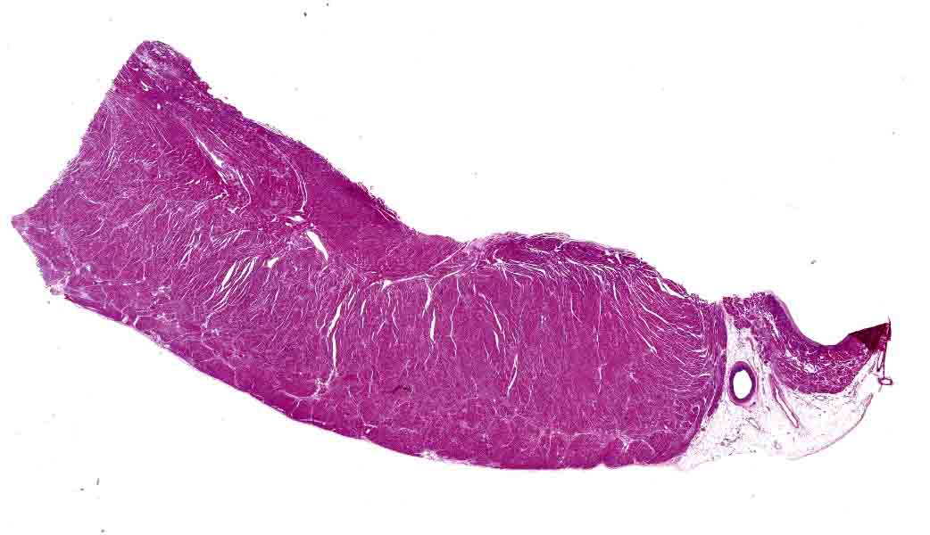
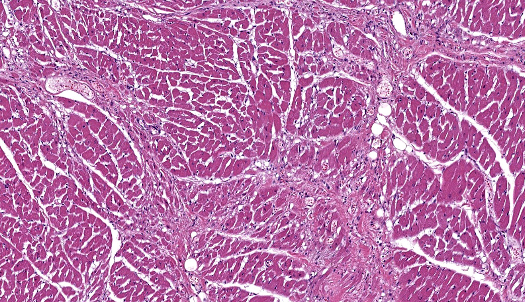
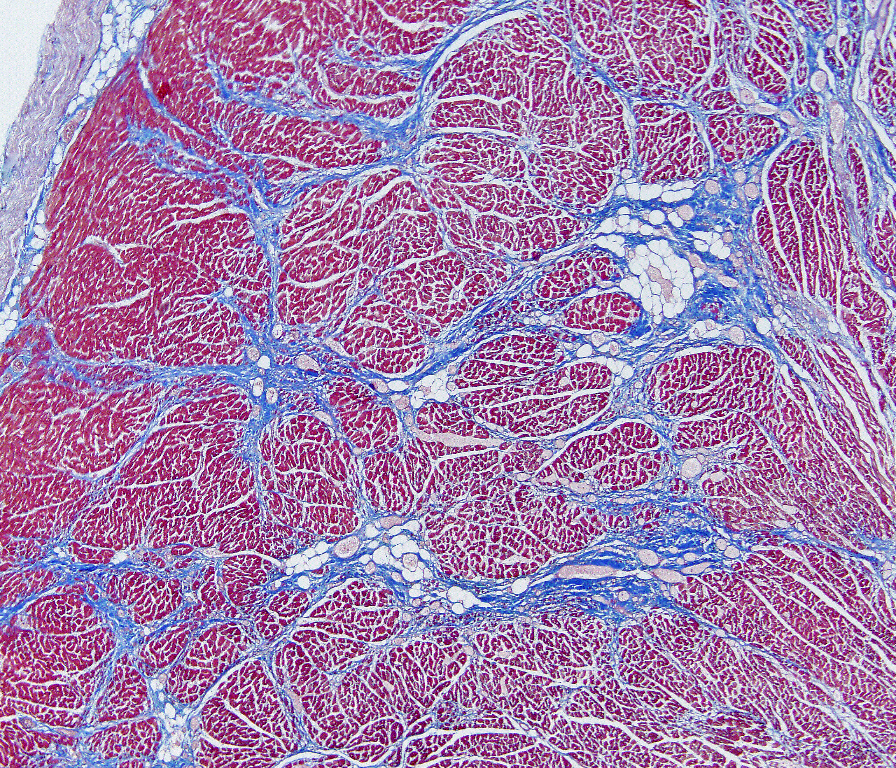
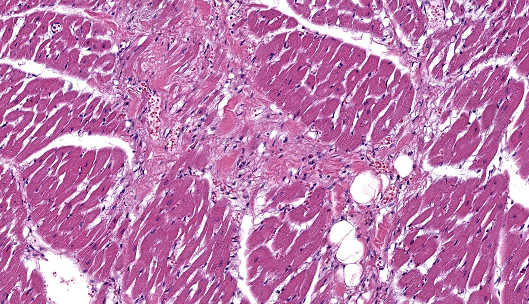
Slides include sections of the interventricular septum and right ventricular free wall or left ventricular free wall. Replacing approximately 20% of the myocardium are multifocal to coalescing areas of fibrosis associated with low numbers of lymphocytes and plasma cells. Some areas of fibrosis contain adipocytes. There is degeneration, necrosis, and rarely mineralization (not present in all sections) of entrapped myofibers. Small groups of myocytes distant from areas of fibrosis are also necrotic. Trichrome staining of the affected myocardium is a beautiful thing.
Contributor's Morphologic Diagnoses:
Marked multifocal myocardial necrosis and fibrosisContributor's Comment:
The signalment and postmortem findings are consistent with juvenile cardiomyopathy of Toy Manchester Terrier dogs.1 In the pub-lished series of 14 cases, the oldest was 13.5 months and the other 13 cases were less than 10 months old. This dog was 18 months old, so slightly older than expected. Sudden death without clinical signs is a common history for these puppies.1 The puppy in this report had food in her stomach indicating a good appetite prior to death. There were no gross or microscopic lesions suggesting pre-existing congestive heart failure. The heart in this dog was not visibly dilated, but gross evidence of dilation is milder in the Toy Manchester Terriers than in Portuguese Water Dogs with juvenile dilated cardiomyopathy.1 The left atrial appendage was larger than the right in our case, a finding that was also described in some of the published cases, most likely due to mitral valve insufficiency resulting from dilation.
Contributing Institution:
University of Tennessee College of Veterinary Medicine
Department of Biomedical and Diagnostic Sciences
Biomedical and Diagnostic Sciences
JPC Diagnoses:
Heart, ventricular free wall: Fibrosis, chronic, multifocal to coalescing, marked with cardiomyocyte degeneration, necrosis, and loss.
JPC Comment:
The term "cardiomyopathy" is used for diseases of the myocardium for which a pre-existing cause, such as a congenital heart defect, valvular disease, hypertension, or other abnormalities, is not previously diagnosed. In humans, the most common five types of cardiomyopathy based on gross and histologic changes of the affected heart include dilated, hypertrophic, arrhythmogenic, restrictive, and non-compaction2; all except for non-compaction have been diagnosed in one or more domestic species.
In the dog, dilatative cardiomyopathy is the second most common cardiac disorder follow-ing degenerative valvular disease.3 Two main forms of DCM exist in the dog &emdash; those with a dietary basis and those with a genetic basis. Dietary forms of DCM in the dog are associated with the feeding of grain-free, legume-rich diets with abundant potatoes and a lack of more traditional feed grains including rice and corn, although a direct causal line has not yet been elucidated. Other diet-driven causes of DCM include diets lacking in taurine and/or carnitine.3
Genetic causes of canine DCM often display a combination of breed predispositions and often cluster in areas where related animals live. Commonly affected breeds include Doberman Pinschers (it is estimated that up to 58% of Dobermans may develop DCM), Airedales, Cocker Spaniels, Boxers, Bulldogs, Great Danes, Golden retrievers, and a number of others.3 It tends to occur more commonly in middle-aged and older dogs, however, in some breeds, such as the Toy Manchester Terrier (the subject of this case) and the Portuguese Water Dogs, it is frequently diagnosed before a year of age.1,3 One or more genetic mutations has been identified in many, but not all breeds, but is largely beyond the generalized scope of this discussion, with the exception of ABCC9, whose missense variant which, when homozygous, results in sudden death in Toy Manchester terriers before the age of two years. The ABCC9 protein is a regulatory sub-unit of cardiac KATP channels; cardiomyocytes lacking functional ABCC9 have reduced fatty acid oxidation and oxygen consumption and are unable to respond to cell stress, leaving affected animals to die suddenly under stressful conditions.
DCM is characterized by systolic dysfunction (heart failure), dilation of both atria and both ventricles (although the diagnosis of DCM is typically predicated on dilation of the left side of the heart), and attenuation and eccentric mural hypertrophy (as opposed to concentric hypertrophy, which is seen in cases of hypertrophic cardiomyopathy, a far less common is-sue in the dog).3
The histologic hallmarks of cardiomyopathy in all species are relatively straightforward: myofiber loss and fibrosis (both of which contribute to the lack of contractility of the failing myocardium. The fibrous connective tissue component of the degenerating heart may be associated with fatty infiltration as well (which is discussed in more detail in the next case).
The fibrosis identified in cardiomyopathy most commonly takes one of three forms: re-placement fibrosis, reactive/interstitial fibrosis, and perivascular fibrosis.2 In these cases, the delicate balance between synthesis and degradation of the extracellular matrix is significantly altered toward the former by the activation and differentiation of cardiac fibroblasts into myofibroblasts, and the secretion of higher amounts of extracellular matrix under the influence of transforming growth factor-beta.
The histologic appearance of fibrosis can be classified into three different subtypes, all of which are present in this particular case.
- Replacement fibrosis occurs following the death of cardiomyocytes, and the histologic appearance is that of com-pacted bands of densely packed fibrous connective tissue which truly replaces cardiomyocytes.
- Interstitial fibrosis is characterized by the deposition of collagen as well as increased extracellular matrix, which imparts a more loosely packed appearance to the fibrous connective tissue. It is present in areas in which there is no loss of cardiomyocytes, and often requires histochemical stains, including Masson's trichrome or Movat pentachrome to identify it. It may be associated with aging, inflammation, or the initial stages of pressure overload.
- Finally, perivascular fibrosis, as the name implies, is seen primarily in the areas of the coronary arterioles, and is typically seen with hypertension.
References:
Legge CH, Lopez A, Hanna P, et al His-tological characterization of dilated cardi-omyopathy in the juvenile toy Manches-ter terrier 2013;50(6):1043-1052.
Eijgenraam TR, Sillje HHW, de Boer RA. Current understanding of fibrosis in genetic cardiomyopathies. Trends Cardio-vasc Med. 2020 Aug;30(6):353-361. doi: 10.1016/j.tcm.2019.09.003.
Donovan TA and Kelly, KM. Cardivascu-lar System. In: Jubb, Kennedy and Palmer's Pathology of Domestic Ani-mals, , 7th ed, Vol. 3, Elsevier Publishing, 2026, pp. 59-65
Furrow E, Tate N, Minor K, Martinson S, Larrabee S, Anttila M, Sleeper M, Henthorn P. An ABCC9 Missense Variant Is Associated with Sudden Cardiac Death and Dilated Cardiomyopathy in Juvenile Dogs. Genes (Basel). 2023 Apr 27;14(5):988. doi:10.3390/genes14050988. PMID:37239348; PMCID: PMC10218235.