Signalment:
9-year-old, spayed, female, domestic shorhair cat (Felis catus)
History:
The cat had a history of vomiting, decreased appetite, and pale mucus membranes. Hemoparasitism was suspected. Blood parasites were not observed in smears from peripheral blood. There was moderate lymphocytosis with high differential count for lymphocytes, and monocytes and a low differential count for neutrophils (neutropenia). Low RBC, HGB, and HCT values (anemia) were recorded. RBC morphology was normocytic normochromic. Due to poor response to palliative / symptomatic treatment, and poor prognosis the cat was euthanized.
Gross Pathology:
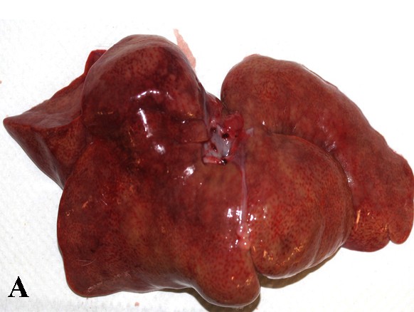
On gross examination, the cat was found in very good nutritional condition with abundant subcutaneous and body fat. The spleen was markedly enlarged and weighed 210 grams. The liver was moderately enlarged, weighed 184 grams and pale yellowish in color. Various body lymph nodes were also moderately enlarged.
Laboratory Results:
Fluorescent antibody test for Feline leukemia virus and Feline parvovirus was negative.
Histopathologic Description:
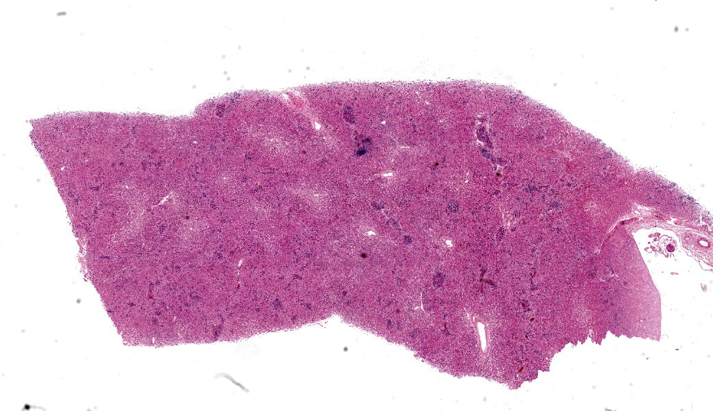
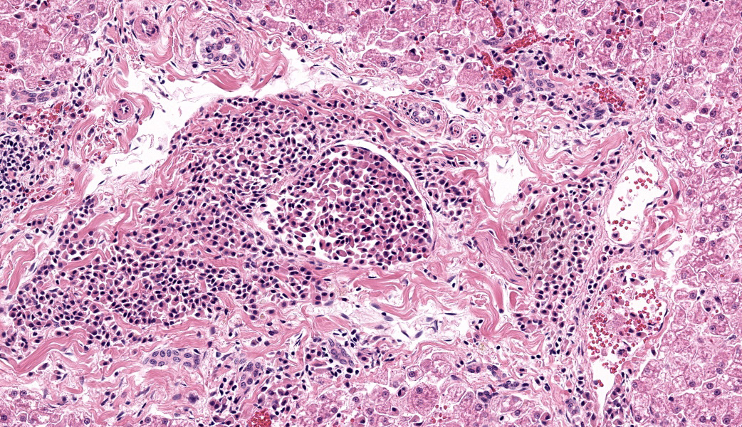
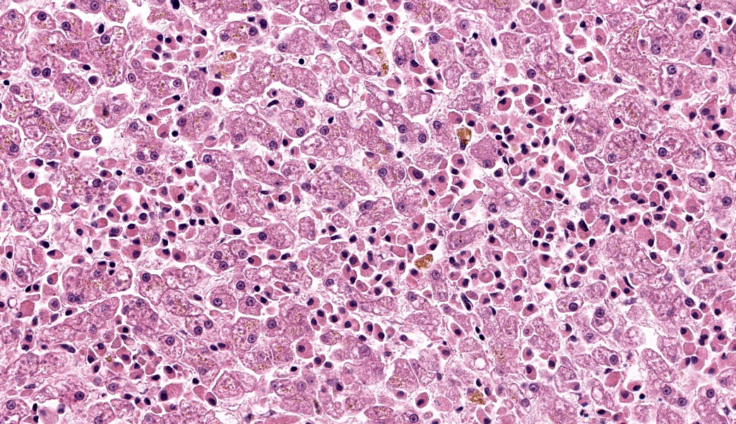
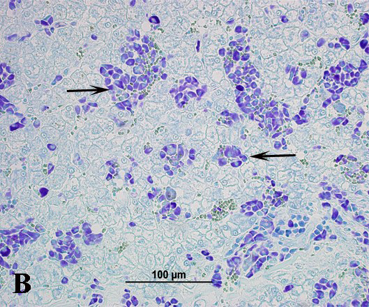
Large numbers of round cells diffusely infiltrated the hepatic sinusoids, periportal areas and formed varisized aggregates. Similar
Contributor's Morphologic Diagnoses:
mastocytosis (mast cell leukemia), liver
Contributor's Comment:
Diffuse infiltrative mast cells in spleen, liver, and various lymph node with vascular invasion is consistent with mast cell leukemia (mastocytosis / mastocytaemia). Given the se-vere splenomegaly observed grossly, and dif-fuse infiltration of splenic parenchyma by mast cells, the mast cell tumor apparently originated from the spleen and invaded other
tissues including liver. The initiating factors that induce mast cell proliferation and mast cell leukemia are largely unknown, but in hu-mans and dogs a derangement of the c-kit receptor and /or its ligand likely plays a primary role in the disease process. Marked splenomegaly is the characteristic gross lesion of mastocytosis in the cat.4
Cats with mastocytosis exhibit signs of systemic illness such as depression, anorexia, lethargy, weight loss and vomiting1,3 as was observed in this case. Mastocytemia in cats, unlike in dogs, is distinctively observed in cats with mast cell tumors and is frequently asso-ciated with the splenic mast cell tumor. Mas-tocytaemic cats with mast cell tumor have a lower hematocrit (HCT) value (anemia) than those without mastocytaemia.5 Anaemia of chronic disease, blood loss due to gastrointestinal ulceration, splenic erythrocyte sequestration, removal of antibody-coated erythrocytes by splenic macrophages and erythrophagocytosis by neoplastic mast cells are suggested to underlie mast cell tumor associated anemia.1,2
Mastocytaemia may be detected by examination of peripheral blood smear, bone marrow aspirate, or a buffy-coat (BC) layer.1 Buffy coat examination is the preferred method and should be mandatory in cases of suspected
mast cell tumor or cats with splenomegaly or vomiting of undetermined cause.5 Cytology on fine needle aspirates of affected organs is usually diagnostic and histopathology of biopsy from affected tissue is confirmatory3. Splenectomy renders long survival time in cats with splenic mast cell tumor and mastocytosis.1
Contributing Institution:
The University of Georgia, College of Veterinary Medicine, Department of Pathology, Tifton Veterinary Diagnostic and Investigational Laboratory
Tifton, GA 31793, http://www.vet.uga.edu/dlab/tifton/index.php
JPC Diagnoses:
Liver: Mast cell tumor.JPC Comment:
While we essentially agree with the contributor's diagnosis of mastoleukemia in this case (due to the presence of neoplastic cells within blood vessels and the hepatic sinusoids), we prefer the diagnosis of hepatic mast cell tumor in this case. Although this section shows numerous mast cells within blood vessels, and the clinical history includes anemia (suggesting the possibility of bone marrow infiltration), the lack of peripheral blood or bone marrow samples for evaluation makes an evidence-based diagnosis of "mastoleukemia" somewhat problematic. That small quibble aside, this section is an excellent example of a visceral mast cell tumor in a cat.
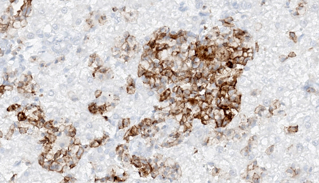
Visceral mast cell tumors are less common than their cutaneous counterparts, comprising up to 50% of all feline MCTs)6,7 and are most commonly seen in one or more of the following: spleen, liver, gastrointestinal tract, lymph nodes, and bone marrow.6 These tumors are reported to account for 15-25% of all feline splenic tumors, and 4% of all intestinal neo-plasms.7 They may be seen in association with cutaneous neoplasms, but there is no definitive evidence that they are the result of visceral metastasis from a cutaneous primary.7 Mastocytemia may be seen with both cutaneous and visceral mast cell tumors; evaluation of peripheral blood smears, bone marrow aspirates, or buffy coat smears is required for the definitive diagnosis of mastocytemia. The gross appearance of visceral mast cell tumors typically includes organomegaly and pallor of the affected organ, particularly the spleen. In the intestine, they are typically non-circumferential and result in a loss of mural layering on ultrasound evaluation.7 Unlike cutaneous mast cell tumors in cats, which may demonstrate any of three well-documented morphologies (mastocytic, histiocytic and pleomorphic), visceral mast cell tumors in cats tend to be populated with cells resembling those of the cutaneous mastocytic type ? round cells with few granules and minimal cellular atypia.7 The lack of granules on HE section is typical of feline mast cell tumors, and are reminiscent of formalin-sensi-tive mucosal mast cells in many species; the use of alcohol fixatives such as Carnoy's fixative or alcohol based cytologic stains such as Wright-Giemsa will often demonstrate these granules which are nearly invisible in formalin-fixed tissue.7 Visceral mast cell tumors, like their cutaneous counterparts, typically have few infiltrating eosinophils, but may have aggregates of lymphocytes scattered throughout the neoplasm.CD117 immunohistochemistry appears to be of more limited utility in the cat than the dog, with one study reporting that 35% of splenic MCTs and 33% of enteric MCTs were positive for this immunomarker.9 Fortunately, most visceral MCTs are diagnosable on HE section, with a relative few requiring immunomarkers to exclude lymphoma for the differential diagnosis. In a study of feline gastrointestinal mast cell tumors, CD117 immunostaining did not appear to correlate with survival outcomes.11
Finally, all visceral mast cell tumors should be considered high-grade aggressive tumors, unlike their cutaneous counterparts. Splenectomy of cats with splenic MCTs has been associated with median survival times of 12-19 months,6,10 and is almost three times that of non-splenectomized cats.10
References:
- Allan R1, Halsey TR, Thompson KG. Splenic mast cell tumour and mastocytaemia in a cat: case study and literature review. N Z Vet J. 2000 Aug; 48(4):117-21.
- Antognoni MT, Spaterna A, Lepri E, Fruganti A, Laus F. Characteristic Clinical, Haematological and Histopathological Findings in Feline Mastocytoma. Veterinary Research Communications, 27 Suppl. 1 (2003) 727-730.
- Hyland JE, Sartin EA, Spangler EA. Pathology in practice. Splenic mast cell tumor with metastasis to the liver in a cat. JAVMA 242(5):627-629 (2013).
- Jacobs RM, Messick JB, Valli VE. Tumors of hemolymphatic system: Mast cell leukemia. In Meuton DJ (edit.), Tumors in domestic animals, 4th edition, Iowa State Press, 2002; pp 190-192.
- Skeldon NC1, Gerber KL, Wilson RJ, Cunnington SJ. Mastocytaemia in cats: prevalence, detection and quantification meth-ods, haematological associations and potential implications in 30 cats with mast cell tumours. J Feline Med Surg. 2010; 12(12):960-6.
- Blackwood, L., Murphy, S., et al. (2012) European consensus document on mast cell tumours in dogs and cats. Vet Comp Oncol 10:e1-e29.
- Williams BH, et al. Mast cell tumors. In: Meuten DJ, ed. Tumors in Domestic Animals. Wiley Blackwell; 2026.
- Sato, A.F., Solano, M., et al. (2004) Ultrasonographic findings in abdominal mast cell disease: a retrospective study of 19 patients. Vet Radiol Ultrasound 45:51-57.
- Mallett, C.L., Northrup, N.C., et al. (2013) Immunohistochemical characterization of feline mast cell tumors. Vet Pathol 50:106-109.
- Evans, B.J., O'Brien, D., et al. (2017) Treatment outcomes and prognostic factors of feline splenic mast cell tumors: a multi-institutional retrospective study of 64 cases. Vet Comp Oncol 16:20-27.
- Barrett, L.E., Skorupski, K et al. (2017) Outcome Following treatment of feline gastrointestinal mast cell tumours. Vet Comp Oncol 16:188-193.