Cofnerence 19, Case 4:
Signalment:
Mouse (Mus musculus), female, 129S/SvEv background, >6 months of age
History:
These transgenic mice are from a 129S/SvEv genetic background. The colony is showing a high {>50%) incidence of ocular lesions manifesting clinically as crusted eyes with conjunctival swelling and ocular discharge.
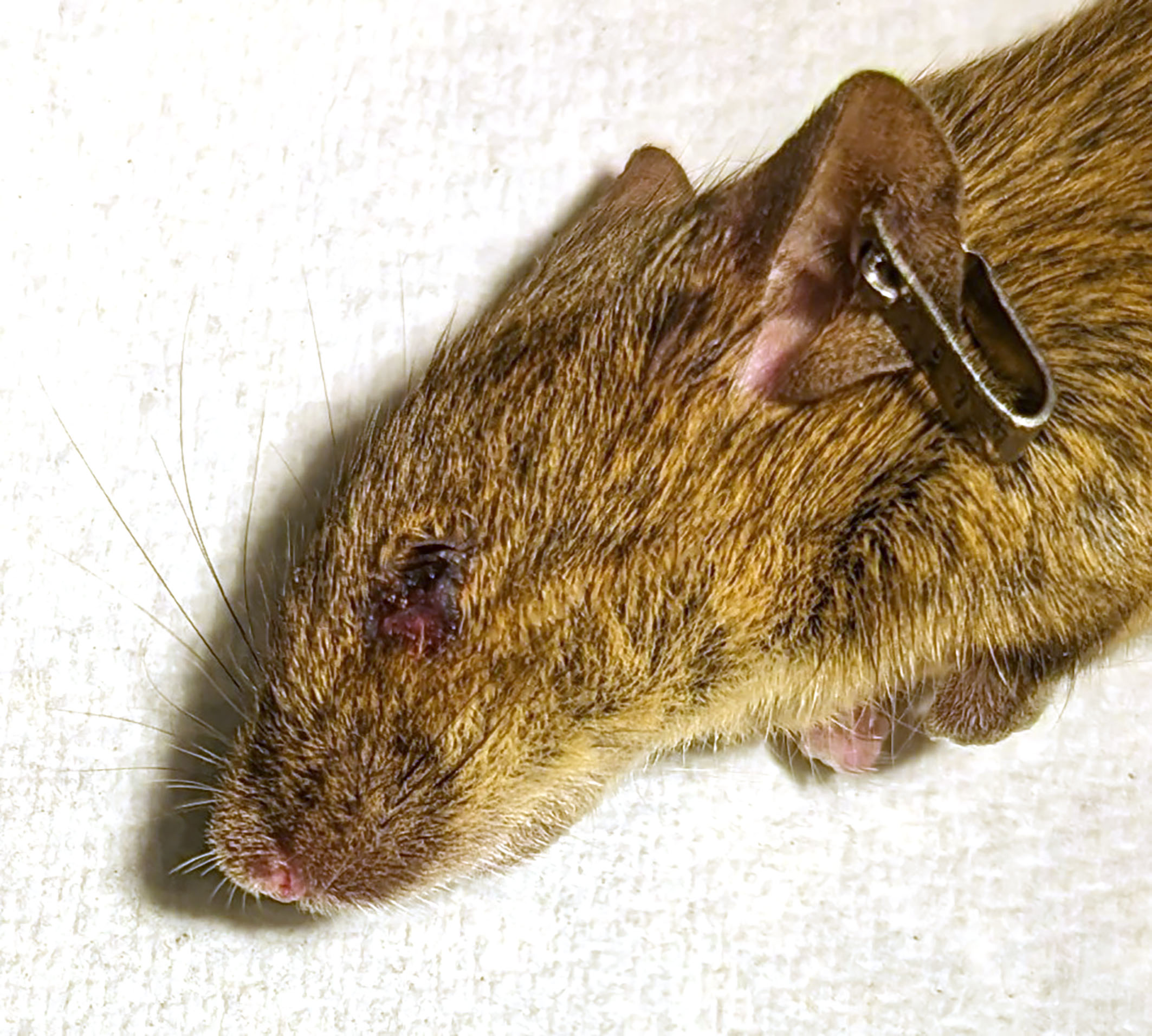
Gross Pathology:
Both mice showed serous ocular discharge, porphyrin staining, medial canthus swelling, and blepharospasm bilaterally. No experimental history of eye manipulation was reported. One mouse had been treated with an ocular antibiotic ointment.
Laboratory Results:
N/A.
Microscopic Description:
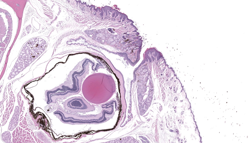
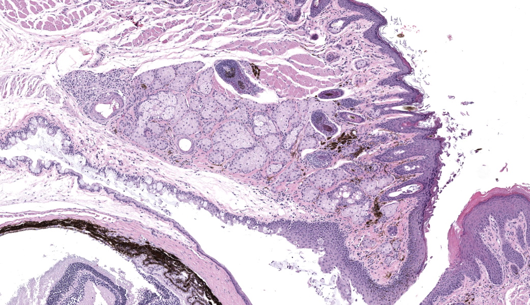
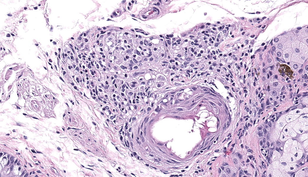
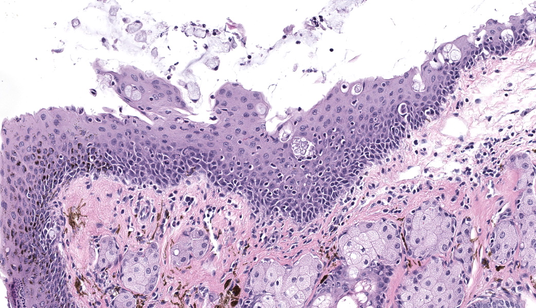
Bilaterally or unilaterally*, the eyelids contained locally extensive epidermal acanthosis and ortho to parakeratotic hyperkeratosis of varying severity. This was accompanied by epithelial erosion and mild serocellular crusting. There was mild to moderate mixed neutrophilic to lymphocytic inflammation within the conjunctiva and eyelid sebaceous glands (Meibomian glands). Focally there was occasional dilation and mild degeneration of Meibomian gland acini or ductules. The cornea, lens, iris, retina, optic nerve and other interior structures of the eyes were normal.
The Harderian gland was mildly enlarged (section plane-dependent) and glandular lumens contained occasional brown pigmented inspissated material (porphyrin secretions). Glandular epithelium lacked atypia and there was no compression or invasion as might be seen with adenoma or carcinoma.
*Grossly, the lesion was present bilaterally in affected mice but, due to section plane and variable severity, it is microscopically more evident unilaterally in the submitted examples.
Contributor’s Morphologic Diagnoses:
- Eyelids: Blepharoconjunctivitis, neutrophilic, with epidermal acanthosis, erosions, and ortho to parakeratotic hyperkeratosis, unilateral to bilateral, moderate, chronic.
- Harderian gland: Hyperplasia with inspissated porphyrin secretions, bilateral, mild, chronic.
Contributor’s Comment:
Blepharoconjunctivitis occurs with high frequency in aging 129S/SvEv, 129P, and other 129 origin substrains.2,3 An incidence of >90% was reported in one lifetime survey of pathology in 129S6/SvEvTac mice (older nomenclature for 129S/SvEv). Findings ranged from suppurative to mononuclear inflammation and epidermal changes included acanthosis and focal erosion or ulceration.2 In this submission, the epidemiology of the lesion presentation and its similarity to the literature descriptions of blepharoconjunctivitis supported that its occurrence was related to the underlying background strain and not to the knockout gene.
The underlying cause for blepharitis or blepharoconjunctivitis in 129S substrains is unknown. Underlying age-related structural and functional changes in the position and function of lacrimal glands are believed to play a role, possibly by impacting mucocutaneous barrier function. Age-associated anterior migration of the palpebral mucocutaneous junction in mice has been cited as contributing to eyelid laxity and poor Meibomian gland function.2Corynebacterium species biochemically similar to C. urealyticum, C. pseudodiphtheriticum, and C. bovis have been cultured from affected eyelids. However, these agents were not identified in all affected animals and are also commensally found on the eyelids of normal mice. Additionally, rederivation into SPF housing was not sufficient to prevent occurrence.2 In the submitted case, the attending clinical veterinarian reported that treatment with topical ocular antibiotic ointment appeared to decrease lesion severity and prolong the time until the animal reached humane endpoints for euthanasia, but did not fully resolve the lesion.
Harderian gland adenomas have been linked to blepharoconjunctivitis in 129S6/SvEvTac mice, but these were believed to be a concurrent aging lesion that played an exacerbating rather than a causative role.2
Nomenclature of 129 mice can be confusing. 129 mice are made up of 3 major subfamilies with enough genetic variation between them that they are considered separate strains. As such, these subfamilies have been designated as the parental strain (129P), the Steel strain (129S), and the teratoma strain (129T). There are also several substrains within each of these three. 129S/SvEv mice, the strain in this submission, are widely commercially available and are frequently utilized to generate embryonic stem cells (ESCs) for the production of mice with targeted mutations (knockouts). They were originally bred from 129S mice received from Dr. Martin Evans, giving them their substrain designation.4
Contributing Institution:
In Vivo Animal Core, Unit for Laboratory Animal Medicine
University of Michigan
2800 Plymouth Road
Ann Arbor, Ml 48109
(https ://animal care .um ich .edu/ business-services/vivo-animal-core)
JPC Diagnoses:
- Eyelid: Blepharoconjunctivitis, unilateral, lymphocytic and neutrophilic, chronic, diffuse, mild, with epidermal hyperplasia and erosion.
- Eyelid, Meibomian gland: Adenitis, lymphoplasmacytic, chronic, focal, mild.
JPC Comment:
Although seemingly a not-so-exciting lesion, this is one that needs to be able to be recognized by the pathologist, especially in 129 mice. It is a common finding in 129 substrains! The contributor did a great job discussing this lesion in their comment which, again, covered most of this case’s conversation. Some participants were thrown off initially by the size discrepancy between the two lenses, but this is largely due to a function of cut during processing and is not a lesion. The contributor mentioned that this was a grossly bilateral lesion but did acknowledge that this is not seen histologically due to sectioning. In short, don’t be thrown off if the lesion isn’t obvious on both eyelids despite the gross description.
Although not in the contributor’s description, a few participants astutely perceived the presence of a sequestrum characterized by necrotic bone with surrounding osteoclastic activity within the right side of the maxillary bone. This sequestrum is surrounded by moderate inflammation and fibrosis, and there is an underlying neutrophilic and lymphocytic gingivitis. Most participants thought this may have been related to some form of oral trauma that resulted in a penetrating injury to the hard palate. Gingivitis is a common background lesion in laboratory mice, and the sequestrum was a neat secondary lesion to find! The JPC did not include the sequestrum and gingivitis in the morphologic diagnoses, however, to ensure clarity on the primary blepharoconjunctivitis lesion in this case.
Conference attendees were also quizzed on additional common background lesions of 129 mice, which include a reduced or absent corpus callosum (can be up to 80% in some groups of 129 mice), spontaneous testicular teratomas and lung tumors, eosinophilic crystalline pneumonia (also known as acidophilic macrophage pneumonia), hyalinosis of the glandular stomach, respiraoty tract, and gallbladder, urolithiasis, megaesophagus, polyarteritis, and age-related hearing loss in 129P3/J mice. This particular lesion is caused by a Cdh23ahl mutation that results in progressive hearing loss by 3 months of age.1 In contrast, 129S/SvEv mice are resistant to noise-related hearing loss.5
References:
- Burghard AL, Morel NP, Oliver DL. Mice heterozygous for the Cdh23/Ahl1 mutation show age-related deficits in auditory temporal processing. Neurobiol Aging. 2019;81:47-57.
- Radaelli E, Castiglioni V, Recordati C, et al. The pathology of aging 129S6/SvEvTac mice. Vet Pathol. 2016;53(2):477-92.
- Sundberg JP, Brown KS, Bates R, Cunliffe-Beamer TL, Bedigian H. Suppurative conjunctivitis and ulcerative blepharitis in 129/J mice. Lab Anim Sci. 1991;41:516-18.
- Mouse strain 129 substrain nomenclature. Mouse Genome Informatics Database at the Jackson Laboratory, Bar Harbor, ME. Accessed September 13, 2023.
- Yoshida N, Hequembourg SJ, Atencio CA, Rosowski JJ, Liberman MC. Acoustic injury in mice: 129/SvEv is exceptionally resistant to noise-induced hearing loss. Hear Res. 2000;141(1-2):97-106.