Wednesday Slide Conference, 2025-2026, Conference 7, Case 4
Signalment:
3-year-old male beagle (Canis lupus familiaris) dogHistory:
All procedures performed on animals were in accordance with regulations and established guidelines and were reviewed and approved by an Institutional Animal Care and Use Committee or through an ethical review process.Gross Pathology:
N/ALaboratory Results:
N/AMicroscopic Description:
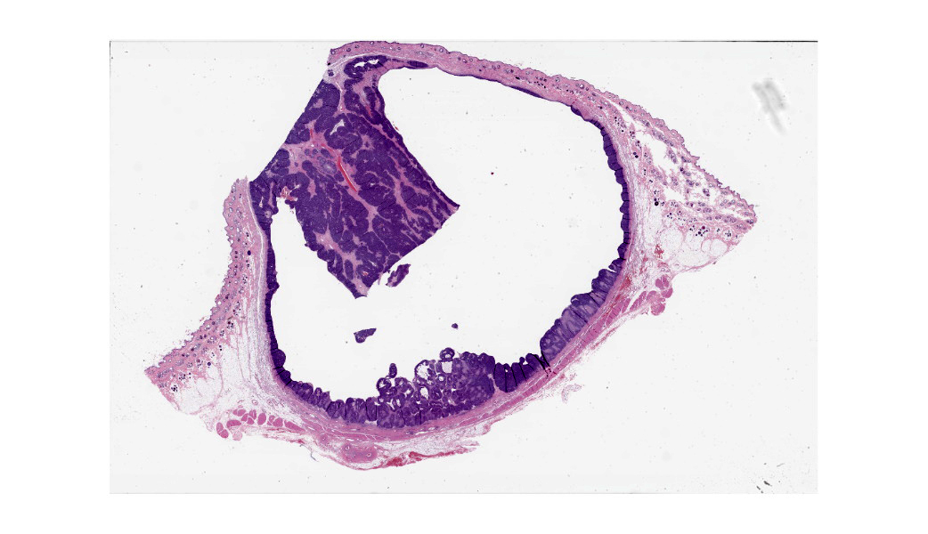
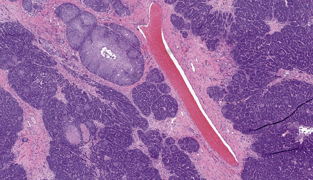
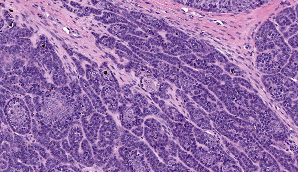
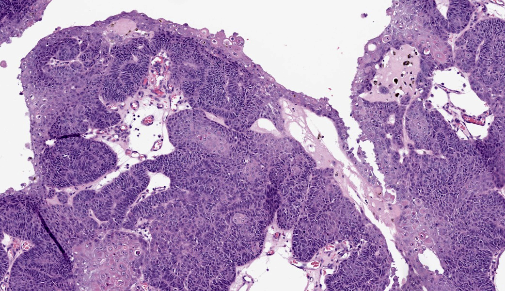
Haired skin: Extending from the dermis deep into the subcutis is an approximately 2 x 2 cm well-demarcated cystic mass lined by neoplastic undifferentiated basaloid epithelial cells. The neoplastic cells are arranged in lobules of dense anastomosing and palisading cords or small tightly packed nests, supported by scant fibrovascular stroma. There are multifocal papillary projections of neoplastic lobules into the lumen of the large cystic cavity. Multifocai lobules have smaller central cyst formation. The neoplastic basaloid cells are cuboidal with scant basophilic cytoplasm and round to oval, hyperchromatic nuclei, with two or more prominent nucleoli. There is minimal anisocytosis and moderate anisokaryosis. There is multifocal abrupt squamous differentiation and necrosis and approximately 5 mitotic figures per high power field. Brown granular pigment, most consistent with melanin, is present throughout the periphery of the mass; multifocal melanomacrophages infiltrate neoplastic lobules. Neoplastic cells extend to within 2 mm of the surgical margin; however, there are multifocal foci suspicious for local invasion.Contributor's Morphologic Diagnoses:
Haired skin, basosquamous cell carcinomaContributor's Comment:
In dogs and cats, cutaneous neoplasms are common. Some of the most common tumor types reported in canine patients include mast cell tumors, lipomas, hair follicular and/or sebaceous gland tumors, histiocytomas, soft tissue sarcomas, and melanocytic tumors.2,5,10 However, in cats, basal cell tumors, mast cell tumors, squamous cell carcinoma, and fibrosarcomas are the most common skin tumors reported.6 Tumors of the epidermis include papillomas and papillomavirus-induced tumors/tumor-like lesions, squamous cell carcinoma, basal cell carcinomas, and basosquamous carcinomas.4Arising from epidermal basal cells, basosquamous carcinomas share features with squamous cell carcinomas and basal cell carcinomas and are considered low-grade malignancies.4 Although rare in dogs and cats, along with other domestic animals, these tumors are more commonly reported in dogs.1 Canine breeds reported to have an increased risk of developing basosquamous carcinomas, include Saint Bernard, bloodhound, Samoyed, and old English sheepdog.1 These tumors are more likely found on the head, neck, and limbs.1
Histologically, basosquamous carcinomas are predominately composed of intradermal to subcutaneous lobules of undifferentiated basal cells with central foci of abrupt squamous differentiation.1 Keratinocytes at the center of neoplastic lobules have mild “nuclear pleomorphism, mitotic activity and dyskeratosis.”1 Unlike other epidermal neoplasms, connection to the overlying epidermis is not a consistent diagnostic feature.4 However, cyst formation and melanization, as seen in this case, are common and can often be appreciated grossly.1,4 Keratinocytes, present in the foci of abrupt and atypical keratinization, that display features of malignancy, along with “the lack differentiation of the follicular isthmus or bulb are features that help differentiate basosquamous carcinomas” from other tumors.4
As basosquamous carcinomas share features with squamous cell carcinomas and basal cell carcinomas, these neoplasms represent possible differential diagnoses for basosquamous carcinomas.1,4 Adequate surgical excision is usually curative for these slow growing malignancies.1
Contributing Institution:
Pfizer Research and Development, 455 Eastern Point Rd., Groton, CT 06340 www.pfizer.comJPC Diagnoses:
Haired skin: Basosquamous carcinJPC Comment:
Basosquamous carcinoma (BSC) was first described in 1910 by Dr. Henry MacCormac.10 He called this particular type of growth, which he astutely related to squamous cell carcinoma, a “rodent ulcer”, which was a once-popular term used to describe ulcerative growths that had a “rat bite” appearance. Although this term has long since fallen out of use, BSC remains a controversial entity that has histologic features of both squamous cell carcinoma (SCC) and basal cell carcinoma (BCC) yet represents its own diagnosis. Whether or not this diagnosis is part of a continuum of changes seen in various stages of either BCC or SCC is currently unclear, and the diagnoses of BSC is highly contested within both human and veterinary medical circles.In the current academic and clinical environment, BSCs are considered rare cutaneous neoplasms in both humans and veterinary species. While thought of as a low-grade malignancy with low metastatic potential in dogs and cats, this is not the case in humans, where basosquamous carcinomas are considered aggressive with more common metastases and a high rate of recurrence, even with wide surgical margins.3 Behaviorally, BSC is more akin to SCC than to BCC in humans. There are rare reports of metastatic BSC in dogs, as well, but this incredibly uncommon for this entity in dogs and cats.9
In human literature, it is currently believed that the BSC initially arises as a BCC that then undergoes genetic and epigenetic alterations, ultimately leading to squamous differentiation through basal-to-squamous transition (BST). As such, the BSC was classified as a BCC variant by the WHO in 2023.7 To further complicate matters, there are also “basaloid SCC” and “keratotic BCC” diagnoses in humans, as well as BCC/SCC collision tumors.7 Participants were shown histologic images of what the moderator currently uses to differentiate between a keratinizing BCC, a basaloid SCC, and a BSC. The basaloid SCC is typically described as having comedonecrosis with squamous differentiation at the edges of lobules and is NOT classified as a skin tumor. Rather, it is found in the oral cavity, cervix, lung, etc.7 The keratotic BCC variant is composed of horn cysts, parakeratotic cells, and abrupt keratinization.7 BSC is identified by abrupt squamous differentiation (not abrupt keratinization) with dyskeratotic squamous cells within the center of lobules of basal cells.7 Both the basal cells and keratinocytes have mitotic activity. Considering these factors, a diagnosis of BSC was favored by participants. Ultimately, though, there seems to be substantial disagreement on how to define the BSC in veterinary literature, even between prominent tumor texts and fascicles.
To throw yet another wrench in this entity, it is well-known that papillomaviruses can induce squamous cell neoplasms in numerous species. Common examples include bovine papillomaviruses 1 and 2 causing equine sarcoids, bovine papillomavirus-4 resulting in squamous neoplasms of the alimentary tract in cattle, and canine papillomavirus-2 causing cutaneous papillomas in dogs.4 Any of these can undergo malignant transformation when the right conditions occur. In recent years, Felis catus papillomavirus-2 (FcaPV-2) was detected within neoplastic cells of a multicentric cutaneous basosquamous carcinoma in a cat.8 Although not generally thought to be associated with a papillomavirus, this was the first case report of a BSC associated with FcaPV-2.8 Although better understood in humans, much has yet to be elucidated about this rare neoplasm in veterinary species and its clinical behavior, survivability, and potential correlations to papillomaviruses in certain cases.
References:
- Goldschmidt MH and Goldschmidt KH, Epithelial and Melanocytic Tumors of the Skin. In: Meuten DJ, ed. Tumors in Domestic Animals, 5th ed. 2017; Ames, IA, John Wiley & Sons, Inc., p.88-141.
- Graf R, Pospischil A, Guscetti F, et al. Cutaneous Tumors in Swiss Dogs: Retrospective Data from the Swiss Canine Cancer Registry, 2008-2013. Vet Pathol. 2018;55(6):809-820.
- Lima NL, Verli FD, de Miranda JL, Marinho SA. Basosquamous carcinoma: histopathological features. Indian J Dermatol. 2012;57(5):382-3.
- Mauldin EA and Peters-Kennedy J, Integumentary System. In: Maxie MG, ed. Jubb Kennedy and Palmer’s Pathology of Domestic Animals, 6th ed. 2016; St. Louis MO, Elsevier Press, vol 1, p.703-736.
- Martins AL, Canadas-Sousa A, Mesquita JR et al. Retrospective study of canine cutaneous tumors submitted to a diagnostic pathology laboratory in Norther Portugal (2014-2020). Canine Medicine and Genetics. 2022;9(1):2.
- Miller MA, Nelson JR, Turk L, et al. Cutaneous Neoplasia in 340 Cats. Vet Pathol. 1991;28:389-395.
- Murgia G, Denaro N, Boggio F, Nazzaro G, Benzecry V, Bortoluzzi P, Passoni E, Garrone O, Marzano A. Basosquamous Carcinoma: Comprehensive Clinical and Histopathological Aspects, Novel Imaging Tools, and Therapeutic Approaches. Cells. 2023;12(23):2737.
- Oh YI, Cheon DS, Lee JK, Choi MH, Hwang SY, Kim HW, Kang BJ, Youn HY. Detection of Felis catus papillomavirus type 2 within multicentric basosquamous carcinoma in a domestic cat. J Vet Med Sci. 2018;80(9):1445-1449.
- Shin SK, Kim TW, Youm SY, Kim G, Na KJ, Chang D, Ahn B. Basosquamous carcinoma with systemic metastasis in a miniature Pinscher. Jpn J Vet Res. 2011;59(4):173-9.
- Villamil JA, Henry CJ, Bryan JN, et al. Identification of the most common cutaneous neoplasms in dogs and evaluation of breed and age distributions for selected neoplasms. JAVMA. 2011;239(7):960-965.
- Wermker K, Roknic N, Goessling K, Klein M, Schulze HJ, Hallermann C. Basosquamous carcinoma of the head and neck: clinical and histologic characteristics and their impact on disease progression. Neoplasia. 2015;17(3):301-5.