CASE I: N2018-0119 (JPC 4153147)
Signalment:
Adult, female, Sambava tomato frog (Dyscophus guineti)
History:
This Sambava tomato frog was found dead during routine morning rounds with no adverse clinical history.
Gross Pathology:
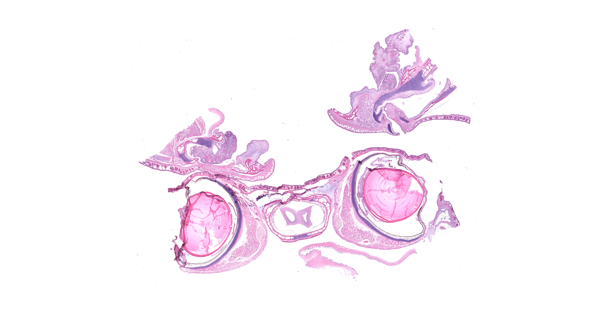
Extending from the rostral maxillary mucosa of the left internal naris is an approximately 3 mm thick x 2 mm diameter, pink and smooth mass.
Laboratory results:
No laboratory findings reported.
Microscopic Description:
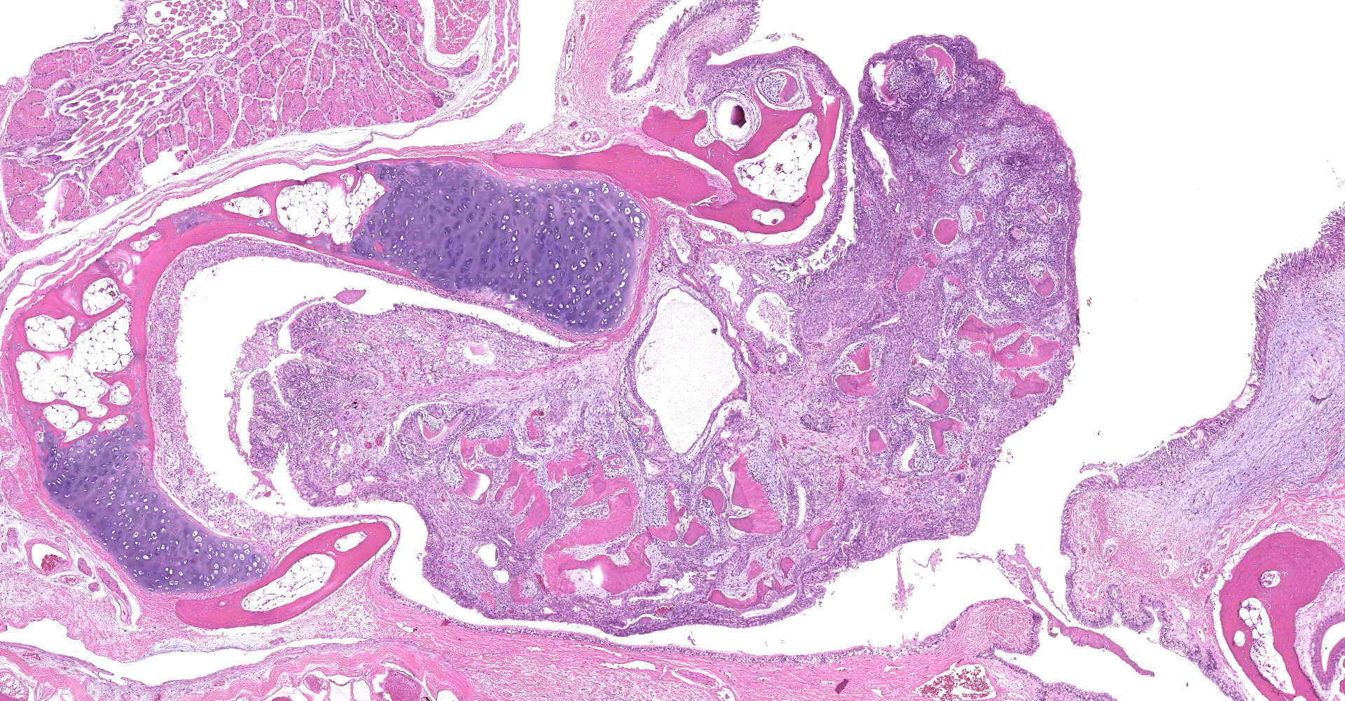
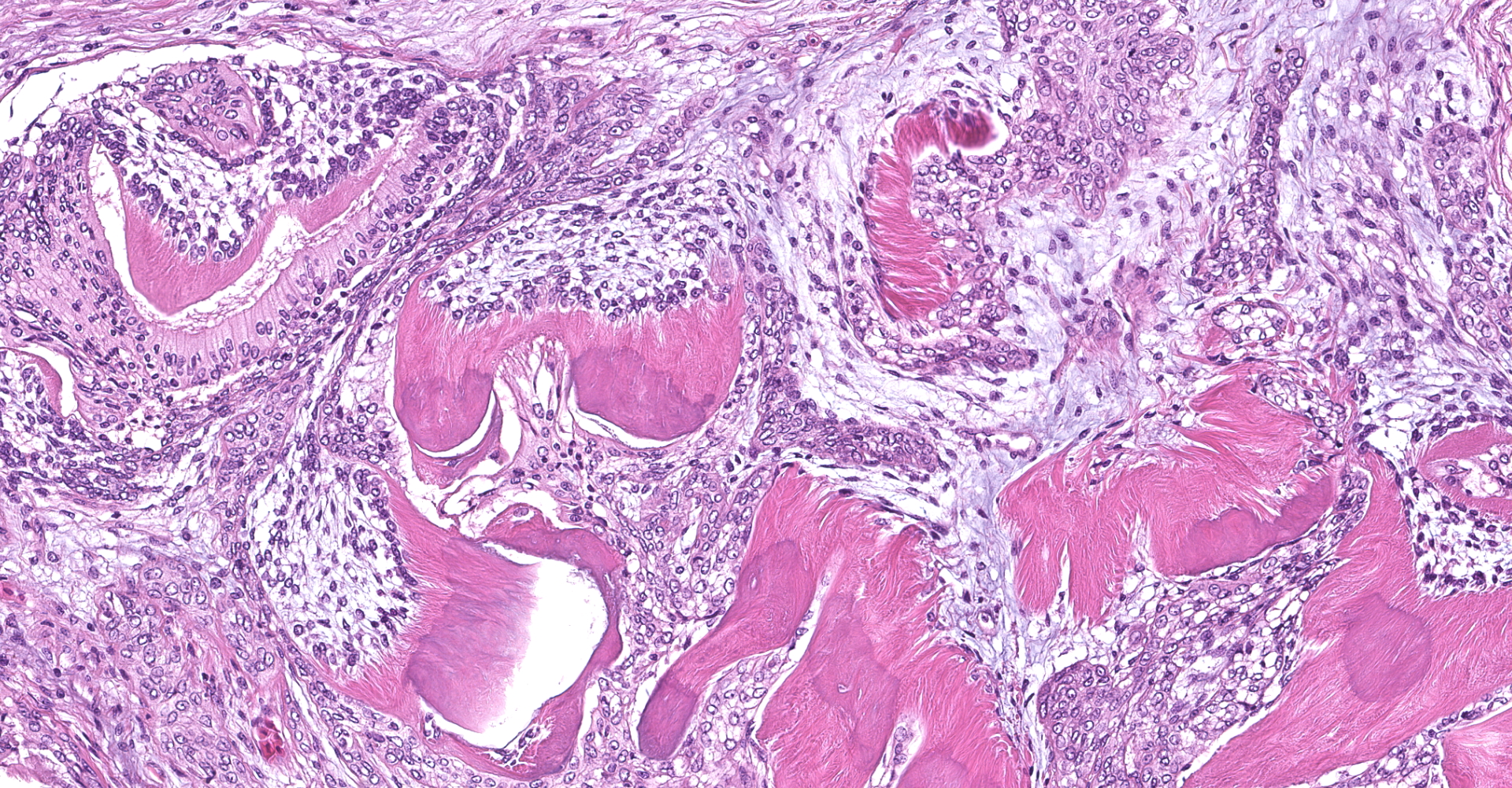
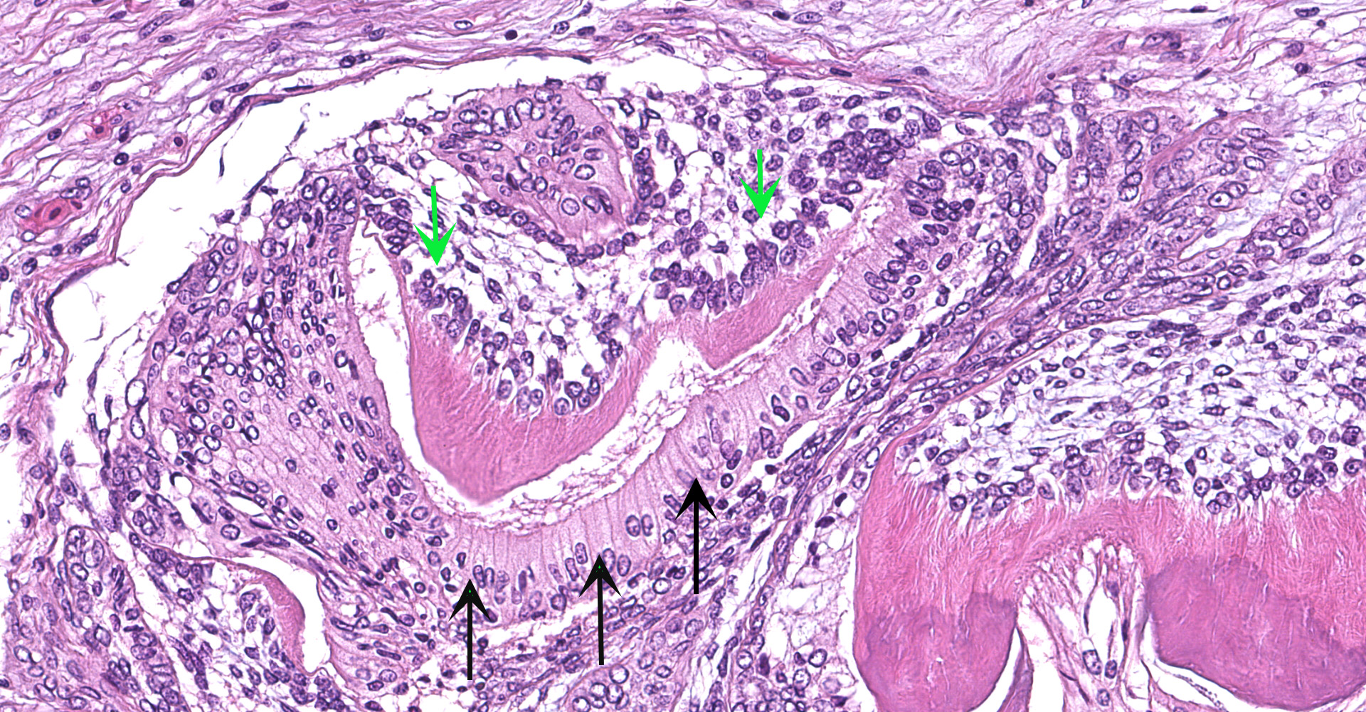
Extending from two representative sections of the left maxillary oral mucosa is an unencapsulated, variably well-demarcated, exophytic, and moderately cellular mass composed of two distinct populations of epithelial and mesenchymal cells forming multiple tooth-like structures (denticles) embedded within a pre-existing stroma. Denticles range from 50 to 250 microns in diameter and are composed of a combination of eosinophilic, homogenous to fibrillar material (dentin), palisading epithelium with apical nuclei and basilar cytoplasmic clearing (odontogenic epithelium; ameloblasts), and proliferating mesenchymal cells with small and hyperchromatic nuclei (odontoblasts). Central arcs of dentin are frequently lined by ameloblasts along their convex surface and odontoblasts along their concave surface. No mitoses are identified, and anisocytosis and anisokaryosis are minimal. Rare individual lymphocytes and plasma cells multifocally infiltrate the mass.
Contributor's Morphologic Diagnoses:
Gingival
mass: Odontoma, compound type
Contributor's Comment:
Odontomas are odontogenic tumors with variably organized dental elements; they are designated as complex or compound based on how closely they parallel tooth embryogenesis. Familiarity with normal early tooth development is thus essential in accurate classification of odontomas, in addition to odontogenic lesions in general.
Two tissue types are involved in mammalian odontogenesis: odontogenic epithelium and ectomesenchyme. The process initiates as ectoderm-derived oral epithelium extends into underlying connective tissue, forming a bud of epithelium referred to as the dental lamina (bud stage).8 As the dental lamina expands, it induces adjacent neural crest-derived ectomesenchyme to proliferate and differentiate into the dental papilla, which organizes along a concavity of the dental lamina and is the future source of dental pulp and odontoblasts (cap stage). Next, a bell-like structure forms from continued proliferation of oral epithelium, which differentiates into the enamel organ, composed of central stellate reticulum, inner ameloblasts and stratum intermedium, and outer ameloblasts (bell stage). The underlying ectomesenchyme condenses and forms a dental follicle; the precursor to the periodontal ligament. As time progresses, production of dental matrix begins by differentiated odontoblasts of the dental papilla, which produce predentin that calcifies to dentin. This then leads to the crown stage, where ameloblasts produce overlying enamel.
Complex odontomas are characterized by disorganized conglomerates of odontogenic epithelium, ectomesenchyme of the dental papilla, and dental hard substances (dentin and enamel). Compound odontomas form distinct, tooth-like structures called denticles, where the order of tissues in tooth embryogenesis is generally maintained.6 A combination of compound and complex structures often exists within a single tumor.7 Odontomas are uncommon in veterinary species, despite being the most common odontogenic tumor reported in humans, especially children and young adults.11 They occur most frequently in young dogs and horses, with fewer case reports in other species.2,4,7 Associated clinical signs generally include unilateral swelling of the face or mandible and displacement of teeth. Although generally benign, they can be locally destructive. Complete surgical excision is typically curative.9
Recently, odontomas were reported in three frogs: a Sambava tomato frog (also known as a false tomato frog [Dyscophus guineti]), an African clawed frog (Xenopus laevis), and a tomato frog of undetermined species.5 The tumor extended from the maxillary gingiva in all three cases, as occurred in this case. Histologically all tumors formed orderly denticles with ameloblasts, dentin, and odontoblasts, characterizing them as a compound subtype. In one case, the frog was euthanized due to the presence of the odontoma and the outcomes of the remaining two were unknown (lost to follow-up).
With the exception of members of the family Bufonidae who lack teeth, the teeth of anurans are present only on the maxilla.10 This may explain the predilection for odontomas arising from exclusively the maxillary mucosa in this group. Anurans have polyphyodont and homodont teeth and lack both cementum and a periodontal ligament.5 Tooth formation in amphibians is similar to that of mammalians, with a few key differences. The posterior teeth may be derived from foregut endoderm, rather than ectoderm.1 Additionally, prior to metamorphosis in some amphibians, odontoblasts produce enamaloid, a dentin-like matrix, that is overlain with enamel by ameloblasts.3 The dental follicle is not histologically apparent in amphibians, which is thought to be related to tooth attachment occurring at the base, rather than the sides, of the teeth.1 Additionally, the enamel organ in amphibians is described to be comprised of two cell layers ? the inner and outer dental epithelium.3 Whether or not a stellate reticulum and stratum intermedium are additionally present is unclear; reptiles appear to develop a stellate reticulum, but the latter is often absent in bony fish.1
The pathogenesis of odontomas is unknown. Some consider them a type of hamartoma rather than a neoplastic process, and all odontomas are classified as hamartomas in human medicine as of 2017.7 In the recent report of frogs with odontomas, trauma to the dental lamina leading to de novo dental element formation was proposed.5
In the current case, the presence of an odontoma was not considered likely to have contributed to mortality. The animal was in good body condition and the tumor was small enough to not interfere with eating. Based on the identification of odontoma development in multiple frogs, this neoplasm should be considered as a differential diagnosis in cases of oral tumors in anurans.
Contributing Institution:
Wildlife Conservation Society, Zoological Health Program; https://oneworldonehealth.wcs.org
JPC Diagnosis:
1. Gingiva: Compound odontoma.
2. Salivary duct: Larval nematodes, numerous.
JPC Comment:
This case is an excellent example of an entity more frequently described in domestic species that also occurs in exotics with similar histologic features. In addition, the contributor provides a thorough review of both mammalian and anuran odontogenesis, odontoma classification, and the unique anatomic features associated with of anuran dentition.
As noted by the contributor, the majority of anurans have teeth restricted to the upper jaw, with exception of some hylids in which teeth are also found on the dentary (anterior bone of the lower jaw). Teeth are arranged in a single row on the paired premaxillaries, maxillaries, and vomers. Using the northern leopard frog (Rana pipiens) as a model, most anurans have homodont bicuspid teeth that are very small, measuring less than 1.0 mm long but numerous, with up to 184 functional teeth, which are each associated with a replacement tooth, resulting in an average of 368 teeth in this species.3
Tadpoles (the aquatic larvae of anurans) do not have truly mineralized teeth. Xenopus laevis (African clawed frog) is technically an exception and will be discussed further below. Instead, most tadpoles have horny labial teeth on the upper and lower beak composed of columns of keratinized epithelial cells. Both the number of teeth and the size of the beak grow throughout larval life, until they're destroyed by autolysis during metamorphosis.3
Odontogenesis in most anurans begins during metamorphosis, near the end of hindlimb organogenesis, with functional teeth present after approximately 25 days. Using R. pipiens as a model, the six dental laminae correspond to the six dentigerous bones (paired premaxillaries, maxillaries, and vomers), and give rise to enamel organs.3
In contrast to other anurans, X. laevis differs in that true teeth develop in the upper jaw of tadpoles during the last stages of larval life. However, these teeth do not erupt until the end of metamorphosis. The teeth are morphologically similar between larvae and adults but differ size. While X. laevis teeth are similar to other anurans in that they form a single row on the upper jaw and are homodont, they're also conical, monocuspid, and slightly curved at the tip.3
As noted by the contributor, the cause of odontoma formation in anurans is unknown, although trauma to the dental lamina has been hypothesized.5 The dental lamina is a specialized segment of the embryologic gingival epithelium that invaginates into the developing jaw and gives rise to the enamel organ, a complex structure of odontogenic epithelium that produces enamel. It includes the outer enamel epithelium, stellate reticulum, and inner enamel epithelium.7 In anurans, the dental lamina is present throughout animal's life due to its polyphyodont dentition (i.e. teeth are replaced throughout the animal's lifetime). Previous studies have demonstrated transplantation of the dental lamina leads to the formation and replication of tooth buds and/or formed teeth, even when transplanted outside the oral cavity.5
During the conference participants discussed common terminology associated with dental tumors, including cemento-osseous matrix versus dental matrix. Cemento-osseous matrix is composed of cementum and/or alveolar bone associated with proliferative lesions arising from the periodontal ligament (e.g. fibrous epulis of the periodontal ligament). In contrast, dental matrix is composed of dentin and enamel. The moderator noted the presence of one dental matrix component indicates the other is also present due to the concept of induction. During odontogenesis, the formation of enamel (amelogenesis) is induced by the initial formation of dentin by odontoblasts. The presence of ameloblasts in turn further induces odontoblasts to continue the production of dentin.
In addition to the odontoma, attendees also noted dozens of larval nematode tangential and cross sections within the salivary duct that are 30µm in diameter with a thin eosinophilic cuticle and a pseudocoelom containing abundant eosinophilic globular material. Unfortunately there are insufficient distinguishing characteristics present within the section for identification.
The following chart outlines various odontogenic tumors in domestic species and common microscopic findings with each:
|
Tumor |
Odontogenic epithelium |
Stroma |
Mesenchyme |
Matrix |
Species affected |
Misc. |
|
Ameloblastoma |
Yes |
Not essential for diagnosis |
None |
None |
Dog, cat, horse |
Keratinization may occur |
|
Amyloid producing odontogenic ameloblastoma |
Yes |
Not essential for diagnosis |
None |
Amyloid |
Dog, cat, horse |
Matrix composed of enamel proteins which are still Congophilic and exhibit apple-green birefringence; IHC + for laminin |
|
Canine acanthomatous ameloblastoma |
Yes |
Stellate fibroblasts in dense collagen; regularly spaced dilated, empty blood vessels |
Periodontal ligament |
None |
Dog |
Interconnected sheets of odontogenic epithelium
|
|
Ameloblastic fibroma |
Yes |
Loose, collagen poor, resembles pulp ectomesenchyme |
Pulp ectomesenchyme |
None |
Young animals, cattle
|
Most common oral neoplasm in cattle
|
|
Ameloblastic fibro-odontoma |
Yes |
Loose, collagen poor, resembles pulp ectomesenchyme |
Pulp ectomesenchyme
|
Dentin or enamel
|
Young animals, cattle |
|
|
Complex odontoma |
Yes |
Well-differentiated dentinal tissue |
Pulp ectomesenchyme |
Dentin, enamel (may be mineralized) |
Dog, rodent, primates, horse |
Horse, rodents produce cementum; "balls of disorganized dental hard substance" |
|
Compound odontoma |
Yes |
Well-differentiated dentinal tissue; dense collagen and vascular connective tissue |
Pulp ectomesenchyme |
Dentin, mineralized enamel |
Young dogs |
Multiple tooth-like structures (denticles) |
|
Table adapted from Munday JS, Lohr CV, Kiupel M. Tumors of the alimentary tract. In: Meuten DJ, ed. Tumors in domestic animals. 5th ed. Ames, IA: John Wiley; 2017:530-542.6,12
|
||||||
References:
1. Berkovitz B, Shellis P. Tooth formation. In: The teeth of non-mammalian vertebrates. 1st ed. Academic Press;2017:235-254.
2. Brounts SH, Hawkins JF, Lescun TB, Fessler JF, Stiles P, Blevins WE. Surgical management of compound odontomas in two horses. J Am Vet Med A. 2004;225(9):1423-1427.
3. Davit-Béal T, Chisaka H, Delgado S, Sire JY. Amphibian teeth: current knowledge, unanswered questions, and some directions for future research. Biol Rev. 2007;82:49-81.
4. Hoyer NK, Bannon KM, Bell CM, Soukup JW. Extensive maxillary odontomas in 2 dogs: diagnosis, pathology, and management. J Vet Dent. 2016;33(4):234-242.
5. LaDouceur EE, Hauck AM, Garner MM, Cartoceti AN, Murphy BG. Odontomas in frogs. Vet Pathol. 2020;57(1):147-150
6. Munday JS, Lohr CV, Kiupel M. Tumors of the alimentary tract. In: Meuten DJ, ed. Tumors in domestic animals. 5th ed. Ames, IA: John Wiley; 2017:530-542.
7. Murphy BG, Bell CM, Soukup JW. Odontogenic tumors. In: Veterinary oral and maxillofacial pathology. 1st ed. Hoboken, NJ: Wiley-Blackwell; 2020:91-127.
8. Murphy BG, Bell CM, Soukup JW. Tooth development (odontogenesis). In: Veterinary oral and maxillofacial pathology. 1st ed. Hoboken, NJ: Wiley-Blackwell; 2020:13-19.
9. Papadimitriou S, Papazoglou LG, Tontis D, Tziafas D, Papaionnnou N, Patsikas MN. Compound maxillary odontoma in a young German shepherd dog. J Small Anim Pract. 2005;46:146-150
10. Pessier AP. Amphibia. In: Terio KA, McAloose D, St. Leger J, eds. Pathology of wildlife and zoo animals. 1st ed. London, UK: Elsevier; 2018:915?944.
11. Weidner N, Matthews K, Regezi JA. Oral cavity and jaws. In: Modern surgical pathology. 2nd ed. Elsevier; 2009:326-362.
12. https://www.askjpc.org/wsco/wsc_showcase2.php?id=bGtKVGNqWVQxc21vdUNQdFpvalYrZz09


