CASE III:
Signalment:
A 5-month-old female Dorset lamb
History:
The lamb showed progressive development of ataxia and incoordination. The owner noticed a small nodule behind the ear at the site of Gudair® (ovine Johne’s disease) vaccination. The owner’s farm had a previous history of copper deficiency in lambs
Gross Pathology:
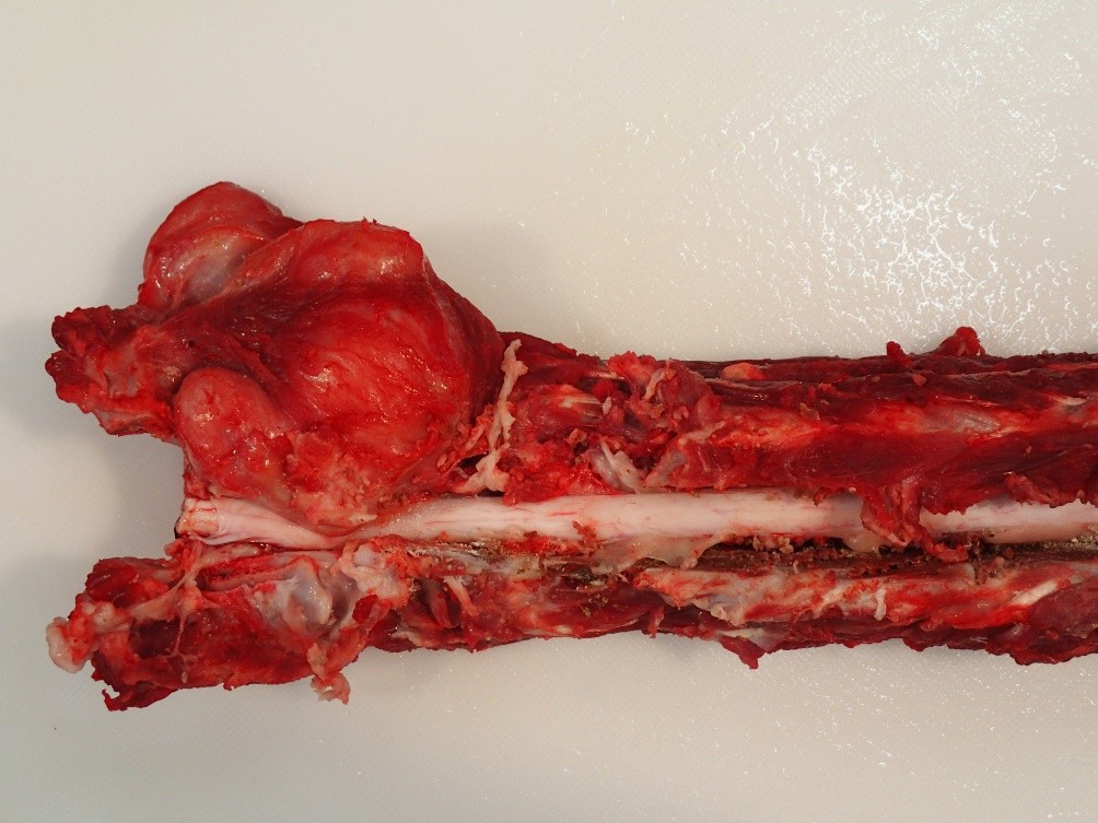
A pale tan to white irregular fleshy mass containing multiple foci of caseous material extended from the right dorsal cervical musculature into spinal canal at the C1–C2 intervertebral space. The mass compressed the underlying spinal cord. Gross examination was otherwise unremarkable.
Laboratory Results:
Serum copper was 12.7 µmol/L (Reference 7.5- 20 µmol/L) and liver copper level was 2.42 µmol/L (reference 0.23 – 3.67 µmol/L).
Microscopic Description:
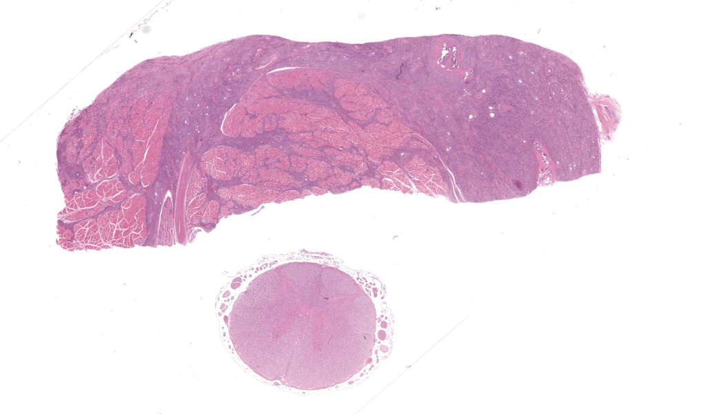
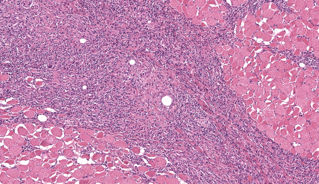
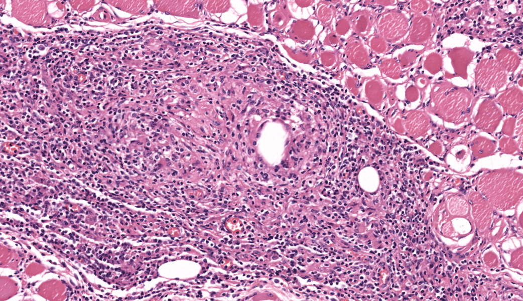
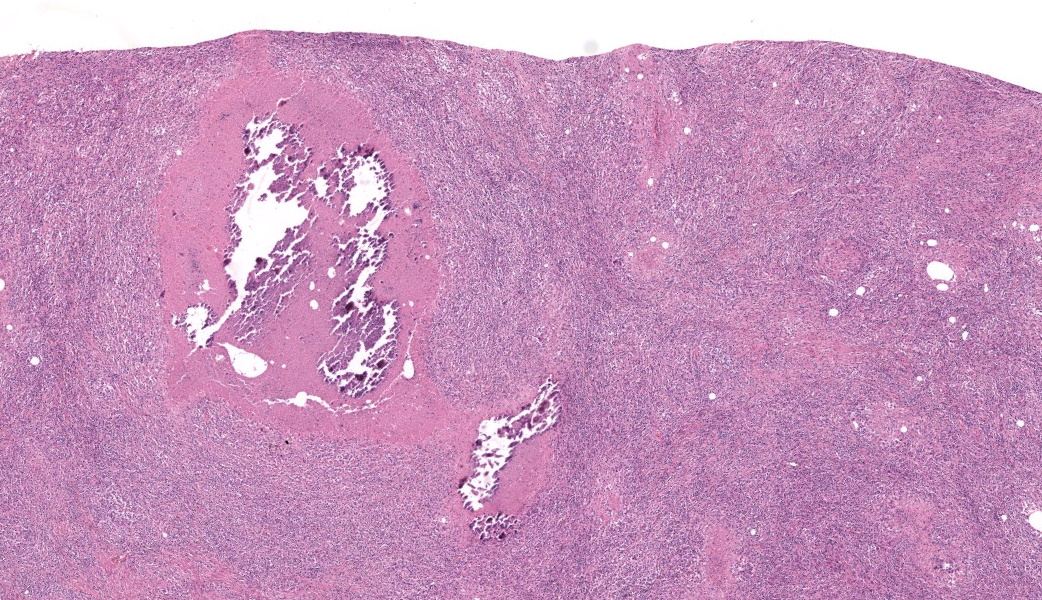
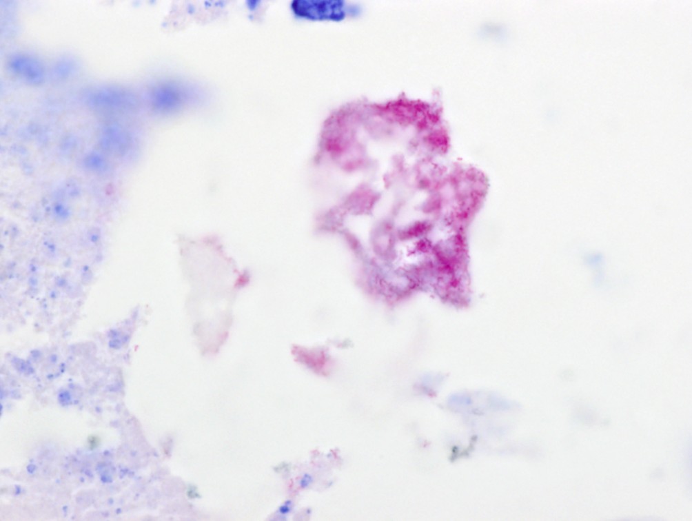
Right cervical mass: Surrounding and extending within the skeletal muscle there are extensive areas of fibrous connective tissue containing large numbers of epithelioid macrophages, lymphocytes, plasma cells and few neutrophils, as well as frequent fibroblasts. Within the fibrous tissue, there are multifocal areas of necrosis often centred on well-defined round clear spaces (lipid or oil), and these foci are surrounded by epithelioid macrophages with giant cell formation. Necrotic areas often display mineralization and occasionally contain aggregates of acid-fast rod-shaped organisms. Adjacent to granulomatous inflammation there are numerous hypereosinophilic myofibres which are often small and angular, and sometimes display centralized nuclei (degeneration and atrophy). Occasional myofibres contain protozoal bradycysts (consistent with Sarcocystis spp.).
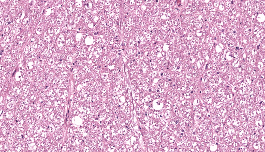
Spinal cord: Within the white matter of the ventral and lateral funiculi there is frequent swelling of myelin sheaths which often contain degenerate gitter cells (digestion chambers). There are also enlarged astrocytes scattered throughout the white matter in these areas.
Contributor’s Morphologic Diagnosis:
Cervical musculature: granulomatous perimyositis, chronic, diffuse, severe with intralesional acid-fast material, marked fibrosis and moderate myofibre degeneration and atrophy
Cervical spinal cord: Degenerative axonopathy, chronic, regional, moderate
Contributor’s Comment:
The submitted case is an example of vaccine-induced compressive myelopathy secondary to injection with Mycobacterium avium subsp. paratuberculosis vaccine (Gudair - imported from CZ Veterinaria, Porrino, Spain by Pfizer Animal Health). Gudair is a killed vaccine containing whole bacteria, paraffin oil, and thiomersal, and is indicated for single-dose subcutaneous injection behind the ear. Granuloma formation is a well-recognized side-effect of vaccine administration, both within the subcutis and also within the underlying cervical musculature if the vaccine is inadvertently delivered into the deeper tissue2,4-5,6,8-9. After introduction of the Gudair vaccine to Australia in 1999, a field trial was undertaken by L. Reddacliff et. al. from 1999 to 2004 on 1200 lambs aged 1-3 months on 3 farms in the central tablelands of New South Wales5. Reddacliff et al. observed that at 2 months post-vaccination, 42% of the vaccinated lambs had nodular vaccine site lesions with a diameter ranging from 3-45 mm3. Only 20 % of the vaccinated lambs had palpable vaccine site lesions by 3-4 years of age, interestingly with no decline in the average diameter of the nodules. Histologically, these lesions were described as multifocal to coalescing necrotizing granulomatous dermatitis and cellulitis4,5. Intralesional mycobacteria, representing residual killed vaccine organisms, were observed in 87.5 % of cases, but these did not grow in aerobic or anaerobic cultures 4,5
Consistent with previous reports, we observed well-defined round clear spaces associated with necrotic areas2. These clear spaces represent the lipid or oil base that acts as the adjuvant of the Gudair vaccine2. Although the mechanism of action of oil is not fully described, it is thought to trap the antigen at the site and release it over an extended period, prolonging immune stimulation. The Gudair vaccine composition of mycobacteria combined with oil essentially mimics Freund’s adjuvant, a substance shown to induce potent cellular immunity that also leads to granulomatous inflammation in different tissues. Granulomatous inflammation is also induced by the oil fraction alone but to a lesser degree compared to the full vaccine3.
Extension of the Gudair vaccine site reactions or granulomatous lesions into the cervical spinal canal has been reported previously6,7. In these reports, incorrect administration of the vaccine induced the formation of a smooth mass protruding dorsally into the cervical spinal cord at the level of C1-C2. Histologically, the cases displayed features of compressive myelopathy, with variable grey matter neuropil necrosis, hemorrhage, Wallerian degeneration of all funiculi, and glia cell infiltration6. These findings demonstrate that incorrect administration of Gudair vaccine intramuscularly or adjacent to the vertebrae may lead to spinal cord compression due to local granulomatous inflammation.
We received 3 more lambs aged 3-5 months from the same farm with the same clinical history. All lambs had similar gross and histopathological findings to those present in the submitted case, including granulomatous inflammation within the lateral subcutaneous and cervical musculature. We also observed well-demarcated clear spaces (presumably oil) associated with necrotic areas within the granulomatous inflammation in cervical musculature and scant bacteria in some areas of necrosis in all the lambs. The neurological issues ceased following training of staff in correct vaccine administration.
Contributing Institution:
Veterinary Anatomic Pathology, Faculty of Veterinary and Agricultural Sciences, The University of Melbourne
JPC Morphologic Diagnoses:
- Skeletal muscle: Myositis , granulomatous and necrotizing , chronic, multifocal to coalescing, marked with lipid vacuoles.
- Spinal nerve root, epineurium: Radiculoneuritis, granulomatous and lymphoplasmacytic, chronic, diffuse, moderate.
- Spinal cord, lateral and ventral funiculi: Axonal degeneration, multifocal, mild.
- Skeletal muscle: Sarcocysts, multiple.
JPC Comment:
The contributor has provided an excellent review of the Gudair vaccine, which is making its debut in the Wednesday Slide Conference. However, the effects of oil-based vaccines are not new to the Wednesday Slide Conference. In the last decade, two WSC cases have involved compressive myelopathy resulting from vaccination of cattle with oil-in-water adjuvanted vaccines against foot-and-mouth disease, a common practice in South America. In both cases, injection of an oil-in-water adjuvanted vaccine extended from a muscle abscess/granuloma through the spinal foramina into the spinal canal, compressing the spinal cord. Typically, these myelopathies affect the white matter, resulting in Wallerian degeneration of axons, while leaving the grey matter The contributor has provided an excellent review of the Gudair vaccine, which is making its debut in the Wednesday Slide Conference. However, the effects of oil-based vaccines are not new to the Wednesday Slide Conference. In the last decade, two WSC cases have involved compressive myelopathy resulting from vaccination of cattle with oil-in-water adjuvanted vaccines against foot-and-mouth disease, a common practice in South America. In both cases, injection of an oil-in-water adjuvanted vaccine extended from a muscle abscess/granuloma through the spinal foramina into the spinal canal, compressing the spinal cord. Typically, these myelopathies affect the white matter, resulting in Wallerian degeneration of axons, while leaving the grey matter unscathed.10 (This is typical of chronic compression, whereas acute compression typically affects the grey matter.)10 The paresis and paralysis seen in affected animals may not only be the result of spinal cord compression, but also of the spinal nerve roots as well, whose thin root sheath and scant endoneurial collagen make them easily compressed by advancing inflammation.10 (This is typical of chronic compression, whereas acute compression typically affects the grey matter.)10 The paresis and paralysis seen in affected animals may not only be the result of spinal cord compression, but also of the spinal nerve roots as well, whose thin root sheath and scant endoneurial collagen make them easily compressed by advancing inflammation. 10
A 2019 article by Asin et al.11 further explores the use of oil-in-water adjuvanted vaccines in small ruminants, documenting the gross lesions, histopathology, and ultrastructure of the granulomas that they cause, as well as briefly describing a systemic autoimmune disease in a subset of vaccinated animals referred to as ovine ASIA (autoimmune/systemic inflammation induced by adjuvants) syndrome. Ovine ASIA syndrome has a significant adverse effect on sheep production following widespread bluetongue vaccination in the late 2010s. Like the oil-in-water vaccines used to vaccinate cattle against FMD, aluminum salts are used as the adjuvant and, while potentiating the vaccine’s antigenic effects, also likely result in granuloma/abscess formation and systemic inflammation in sheep, which are very susceptible to these effects. (In cattle, the highest frequency of granulomas is seen in oil-in-water bacterins, prompting the supposition that endotoxins released from dying bacteria may potentiate tissue damage at the site of vaccination.)10
Ovine ASIA syndrome, as previously stated, was first described following a widespread bluetongue vaccination campaign in the late 2010s. ASIA has both an acute and chronic phase, and in this study, it was reproduced following repeated vaccinations with aluminum-adjuvanted oil-in-water vaccines. The acute phase, which appears 2-6 days following inoculation, results in acute meningoencephalitis and diminished response to external stimuli. The less common (or at least less commonly observed) acute phase appears 2-6 days after an adjuvant-containing inoculation. An acute neurological episode with low response to external stimuli and a high rate of recovery characterizes it. Most animals apparently recover afterward. The chronic phase is seen in a higher proportion of flocks. The chronic phase, seen in a higher percentage of flocks, begins with an excitatory phase, often triggered by low temperatures, and is followed by weakness, extreme cachexia, tetraplegia, and death. The cachexia is characterized by severe muscular atrophy, and microscopic lesions are mostly linked to a neurodegenerative process in both dorsal and ventral columns of the gray matter of the spinal cord. Experimental reproduction of ovine ASIA in a small group of repeatedly vaccinated animals was successful.12
References:
- Billau A, Matthys P. Modes of action of Freund’s adjuvants in experimental models of autoimmune diseases. Journal of Leukocyte Biology; 2001:70: 849-857
- Eppleston J, Windsor PA. Lesions attributed to vaccination of sheep with Gudair™ for the control of ovine paratuberculosis: post farm economic impacts at slaughter. Australian Veterinary Journal; 2007:vol 97 (3)
- Laufer A, Tal C, Behar AJ. Effect of adjuvant (Freund’s type) and its components on the organs of various animal species. A comparative study. The British Journal of Experimental Pathology; 1959:Vol XL:1
- Musk GC, Kershaw H, Tano K, Niklasson A, von Unge M, Dilley RJ. Reactions to Gudair® vaccination identi?ed in sheep used for biomedical research. Australian Veterinary Journal; 2019: vol 97 (3)
- Reddacliff L, EpplestonJ, WindsorP, WhittingtonR, Jones S. Efficacy of a killed vaccine for the control of paratuberculosis in Australian sheep flocks: Veterinary Microbiology;2006: 115: 77-90
- Strugnell BW, Wessels M, Reynolds M, Brown P. Inadvertent intrathecal injection of Gudair vaccine leading to recumbancy and ataxia in replacement gimmers: a case of ‘OJD staggers’ in North East England.Veterinary Record Case Reports; 2018: doi:10.1136
- Watt BR, Lachlan B, Robinson S. Cervical abscessation causing paresis, ‘OJD staggers’ in weaned lambs. Flock and Herd; 2011
- Windsor P. Research into vaccination against ovine Johne’s disease in Australia.Small Ruminant Research; 2015: 62: 139-142
- Windsor PA, Eppleston J. Lesions in sheep following administration of a vaccine of a Freund’s complete adjuvant nature used in the control of ovine paratuberculosis. New Zealand Veterinary Journal; 2006:54:5,237-241
- O’Toole D, McAllister MM, Griggs K. Iatrogenic compressive lumbar myelopathy and radiculopathy in adult cattle following injection of an adjuvanted bacterin into loin muscle: histopathology and ultrastructure. J Vet Diagn Invest 1995;7:237-244.
- Asín J, Molín J, Pérez M, et al. Granulomas Following Subcutaneous Injection With Aluminum Adjuvant-Containing Products in Sheep. Vet Pathol. 2019 May;56(3):418-428.
- Luján L, Pérez M, Salazar E, et al Autoimmune/autoinflammatory syndrome induced by adjuvants (ASIA syndrome) in commercial sheep. Immunol Res. 2013 Jul;56(2-3):317-24.