CASE II:
Signalment:
12-Year-old, Female, Spayed, Miniature Schnauzer, Canis familiaris, canine.
History:
This dog had a history of lens subluxation, glaucoma, hyphema, and corneal neovascularization. The most recent/highest IOP (mmHg) are 30 OD and 26 OS.
Gross Pathology:
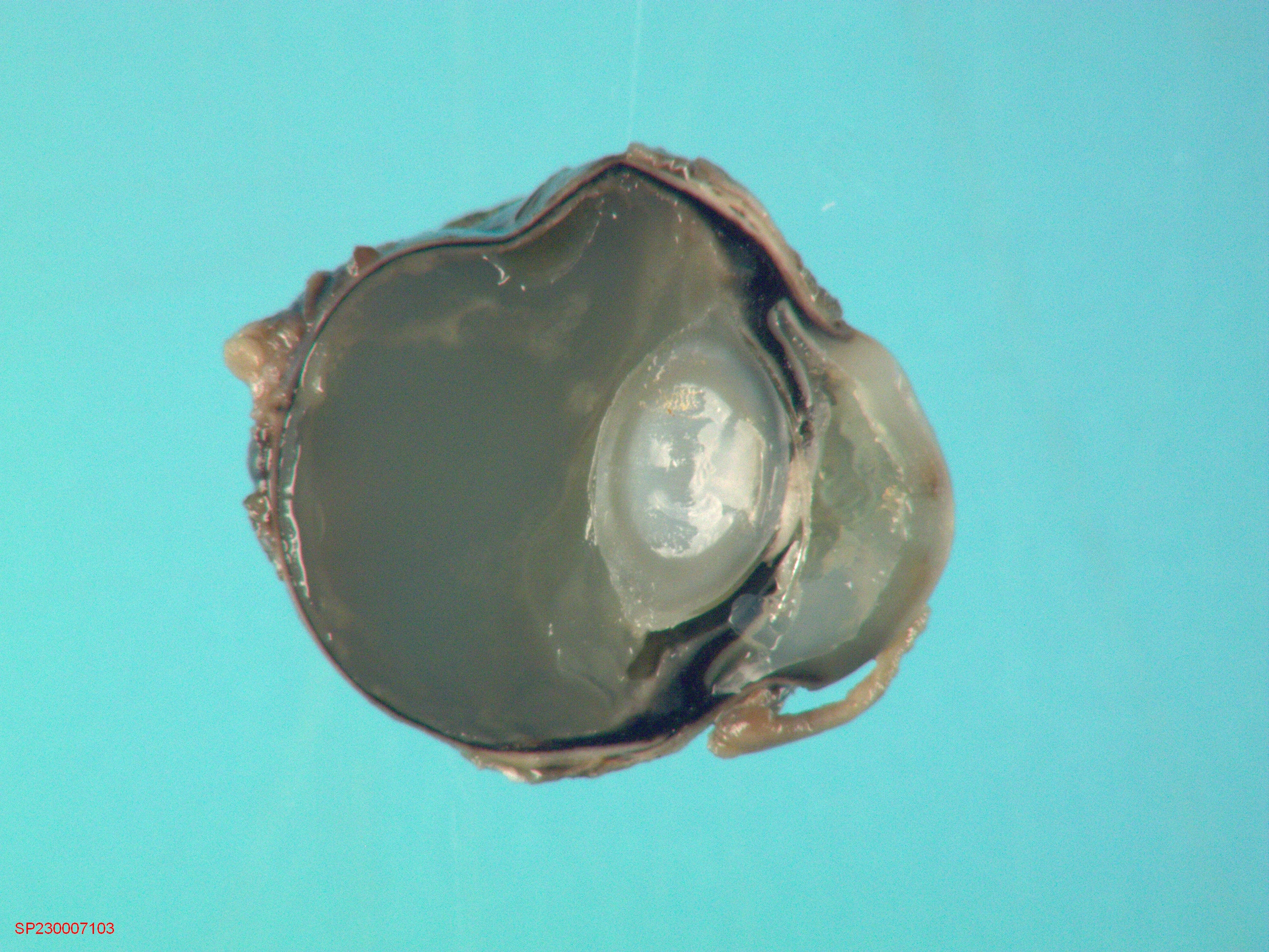
The entire uveal tract was lined by thick bands of white to tan material. All chambers of the globe were filled with translucent gelatinous material.
Laboratory Results:
Not provided
Microscopic Description:
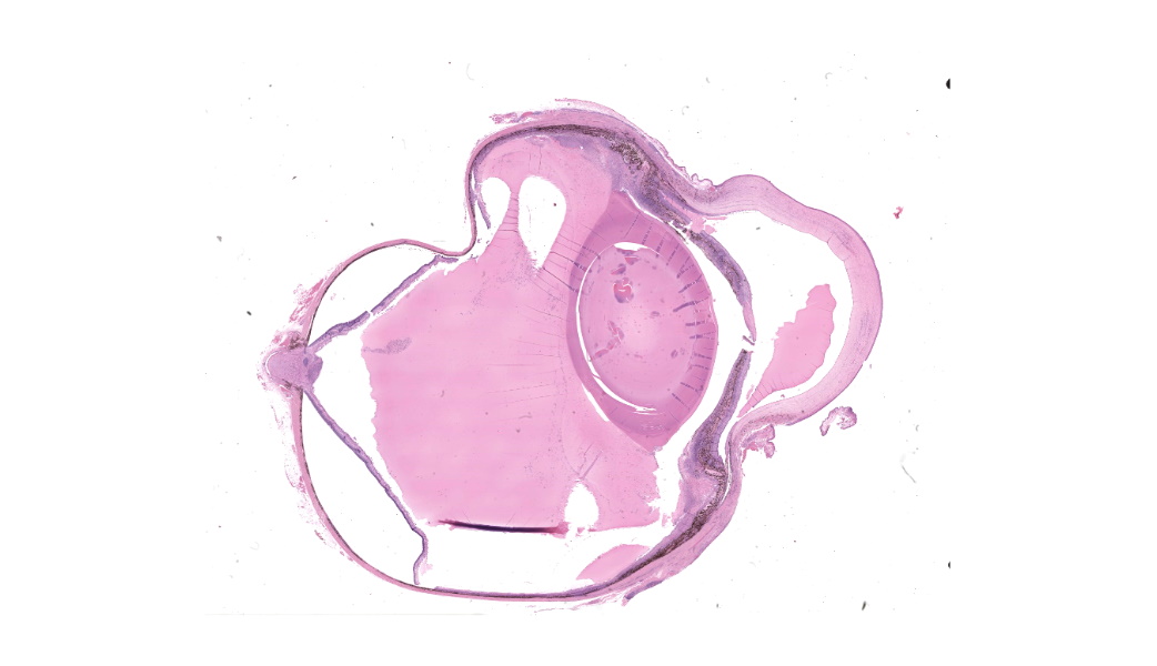
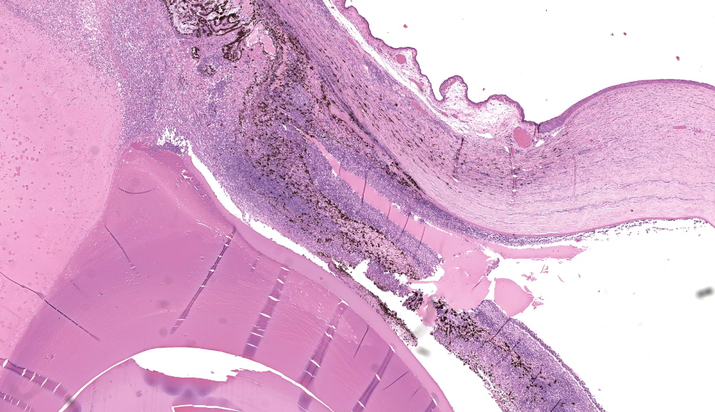
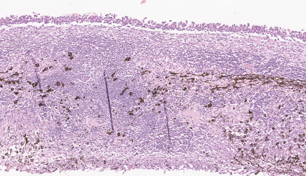
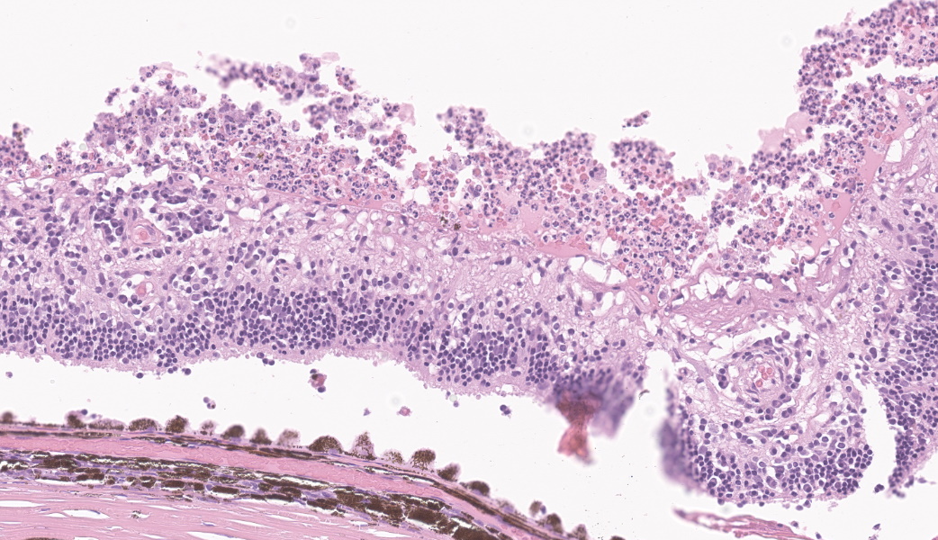
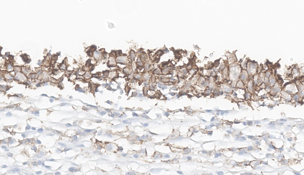
There were thick bands of epithelioid macrophages mixed with neutrophils forming a carpet-like covering over thick bands of fibrovascular stroma that contained plasma cells and lymphocytes and lined the anterior and posterior surfaces of the iris. These epithelioid macrophages occasionally contained variably sized eosinophilic globules, most consistent with lens material. The carpet of epithelioid macrophages also extended to the inner cornea and covered the inner retina and optic nerve head. The bands of fibrovascular stroma bridged from the base of the iris over the collapsed ciliary cleft to the termination of Descemet's membrane, extended around the posterior aspect of the iris and ciliary body surface, and bridged to the lens capsule, resulting in peripheral anterior and posterior synechia.
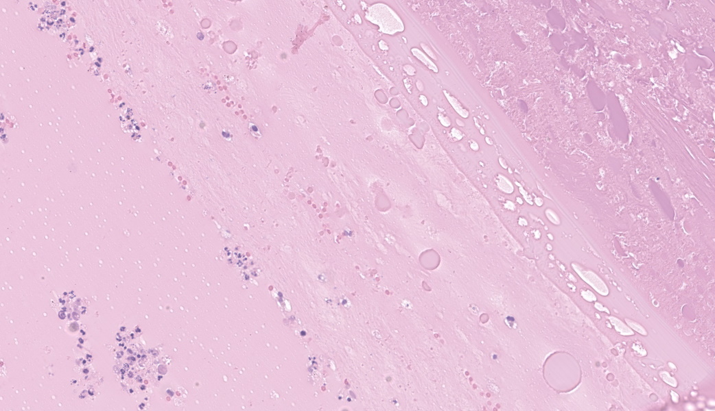
There was moderate cataractous change, characterized by liquefaction and fragmentation of lens fibers, formation of Morgagnian globules and bladder cells, hyperplasia of lens epithelium, and posterior lens epithelial migration. Focally, the lens capsule was ruptured at the equator, and the ruptured lens capsule margin and the exposed lens fibers were infiltrated by degenerate neutrophils and epithelioid macrophages containing intracytoplasmic eosinophilic globules (phagocytized lens protein).
There were perivascular infiltrates of lymphocytes and plasma cells within the iris stroma, choroid, retina, and sclera, and multifocally in the optic nerve. Segmentally, the photoreceptor segments were lost, shortened, and tattered
Focally, there was loss of cellular architecture in the peripheral neural retina and replacement by flocculent eosinophilic material, karyorrhectic debris, high numbers of degenerate neutrophils, and macrophages.
The anterior and posterior chambers and vitreal space were filled with eosinophilic proteinaceous fluid and scattered degenerate neutrophils and macrophages. There were variably sized small eosinophilic globules free in the posterior chamber and vitreal space, particularly surrounding the lens capsule.
There was moderate neovascularization of the corneal stroma accompanied by moderate perivascular infiltrates of neutrophils, lymphocytes and plasma cells. The axial corneal epithelium was moderately hyperplastic and exhibited rare keratinization.
Contributor’s Morphologic Diagnoses:
Eye, OD:
Severe pyogranulomatous and lymphoplasmacytic panuveitis and endophthalmitis with epithelioid macrophage carpeting and intravitreal free lens protein, suggestive of asymmetric uveitis
Preiridal, postiridal, cyclitic, and retrocorneal fibrovascular membranes, with peripheral anterior and posterior synechia
Lens capsule rupture, pyogranulomatous phakitis, and moderate cortical cataractous change
Mild multifocal lymphoplasmacytic optic neuritis and retinitis with focal retinal necrosis
Moderate neutrophilic and lymphoplasmacytic keratitis with stromal vascularization and moderate regional corneal epithelial hyperplasia and keratinization
Contributor’s Comment:
Unlike other organs, the eye protects vision by restricting local immune and inflammatory responses, a concept known as immune privilege.6 Lens-Induced Uveitis (LIU) is a response to leakage of lens material, which is often seen in canine cataracts. 2,5
It had been speculated that small amounts of lens protein escaping from the normal intact lens helps maintain low-dose immunologic tolerance. Increased exposure to lens material, whether from lens trauma, surgery, or cataracts, can overwhelm this tolerance and induce an intraocular or systemic immune response.2 In one human study, they found that anti-lens antibodies were not only present in cataract patients but were also found in other inflammatory eye disease, including uveitis and cyclitis, while patients with lens-induced uveitis
had a high titer.4 This study supports the speculation of low-dose immunologic tolerance and hypersensitivity reaction to lens protein. The inflammation pattern of LIU is typically lymphoplasmacytic to neutrophilic (if there is an infectious agent), while granulomatous inflammation is usually not seen in LIU, except in a condition called “asymmetric uveitis.”1
Diagnosis of asymmetric uveitis is based on histological findings of carpet of epithelioid macrophages covering the surface of the anterior uvea and sometime extending to blanket the inner retina or the inner aspect of Descemet’s membrane, in conjunction with cataract. Phagocytized lens fiber material is often, but not always, seen within epithelioid macrophages. Retinitis and segmental retinal necrosis can also be observed.1 All these characteristic histologic features were present in the submitted case.
Upon communication with the Comparative Ocular Pathology Laboratory of Wisconsin (COPLOW), canine asymmetric uveitis is a disease entity that is suspected to be a hypersensitivity reaction to lens protein. Despite being diagnosed at COPLOW and MSU fairly frequently, this diagnosis has not yet been reported in the veterinary literature. This condition can later develop in the contralateral eye if cataracts develop. The interval between the enucleation of the first eye and the clinical onset in the second eye varies from nearly immediately to four years.1 Therefore, close monitoring of the contralateral eye clinically is recommended.
This disease entity is often associated with diabetic cataracts, but it can be observed with any cataractous change. Miniature Schnauzers, like this case, are predisposed to developing both hereditary and diabetic cataracts, and as a result seem to be over-represented in bilateral, simultaneous asymmetric uveitis.2,3
Contributing Institution:
Michigan State University
Veterinary Diagnostic Laboratory
4125 Beaumont Rd, Lansing, MI 48910
https://cvm.msu.edu/vdl
JPC Morphologic Diagnosis:
Eye: Panophthalmitis, lymphoplasmacytic and histiocytic, chronic, diffuse, marked, with fibrovascular membranes, synechiae, cataractous change, lens rupture, and retinal detachment with necrosis.
JPC Comment:
The contributor provides a concise, but through review of the histologically unique condition of asymmetric uveitis. Although phacoclastic uveitis is not a new entity for the WSC (there are several cases in the WSC archive, to include several traumatic lens ruptures – one by cactus spines, and several in rabbits with intralenticular Encephalitozoon cuniculi), we have not previously had a case with the characteristic carpeting of intraocular tissues by epithelioid macrophages consistent with asymmetric uveitis.
The contributor is correct in that there is limited material available on this particular condition, and that the subject matter experts at the Comparative Ocular Pathology Laboratory of Wisconsin (COPLOW) are the only organization that currently recognizes this as a discrete entity. The Department of Veterinary Pathology of the JPC utilizes this outstanding group of pathologists as our consultant on complex ocular cases, and for many years we have fervently believed in their diagnostic acumen and see no reason to question it over this entity. We are hopeful that this first entry of asymmetric uveitis will alert more pathologists to its existence. Additionally, the first reference, Veterinary Ocular Pathology: A Comparative Review by Richard Dubielzig et al.1, is an excellent, well-illustrated reference texts which reviews (and illustrates) the entities within the COPLOW database at the time of its publication (2010). We live in hope that a new edition with an updated review is forthcoming. COPLOW also maintains an outstanding website (https://www.vetmed.wisc.edu/lab/coplow/) with an online atlas, links to ocular pathology rounds, and many other resources that make it a mandatory, routine place for any ocular-oriented pathologist to visit. COPLOW also maintains an excellent presence on social media, to include their Facebook page.
A few additional details are likely in order on canine asymmetric uveitis for those who may be encountering it for the first time. This is often a disease of small breed dogs with Poodles overrepresented in cases of non-diabetic asymmetric uveitis. Diabetic asymmetric uveitis is seen in Miniature Schnauzers1 and dachshunds8 whose predisposition to the development of diabetic cataracts predisposes them to this particular sequela. The histologic changes in the two variants of this entity are very similar.1
Another important detail is that many affected globes, if serially sectioned, will demonstrate a penetrating injury to the lens1 (a lesion that is very visible in this particular case).
But why is the identification of this condition important? Well, it’s in the name. Often, early identification of the lesion may allow for aggressive anti-inflammatory therapy which may preserve sight in the remaining eye, which is not as severely affected. In the COPLOW collection as published in 2010, 10/23 cases had the second eye ultimately submitted (with similar lesions), with a clinical onset of signs in the remaining eye ranging from a matter or days to over 4 years.
For those who are immunologically inclined, the antigenic substances in the eye were identified in 1977 by Rahi et al in a series of experiments in rabbits. The three crystallins with lens protein (alpha-, beta- and gamma-crystallin) were identified as immunogenic, with their immunogenicity and the appearance of antibodies well correlated to their alphabetized names, with alpha-crystallin being the most immunogenic and resulting in the earliest demonstration of antibody production. These proteins generate a profound antibody response, but a week cell-mediated (T-cell response.)
On a comparative note (and to avoid confusion), the term used in humans for phacoclastic uveitis in human is “phacoanaphylaxis”, which is a bit of a misnomer as previous sensitization to crystallin protein is not required for the manifestation of this disease,8 nor will it cause traditional anaphylaxis in affected individuals.
References:
- Dubielzig RR, Ketring KL, McLellan GJ, Albert DM eds. Veterinary Ocular Pathology: a Comparative Review. Saunders-Elsevier: St Louis MO. 2010:263-266.
- Gelatt KN, Ben-Shlomo G, Gilger BC, Hendrix D V.H., Kern TJ, Plummer CE. Veterinary ophthalmology Vol. 2, 6th ed. Wiley Blackwell. New Jersey; 2021:1277-1278, 1326-1339
- Holder AL, Kennedy LJ, Ollier WE, Catchpole B. Breed differences in development of anti-insulin antibodies in diabetic dogs and investigation of the role of dog leukocyte antigen (DLA) genes. Vet Immunol Immunopathol. 2015;167(3-4):130-8.
- Patel M, Shine B, Murray PI. Antilens antibodies in cataract and inflammatory eye disease: an evaluation of a new technique. Int Ophthalmol. 1990;14(2):97-100.
- Wilcock BP, Njaa BL. Special senses. In Maxie G, ed. Jubb, Kennedy, and Palmer’s Pathology of Domestic Animals Vol 1, 6th ed. Elsevier, Inc. St. Louis; 2016:457-459.
- Zhou R, Caspi RR. Ocular immune privilege. F1000 Biol Rep. 2010;2:3.
- Rahi AH, Misra RN, Morgan G. Immunopathology of the lens. I. Humoral and cellular immune responses to heterologous lens antigens and their roles in ocular inflammation. Br J Ophthalmol. 1977 Mar;61(3):164-76.
- Grahn B, Peiffer R, and Wilcock B. Histologic Basis of Ocular Disease in Animals, Wiley Blackwell.