CASE III:
Signalment:
10-year-old, male-neutered, Maine Coon cat, Felis silvestris, cat
History:
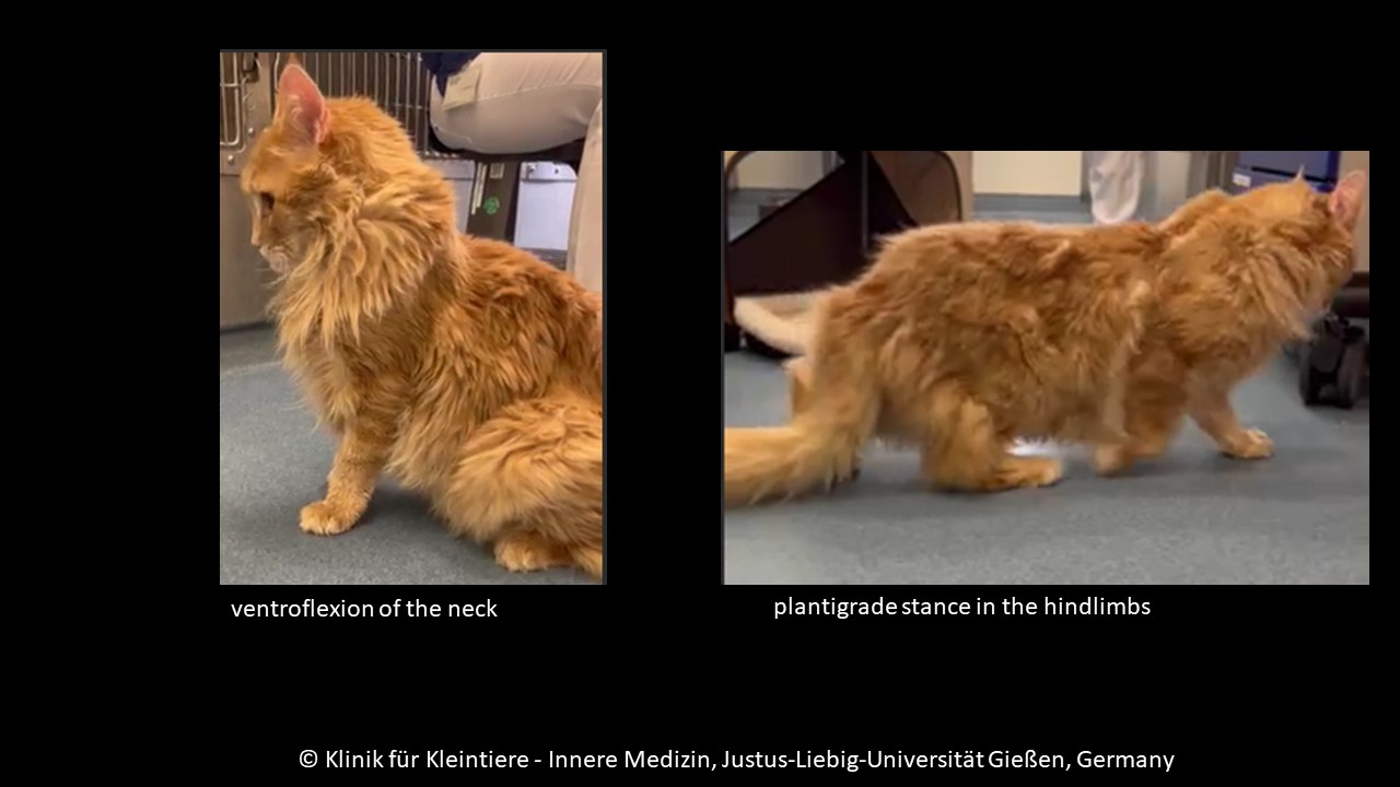
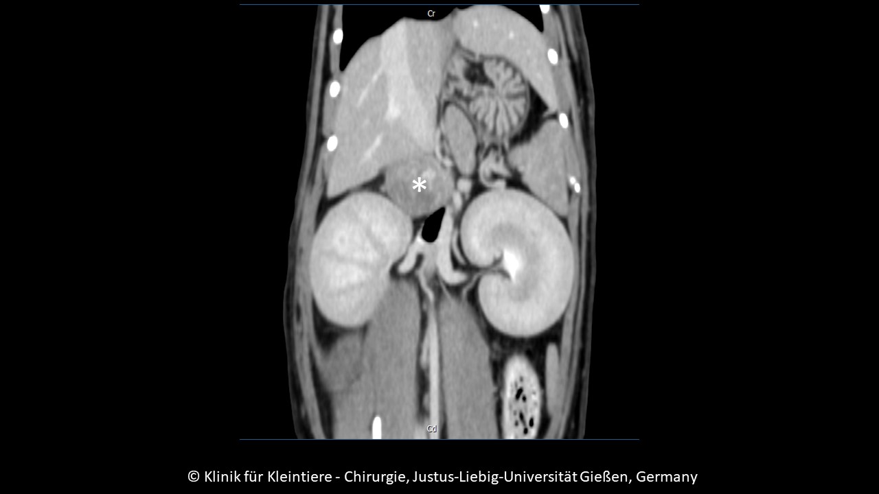
The animal was presented due to apathy, weakness and the suspicion of myopathy. Clinical examination also revealed plantigrade stance of the hind limbs and ventroflexion of the neck. Blood analysis demonstrated severe hypokalemia, hyponatremia, hypocalcemia, hypomagnesemia, and marked hypertension. Creatinine kinase was markedly elevated as was aldosterone (10 times higher than normal!). Based on the interpretation of all findings, clinically manifest hyperaldosteronism was diagnosed. Ultrasonography and CT scan revealed a round mass 3 cm in diameter in the region of the right adrenal gland, compressing the caval vein.
Surgical resection of the mass was attempted unsuccessfully as the caudal vena cava was in too close proximity. The cat was euthanized, the mass was resected post mortem and submitted for histopathological examination.
Gross Pathology:
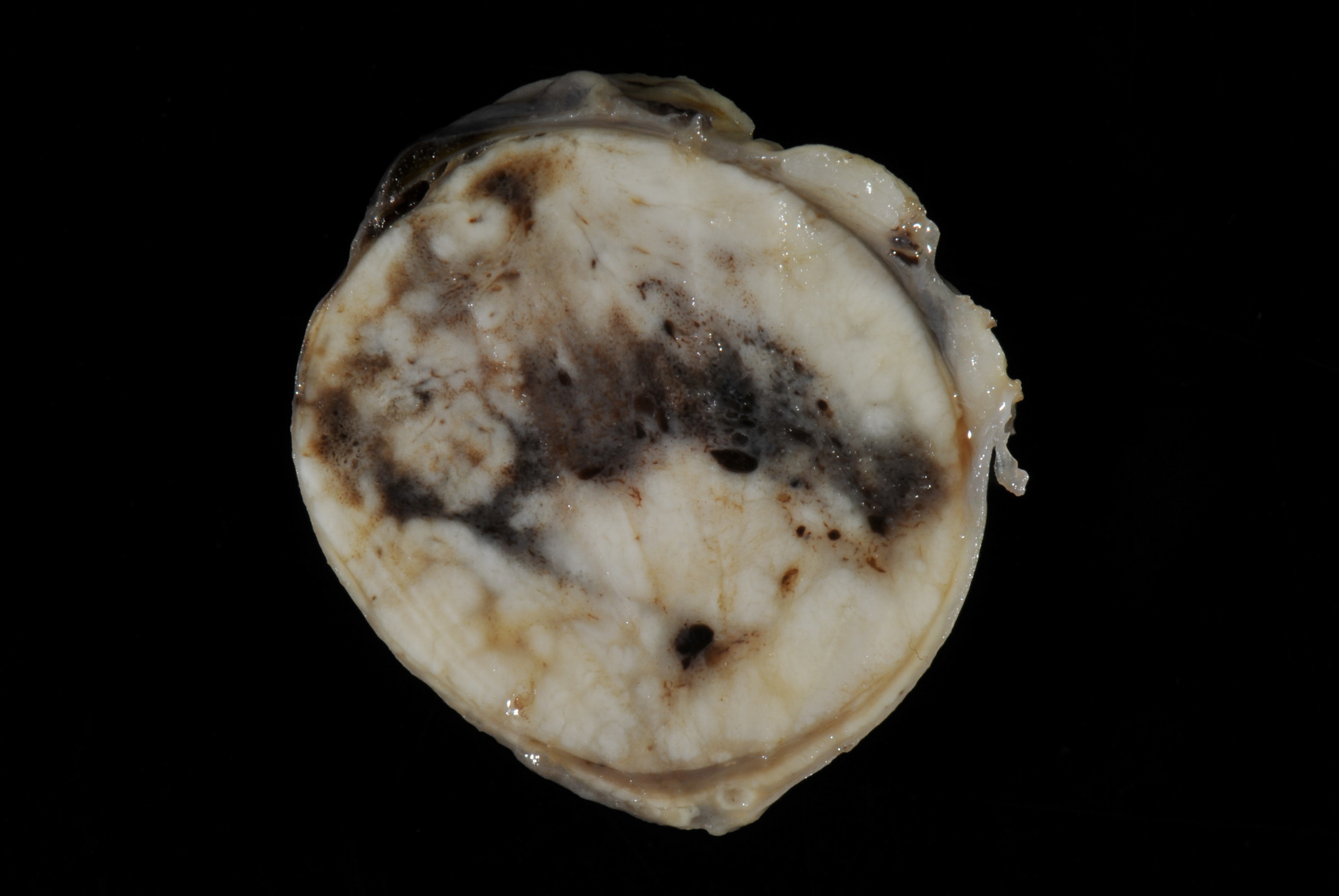
Right adrenal gland (per contributor): 2.5 cm in diameter whitish-brown lobulated mass replacing the normal adrenal gland tissue.
Laboratory Results:
Potassium: 1.8 mmol/l
Sodium: 147.5 mmol/l
Magnesium: 0.29 mmol/l
Calcium: 1.00 mmol/l
Chloride: 116 mmol/l
Creatinine kinase: 71400 U/l
Aldosterone: 6149.40 pmol/l
Immunohistochemistry (neoplasia)
Melan A: negative
Cytokeratin: negative
Synaptophysin: negative
Chromogranin: negative
Microscopic Description:
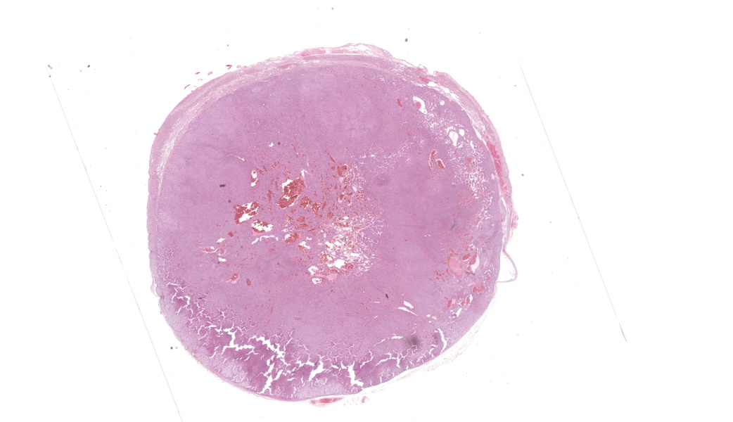
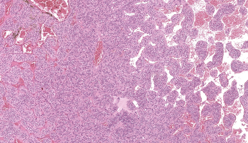
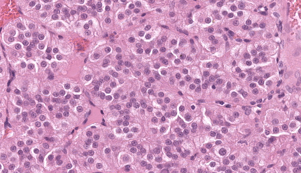
Adrenal gland: Compressing the adrenal tissue leaving only a small rim of adrenal cortex and medulla is a round, approximately 2 cm in diameter, densely cellular, well-demarcated, encapsulated tumor composed of packets, nests and cords of neoplastic cells on a fine fibrovascular stroma. Neoplastic cells are polygonal with distinct cell borders, moderate amounts of often microvacuolated eosinophilic cytoplasm, and a round to oval nucleus with finely stippled chromatin and one distinct nucleolus. In some cells, the cytoplasm is replaced by larger vacuoles (lipoid degeneration). Anisocytosis and anisokaryosis are mild, and the mitotic count is 3 mitoses per 2.37 mm2 (10 high power fields). Within the tumor there are many dilated blood-filled veins (telangiectasis), and focally there is a small area of hemorrhage and necrosis surrounded by hemosiderin-laden macrophages.
Contributor's Morphologic Diagnoses:
Adrenal gland: Cortical adenoma
Contributor's Comment:
Adrenal cortical adenomas are commonly diagnosed in older dogs and rarely in horses, cattle, and sheep. These tumors are often associated with hypercortisolism.4,6,7 In cats, adrenal cortical tumors are very rare, but recent studies have provided more information about the disease in felines.1,3 Hyperaldosteronism associated with functional adrenocortical tumors is more commonly diagnosed in cats than hypercortisolism.1
Conn's syndrome is the synonym for hyperaldosteronism and the disease was named after the endocrinologist Jerome Conn (1907-1994), who first described primary hyperaldosteronism in humans in 1955.3
Cats show muscle weakness, inability to lift the head, plantigrade stance of the hind limbs, or ocular lesions due to hypertension. Severe mineral imbalances with characteristic hypokalemia, increased creatinine kinase activity and elevated aldosterone levels are pathognomonic. Often abdominal masses in the adrenal region can be localized by palpation or diagnostic imaging. The disease appears to be underdiagnosed in cats.1,3
At necropsy, an adrenal mass should be found unilaterally or bilaterally. Tumors may be located in the cortex, well demarcated with a capsule and compressing the normal tissue. The cells are often arranged in broad trabeculae or nests with a delicate fibrovascular stroma. They have abundant lightly eosinophilic or vacuolated cytoplasm. Dilated vessels/telangiectasia and hemorrhages or hemosiderosis may be present.4,6,7 Immuohistochemical demonstration of aldosterone-positive cells has been described5, but was not performed in our case.
Surgical therapy is suggested to be curative in many cases.2 Several complications may occur, and systemic hypertension may lead to chronic vascular lesions and severe circulatory disturbances.5
In our case of Conn's disease, unfortunately, the carcass was not submitted for necropsy. The applied immunohistochemical methods support the diagnosis of adrenocortical adenoma.
Contributing Institution:
Institut fuer Veterinaer-Pathologie, Frankfurter Str. 96, 35392 Giessen, Germany
http://www.uni-giessen.de/cms/fbz/fb10/institute_klinikum/institute/pathologie
JPC Diagnoses:
Adrenal gland: Adrenocortical adenoma.
JPC Comment:
As the contributor notes, primary hyperaldosteronism is the most common disorder of the adrenal cortex in the cat, eclipsing hypercortisolism (which was discussed in case 1 of conference 2 of this Wednesday slide conference year). The contributor has provided a concise review of feline primary hyperaldosteronism above, but a review of aldosterone's effects in the normal animal may be in order.
Whereas cortisol and sex steroids are in all three zones of the adrenal cortex through the effects of cytochrome P450 enzymes on cholesterol, aldosterone is produced only in the zona glomerulosa.8 (While the primary production of aldosterone is within the adrenal cortex, limited amounts are also produced in the brain, heart, and blood vessels, which act in an autocrine mode.)9 Like other steroid hormones, it is metabolized in the liver and excreted in the bile and feces in this species.8
In contrast to the other steroids produced by the adrenal cortex, which are regulated by secretion of ACTH from the pituitary gland, aldosterone release is controlled by the renin-angiotensin system and circulating levels of potassium. The renin-angiotensin system stimulates aldosterone-induced sodium retention during periods of hypovolemia, and decreased secretion of this hormone is seen during periods in which the blood volume is elevated. In addition to the renin-angiotensin system, potassium stimulates aldosterone synthesis and secretion via direct effect on cells of the zona glomerulosa.8 Aldosterone secretion may also be mildly influenced by ACTH, and natriuretic hormone.8
Epithelial cells within the kidney, and salivary glands, possess cytoplasmic receptors for aldosterone. Following its passage through the plasma membrane, it binds to these receptors. In the distal convoluted tubules, the now-intact aldosterone receptor complex stimulates a Na-K ATPase in the cell membrane, resulting in an active increase in sodium reabsorption and passive potassium excretion based on the amount of sodium resorption.8
Elsewhere, beyond its results on circulating blood vessels, aldosterone may act locally on endothelial cells and cardiac tissue to increase total peripheral resistance. This may explain the microangiopathies mentioned by the contributor, especially in long-standing cases of hyperaldosteronism.8
References:
- Daniel G, Mahony OM, Markovich JE, Appleman E, Monaghan KN, Lawrence YA, Fiocchi EH, Weaver K, Johnston A, Barton B. Clinical findings, diagnostics and outcome in 33 cats with adrenal neoplasia (2002-2013). J Feline Med Surg. 2016;18:77-84.
- Del Magno S, Foglia A, Rossanese M, Montinaro V, Cola V, Pisoni L, Rossetti D, Cantatore M, De La Puerta B, Nicoli S, Pisani G, Collivignarelli F, Romanelli G, Cinti F, Olimpo M, Fracassi F. Surgical findings and outcomes after unilateral adrenalectomy for primary hyperaldosteronism in cats: a multi-institutional retrospective study. J Feline Med Surg. 2023;25:1-7.
- Djajadiningrat-Laanen S, Galac S, Kooistra H. Primary hyperaldosteronism: expanding the diagnostic net. J Feline Med Surg. 2011;13:641-50.
- Kiupel M, Capen C, Miller M, Smedley R. Histological classification of tumors of the endocrine system of domestic animals. WHO, Washington, DC: Armed Forces Institute of Pathology; 2008,second series, vol. 12.
- Mau J, Dietert K, Rohwedder T, Deutschland M, Bottcher P, Klopfleisch R Bertram CA. Conn's syndrome in two cats with unusual complications. Prakt Tierarzt. 2020;101:342-358.
- Rosol T, Groene A. Endocrine glands. In: Maxie MG, ed. Jubb, Kennedy, and Palmer's Pathology of Domestic Animals. 6th ed. Philadelphia, PA: Elsevier; 2016; vol. 3:270-357.
- Rosol T, Meuten DJ. Tumors of the endocrine glands. In: Meuten DJ, ed. Tumors in domestic animals. 5th ed. Ames, IA: Wiley Blackwell; 2017:766-833.
- Kooistra HS. Primary hyperaldosteronism in cats: An underdiagnosed disorder. Vet Clin Small Anim 2020; 50: 1053-1063.
- Connell JM, MacKenzie SM, Connell JM, Davies E. Non-adrenal synthesis of aldosterone: a reality check. Mol Cell Endocrinol. 2012 Mar 24;350(2):163-7.