CASE IV:
Signalment
Adult, male, Sprague-Dawley (CD) rat (Rattus norvegicus)
History
A week after arrival from the vendor, the rat had EEG telemetry implantation surgery. The rat was found by the research team quiet, alert, and responsive; cold to the touch; and slightly dehydrated approximately two weeks post-surgery. A veterinary evaluation revealed a decreased body temperature (32°). Heat support & subcutaneous fluids were provided. The rat was found dead in cage later that day. A necropsy was performed due to the unexpected death.
Gross Pathology
This 364 g, male CD rat is in optimum body condition with adequate subcutaneous and visceral adipose, 3/5 BCS, a mildly distended abdomen, eyes mildly sunken, mild dehydration, and in fresh condition. The top of the head was shaved with partial regrowth and a healed linear midline incision. There was illegible writing on the dorsal aspect of the tail. A telemetry device was present in the left flank with wires running cranially through the subcutis to the head and there was fibrous tissue in the subcutis surrounding the telemetry device. There was a foul odor on opening the abdominal cavity. Within the abdominal cavity there was approximately 5-6 mL of cloudy yellow fluid and small amounts of fibrin and few adhesions between the mesentery and viscera (primarily the kidneys and urinary bladder) throughout the abdomen. On the outer/lateral surface of both kidneys there were multifocal 0.5-1.5 mm white-yellow foci. On the caudal and medial surface there were fibrous adhesions. On cut section, the pelvis of the left kidney was mildly dilated and there was a small amount of white purulent material present. The urinary bladder was empty, and the serosal surface was irregular/rough with multifocal areas of dark red discoloration and few foci of adherent fibrin. There was a firm adhesion between the mesentery and the apex of the bladder and one small indented irregular area that may represent a site of rupture. The spleen was diffusely dark and mildly enlarged. The oral cavity was empty. Within the stomach there was an abundant amount of normal ingesta, in the small intestine and cecum there was a moderate amount of normal digesta, and within the colon there were rare formed fecal pellets. There was mild congestion along the transverse and longitudinal cerebral fissures. There were four small EEG screws placed through the dorsal skull with acrylic on surface of screw. All other organs observed &emdash; NSGL.
Laboratory Results
Culture results from abdominal fluid indicate strong growth of Proteus mirabilis.
Cytologic analysis of the fluid from the abdomen showed many degenerate and nondegenerate neutrophils, few macrophages, few rhomboid crystals, small amounts of debris, and moderate numbers of intracellular (within neutrophils) and extracellular coccobacilli on a thick, pale eosinophilic background.
Microscopic Description
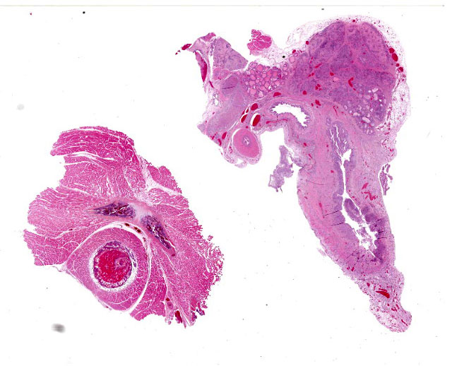
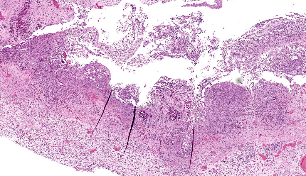
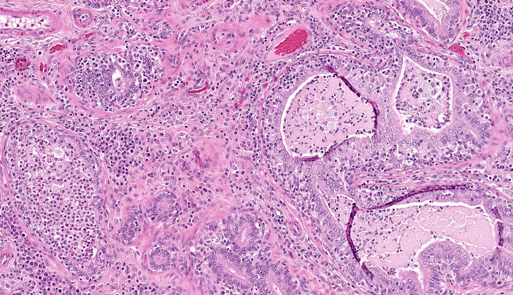
Urinary bladder and prostate: There is ulceration of the bladder mucosa, marked thickening of the serosal surface with fibrosis, and marked fibrosis surrounding the prostate glands. There is submucosal infiltration of neutrophilic inflammation in the ulcerated area of the bladder and filling the bladder lumen. Neutrophils fill the lumen of one gland in the prostate and extend into the surrounding stroma.
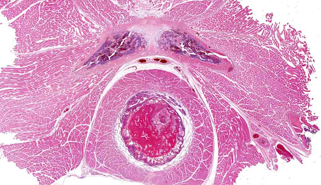
Pelvic urethra (note pubic bone): The lumen is ectatic and expanded and filled by a dark eosinophilic coagulum. The coagulum is tightly associated with the attenuated epithelium; there are a few enmeshed neutrophils in the coagulum and there is edema in the submucosa.
Contributor's Morphologic Diagnoses:
1. Kidney, right and left: Pyelonephritis, neutrophilic, acute on chronic, multifocal to focally extensive, marked with tubular necrosis (infarction), hemorrhage, pelvic erosion/ulceration and fibrosis, many gram-negative colonies of large rod-shaped bacteria and mineralization.
2. Ureter, left: Erosion, acute, multifocal, mild.
3. Urinary bladder: Cystitis, transmural, neutrophilic and mononuclear, acute on chronic, diffuse, marked with ulceration, fibrosis and mineralization, and moderate numbers of individual and small colonies of gram-negative rods, both intra and extra cellular, predominantly in the exudative debris in the lumen and fewer, predominantly extracellular gram-positive cocci.
4. Prostate: Prostatitis, pyogranulomatous and mononuclear, chronic, focally extensive, marked with severe fibrosis and atrophy, and moderate numbers of individual and small colonies of gram-negative rods, both intra and extra cellular, predominantly in the exudative debris in the lumen and fewer, predominantly extracellular gram-positive cocci..
5. Pelvic urethra: Coagulum, adherent, subacute, diffuse, marked with fully occlusive, with urethral ectasia, bacterial colonies, low numbers of neutrophils and epithelial attenuation.
6. Penis: Mucosal hyperplasia, chronic, multifocal, moderate with luminal coagulum.
Contributor's Comment:
The cause of death in this rat is related to urethral obstruction followed by ascending urinary tract infection, pyelonephritis and urinary bladder rupture. The coagulum fully obstructed and expanded the urethral lumen, and in many locations appeared adhered to the mucosa. It was present across multiple transverse sections spanning both the pelvic and penile urethra, indicating its extensive nature. The severe degree of infection in the kidneys and prostate, along with fibrosis in the renal pelvis, prostate and urinary bladder, indicates chronicity, suggesting this was an ongoing and severe condition which spread throughout the urogenital system. Infection in the kidneys was bilateral and characterized by the presence of many bands of neutrophils radiating throughout the cortex as well as abundant tubular necrosis and many colonies of gram-negative rod-shaped bacteria.
Proteus mirabilis (Order: Enterobacterales; Family: Morganellaceae) is a gram-negative bacillus bacteria often associated with urinary tract infection in human patients with long term urethral catheterization. Rats and mice have been used to study this agent as a cause of urinary tract infections. 1 The urinary bladder likely ruptured secondary to mechanical pressure from obstruction, but ongoing infection may have weakened the urinary bladder wall. Urinary obstruction in male rats secondary to urethral coagulum from secondary sex gland secretions is not an uncommon occurrence but is more commonly documented in mice. It does not always progress to urinary bladder rupture and ascending infection, depending on the completeness of obstruction and therapeutic intervention. In some cases, obstruction may resolve spontaneously, particularly if partial, but is often fatal if the urethra is fully obstructed. Certain anesthetic protocols, drugs, and decreased ability to groom have been documented as risk factors.2. A second bacteria (gram-positive) was identified within the urinary bladder and prostate via tissue Gram stain, but this agent was not cultured from the abdominal fluid sample.
Proteus mirabilis is found in soil, water, and sewage, but is also a commensal in human and animal gastrointestinal tracts. It is well known for its urease activity, swarming motility across culture plates in a "bull's eye pattern", and biofilm formation. The enzymatic urease production breaks down urea into ammonia which alkalizes urine and promotes urinary calculi formation (typically struvite or apatite). The swimming and swarming motility is flagella mediated. Fimbriae and other adhesins play a significant role in catheter biofilm formation. Other virulence factors include various cytotoxins such as hemolysin3.
Contributing Institution
Comparative Pathology Department, Research Support Division, U.S. Army Medical Research Institute of Chemical Defense https://usamricd.health.mil/
JPC Diagnoses
1. Urinary bladder: Cystitis, necrotizing and suppurative, chronic, transmural, severe, with mineralization.
2. Prostate: Prostatitis, necrotizing and suppurative, chronic, multifocal to coalescing, severe.
3. Peritoneum: Peritonitis, necrotizing and suppurative, chronic, focally extensive, severe.
4. Pelvic urethra: Seminal plug, chronic, with diffuse mild neutrophilic urethritis.
JPC Comment
The contributor provides a concise review of urethral obstruction and Proteus mirabilis infection in laboratory rodents. A more detailed review of some of the aspects of Proteus spp. ability to cause disease is likely in order.
As the contributor mentioned, the main portal of entry for Proteus mirabilis is the urinary tract, as it is a well-known cause of UTIs and catheter-associated urinary tract infections in humans. It is typically the initial colonist of newly introduced urinary catheters, rapidly forming biofilms which are often polymicrobial. Providencia stuartii is a common microbial partner in catheter-associated urinary tract infections.
Proteus mirabilis benefits from its ability to change its mobility based on the medium in which it finds itself. In a liquid environment, the bacteria "swim", while on a solid surface, such as the transitional epithelium or the urinary tract, or a urinary catheter, the bacilli "swarm", forming large mobile rafts of bacilli.4 This ability, discovered in 1885 by the German pathologist Gustav Hauser, led him to name the bacterium Proteus, after the shape-shifting demigod of Greek mythology.4 Both methods of movement are facilitated by flagella; "swimmer" bacilli possess several peritrichous flagella, but the evolution to a swarmer-type bacterium on agar plates results in the development of literally hundreds to thousands of flagella.1
Flagella production is "first and foremost" when it comes to the virulence factors produced by Proteus mirabilis. "Foremost" in that non-flagella-producing strains are non-pathogenic, and "first" in that the additional virulence factors are produced only after the development of flagella and the assumption of the swarmer morphology.
P. mirabilis also possesses several other virulence factors, including fimbriae, urease, ZapA, iron and zinc scavengers, and multiple types of fimbriae, which make it a potent pathogen of the urinary tract. The bacilli's ability to adhere to epithelium and the surfaces of catheters is mediated by up to 17 different fimbriae.1
Urease is a nickel-based metalloproteinase that splits urea into ammonia and carbon dioxide, which increases pH.1,3 The presence of other uropathogenic co-infective bacteria enhances the production of urease by P. mirabilis. Urease increases the potential for bacterial proliferation and bacteremia, enhances the destructive effects of the inflammatory response, and facilitates urolith formation. Stone formation is likely an evolutionary adaptation of the bacterium, and it may sequester itself and proliferate within stones, evading the host's immune response.5 Another unfortunate expression of urolithiasis is the fact that 50% of humans with long-term catheterization (>28 days) will experience catheter obstruction by crystalline deposits.3
Another interesting virulence factor of P. mirabilis is an IgA-degrading proteinase known as ZapA, which degrades IgA, the main mucosal-associated immunity factor of the mucosal tract. This metalloproteinase is produced during the bacterium's transition from "swimmer" to "swarmer" as it colonizes the solid surface of the urinary tract, where IgA is located.5
Another important flagella-driven activity of P. mirabilis is its ability to invade cells. Murine studies have demonstrated that within 30 minutes of inoculation, bacteria are present within urothelial cells. They may also infiltrate and live intracellularly within renal tubular epithelium in ascending infections (although this facet is less well developed than in other urinary pathogens, such as uropathogenic E. coli, which establishes intracellular "communities" of bacilli).3 This intracellular niche not only protects them from the body's immune system, but may also provide a resident population that contributes to infection chronicity.3 In addition to flagella, P. mirabilis utilizes the AipA autotransporter for internalization into bladder cells. If simple internalization is not enough, P. mirabilis can lyse bladder epithelial cells using a combination of the Proteus toxic agglutinin (Pta) and a hemolysin.3
The submitted slide in this case was a busy descriptive exercise for conference participants, which included two separate tissues with different pathogenesis. The conference moderator, who was also the contributor of this case, did not submit the kidney, which had a very significant pyelonephritis.
The presence of a seminal plug in the pelvic urethra and its relationship to the cystitis, prostatitis, and pyelonephritis is unclear. Conference participants ultimately agreed that the plug was most likely an agonal event, as the chronicity of the inflammatory response in the lower and upper urinary tract likely exceeds the expected lifespan of an animal with urethral obstruction.
References
- Jessica N Schaffer, Melanie M Pearson. Proteus mirabilis and Urinary Tract Infections. Microbiol Spectr. 2015 Oct;3(5):10.1128/microbiolspec.
- Sara Wells, Chris Trower, Tertius A Hough, Michelle Stewart, Michael T Cheeseman. Urethral obstruction by seminal coagulum is associated with medetomidine-ketamine anesthesia in male mice on C57BL/6J and mixed genetic backgrounds. J Am Assoc Lab Anim Sci. 2009 May;48(3):296-9.
- Chelsie E. Armbruster, Harry L. T. Mobley, Melanie M. Pearson. Pathogenesis of Proteus mirabilis Infection. EcoSal Plus 2018: 10.1128.
- Jones B, Young R, Mahenthiralingam E, Stickler D. Ultrastructure of Proteus mirabilis swarmer cell rafts and role of swarming in catheter-associated urinary tract infection. Infect Immun. 2004; 72 (7):3491-3950.
- Coker C, Poore CA, Li X, Mobley HLT. Pathogenesis of Proteus mirabilis urinary tract infection. Microbes Infect. 2000; 2(12):1497-1505.