Wednesday Slide Conference, Conference 16, Case 1
Signalment:
12-day-old, female, collared finchbill (Spizixos semitorques).
History:
A parent-raised chick was found dead on the ground outside of the nest without any premonitory signs. The carcass was covered by ants. The weather conditions on the day before were reported to be very hot.
Gross Pathology:
The chick was in fair body condition with small amounts of visceral adipose tissue. No major gross changes were identified except for some loss of skin, which was presumed to be due to postmortem scavenging.
Laboratory Results:
Bacteria were identified as Clostridium piliforme on the basis of conventional PCR and subsequent sequencing (target bacterial 16S rRNA) utilizing formalin-fixed paraffin-embedded brain tissue.
Microscopic Description:
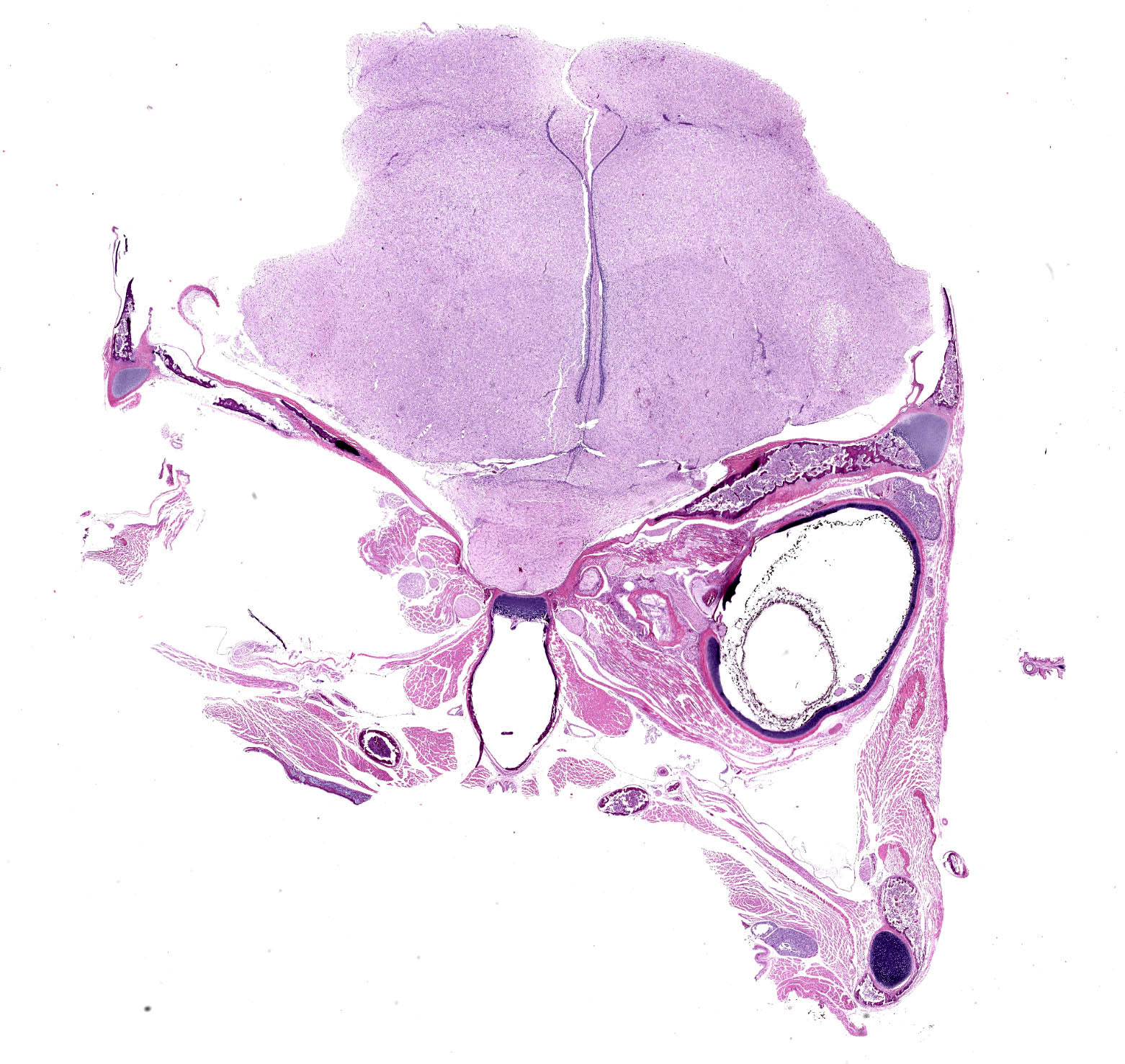
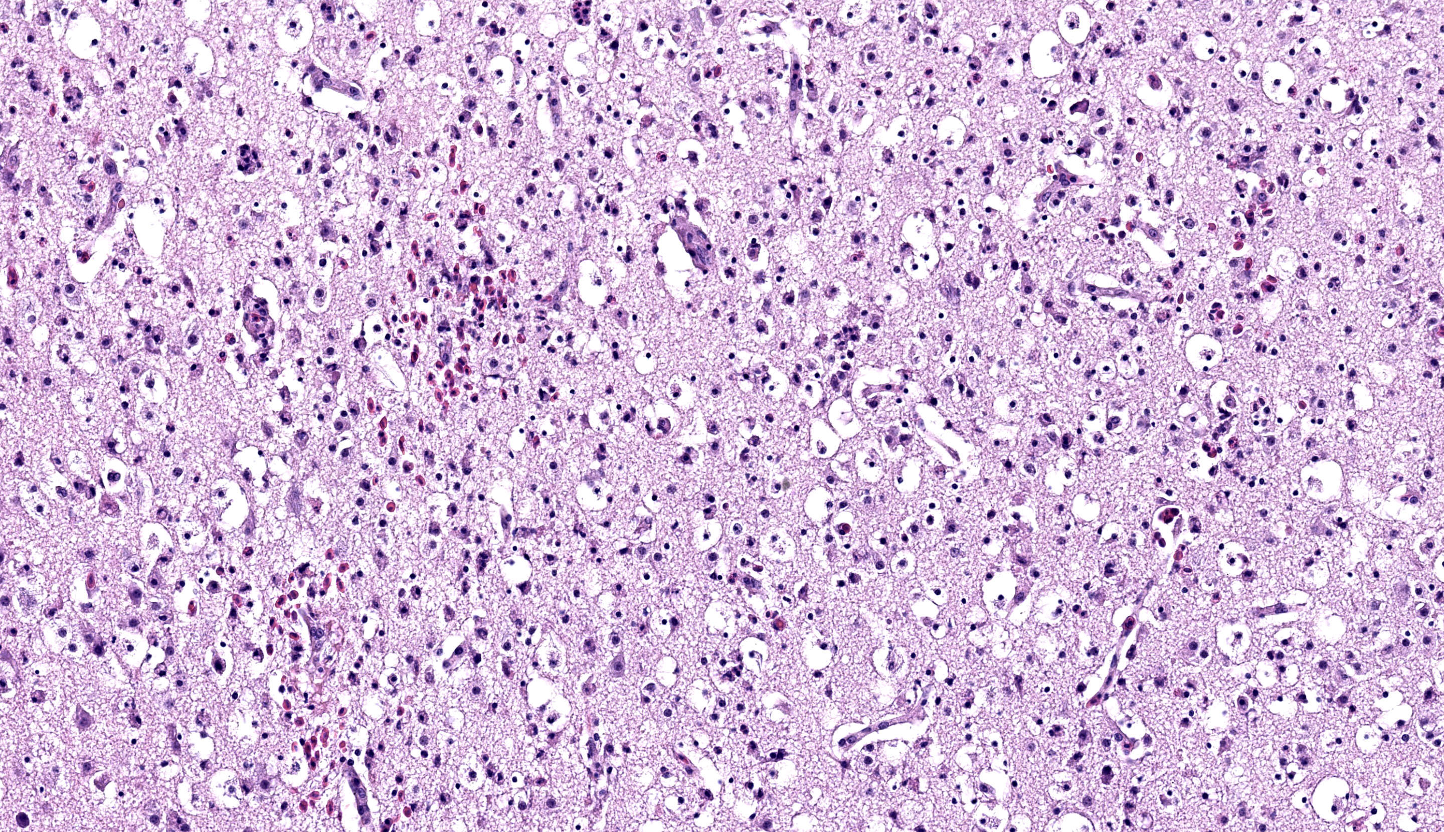
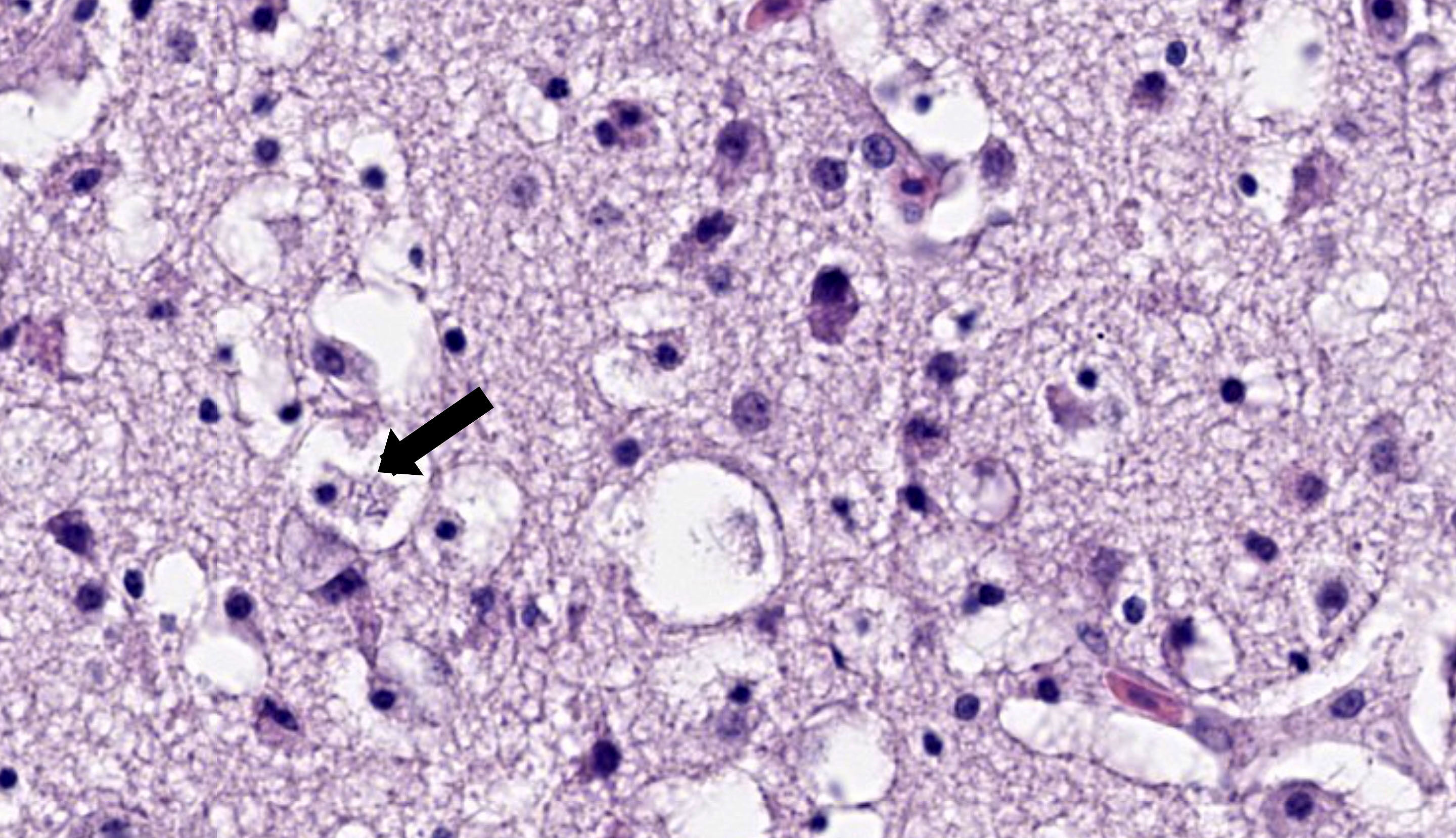
Scattered throughout the cerebrum and affecting approximately 30% of the neuroparenchyma are multiple poorly demarcated to coalescing areas of hypercellularity. These areas have slightly prominent vasculature and are occasionally associated with hypereosinophilia or small lakes of extravasated erythrocytes. The cells infiltrating the neuroparenchyma are with a mix of granulocytes (heterophils), macrophages, and fewer lymphocytes and plasma cells, accompanied by increased numbers of glial cells and some necrotic debris. Occasionally, neurons are hypereosinophilic with smudgy or pyknotic nuclei. Other neurons relatively frequently contain stacks of faint, long, rod-shaped bacteria. Larger blood vessels within the inflammatory foci are frequently cuffed by dense aggregates of mononuclear cells that are up to five cell layers thick. The inflammatory cells additionally involve the overlying leptomeninges.
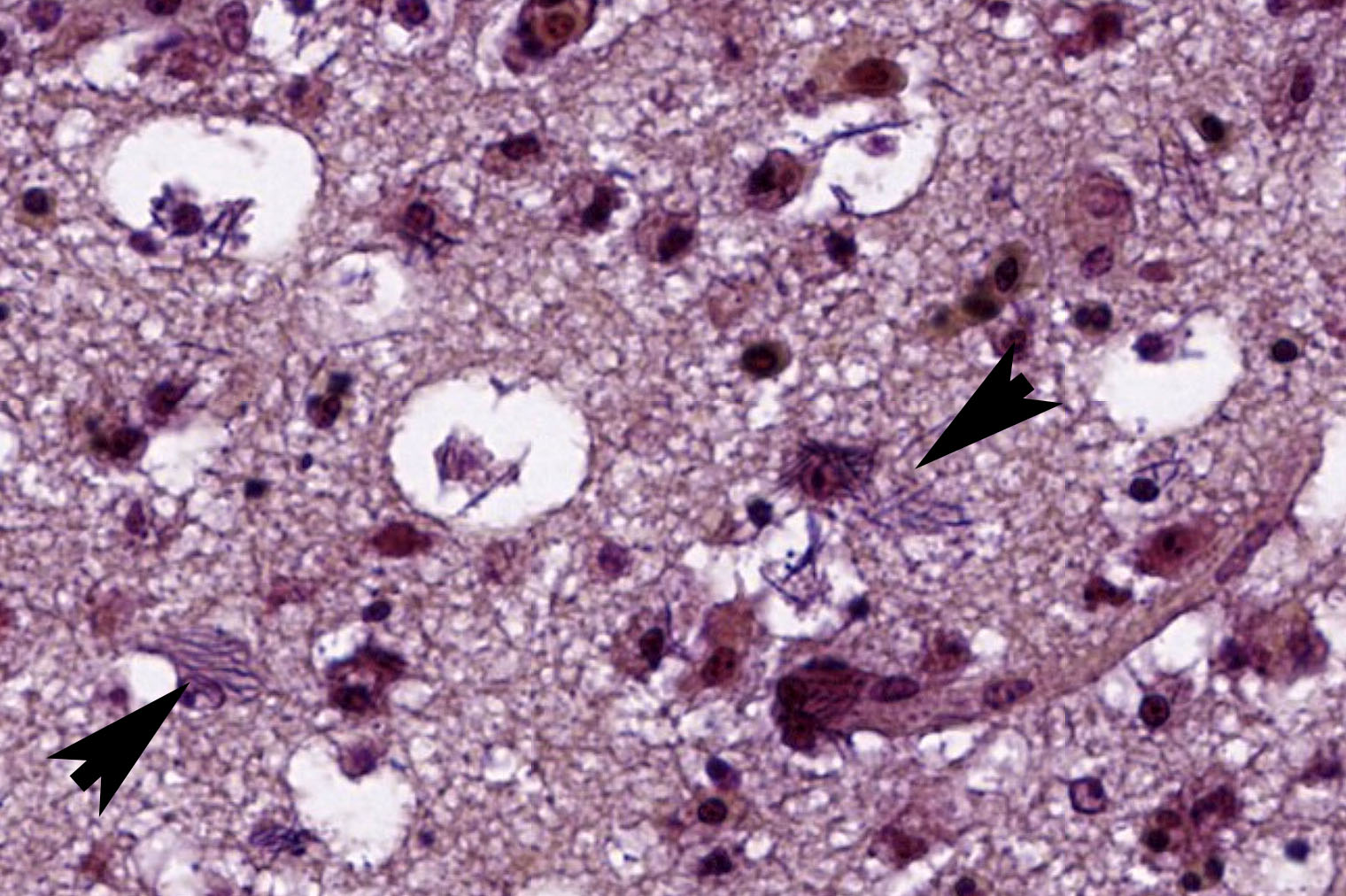
Steiner?s silver and Gram stains were performed to better characterize the intracellular bacteria. Myriads of strongly argyrophilic, curvilinear to filamentous bacilli were highlighted on the silver stain, mapping to the areas of inflammation and necrosis. On the Gram stain, these bacteria were gram-negative.
Contributor?s Morphologic Diagnosis:
Brain, cerebrum: moderate, multifocal to coalescing, subacute heterophilic and necrotizing meningoencephalitis with myriad intracellular argyrophilic and gram-negative filamentous bacilli, consistent with clostridial encephalitis
Contributor?s Comment:
The characteristic histologic appearance of the intracellular filamentous bacilli was highly suggestive of Tyzzer?s disease, caused by Clostridium piliforme. The etiologic diagnosis was further supported by the strongly argyrophilic and gram-negative nature of the bacteria and subsequently confirmed by molecular techniques. There were no bacteria morphologically compatible with C. piliforme or necrotizing inflammation in the other examined organs however.
Tyzzer?s disease has been reported in a wide variety of mammalian species and is a major differential diagnosis for cases of hepatitis, myocarditis, and colitis, especially when this triad of lesions is noted concurrently.7 While C. piliforme is not often thought of as a pathogen affecting birds, encephalitis in young birds is a known manifestation of Tyzzer?s disease.1,2,4,5 The condition is reported to affect both wild and captive birds from several orders, including passerines, psittacines, and piciformes. Affected birds can present with neurologic signs such as head tilt and torticollis. Histopathologic changes are often localized to the brain and characterized by multifocal to coalescing areas of mixed inflammation and necrosis.1-2 Less commonly, birds can present with lesions in the liver, heart, and gastrointestinal tract, similar to mammals.4
At our institution, we have identified seven cases of avian Tyzzer?s disease to date, including five metallic starlings (Aplonis metallica) between 11 to 25 days-old and two other collared finchbills (12 days old and 3 years, 5 months old). Six of these cases had encephalitis with no evidence of argyrophilic bacteria in the other examined tissues. The one case without brain involvement was the only adult bird in this list. This adult finchbill had multifocal random hepatitis with intracellular argyrophilic bacteria. Tyzzer?s disease should be on the list of differential diagnoses in young birds with encephalitis.
Contributing Institution:
San Diego Zoo Wildlife Alliance
Disease Investigations
https://science.sandiegozoo.org/disease-investigations
JPC Diagnosis:
Brain: Meningoencephalitis, heterophilic and necrotizing, subacute, multifocal, moderate with intracytoplasmic filamentous bacilli.
JPC Comment:
This week?s moderator was Dr. Francisco (Paco) Uzal, Distinguished Professor of Veterinary Diagnostic Pathology at UC Davis and a in gastrointestinal and clostridial diseases.
This first case was recently shared at the Davis-Thompson Foundation?s 2024 Northeast Veterinary Pathology Conference and we are pleased to share it with a wider audience. Conference participants homed in on the prominent cuffing of vessels and heterophilic inflammation within the neuroparenchyma and meninges. The increased cellularity is notable, even for an avian brain (which are typically more cellular than their mammalian counterparts.)Differential diagnoses from the group favored viral etiologies (e.g. highly pathogenic avian influenza) and avian chlamydiosis. Dr. Uzal noted that visualizing intracytoplasmic filamentous bacteria was difficult on H&E, though either a Gram or argyrophilic stains were helpful for making a definitive diagnosis.
The contributor nicely summarizes the current literature on avian cases of Tyzzer?s disease and adds several observations from their own collection. We have covered C. piliforme many times in the WSC, most recently in a horse in Conference 4, Case 1, 2023-2024. Neurologic involvement remains rare,although there is a recent case report in a cat of systemic involvement with cutaneous and neurologic infection.3 In this report, immunosuppression by concurrent infection with feline panleukopenia virus and trafficking of bacteria by macrophages aided dissemination of C. piliforme. This is not a feature of the present case. Finally, there is a single case report (from the archives of the AFIP) of zoonotic transmission of Tyzzer?s disease to a human patient infected with HIV-1 with cutaneous involvement,6 though this lesion was benign.
References:
- Mete A, Eigenheer A, Goodnight A, Woods L. Clostridium piliforme encephalitis in a weaver bird (Ploceus castaneiceps). J Vet Diagn Invest. 2011;23(6):1240?1242.
- Mete A, Rogers KH, Woods L. Tyzzer?s disease in free-ranging passerine birds in California, USA. J Wildl Dis. 2017;53(4):938?941.
- Oliveira ES, Queiroz CRR, Santos DO, et al. Neurologic and cutaneous infection by Clostridium piliforme in a kitten with systemic Tyzzer disease. J Vet Diagn Invest. 2023 May;35(3):322-326.
- Raymond JT, Topham K, Shirota K, Ikeda T, Garner MM. Tyzzer?s disease in a neonatal rainbow lorikeet (Trichoglossus haematodus). Vet Pathol. 2001;38(3):326?327.
- Saunders GK, Sponenberg DP, Marx KL. Tyzzer?s disease in a neonatal cockatiel. Avian Dis. 1993;37(3):891?894.
- Smith KJ, Skelton HG, Hilyard EJ, et al. Bacillus piliformis infection (Tyzzer's disease) in a patient infected with HIV-1: confirmation with 16S ribosomal RNA sequence analysis. J Am Acad Dermatol. 1996 Feb;34(2 Pt 2):343-8.
- Uzal FA, Plattner BL, Hostetter JM. Alimentary system. In: Maxie MG, ed. Jubb, Kennedy & Palmer's Pathology of Domestic Animals. 6th ed. Vol. 2. Elsevier; 2016:1-257.