Signalment:
Gross Description:
Histopathologic Description:
Periodic acid-Schiff and Gomori methenamine silver stains revealed no fungi.
Morphologic Diagnosis:
Lab Results:
Condition:
Contributor Comment:
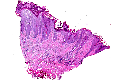
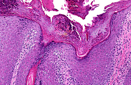
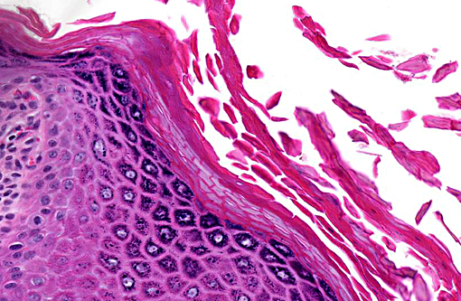
Gross lesions of zinc-responsive dermatosis are similar in all species and include hyperkeratosis, alopecia and erythema. The distribution is generally, but not always, symmetrical and can involve the periocular and perioral skin, the pinnae and the nasal planum. In large animals the distal legs and coronary bands may develop crusts and fissures. In pigs, the ventral abdomen may be affected. Low or low normal serum alkaline phosphatase levels have been described in ruminants, and serum zinc levels, as a rule, do not correlate with gross or histological lesions.(7)
Zinc deficiency in domestic animals is classified as hereditary or dietary. Hereditary zinc deficiency occurs in three forms: 1) lethal trait A46 in Black Pied Danish and Friesian calves; 2) lethal acrodermatitis in bull terriers (hypothetically due to zinc deficiency); and 3) inherited zinc responsive dermatosis in Northern breed dogs. Lethal acrodermatitis in bull terriers is not responsive to zinc supplementation, and its association with zinc deficiency is based largely on clinical similarities to inherited zinc deficient dermatoses in cattle and humans. Dietary zinc deficiency is the predominant form in production animals and large breed puppies.
The most severe inherited forms are lethal trait A 46 or bovine hereditary zinc deficiency in Black Pied Danish and Freisian calves and lethal acrodermatitis in bull terrier pups. These diseases are clinically similar to acrodermatitis enteropathica in humans, caused by a defect in the SLC39A4 gene that encodes the Zip4 protein, a zinc transporter protein distributed along the apical border of duodenal and jejunal enterocytes. Similar mutations in the bovine ortholog of SLC39A4 have been identified in affected calves.(7) In bull terriers the disease is not genetically characterized, but the pathogenesis of the disease has recently been attributed to increased oxidative stress secondary to hepatocelluar metabolic dysfunction. Symptoms begin in the first few weeks of life, and can include acrodermatitis, generalized alopecia, growth retardation, diarrhea, small or absent thymus, defective T-lymphocyte function, and chronic infections. Without treatment affected humans and animals invariably succumb to secondary infection. Humans and calves are responsive to oral zinc supplementation. The disease is currently untreatable in bull terriers.(10)
Northern breed dogs, Siberian huskies and Alaskan malamutes, are genetically predisposed to a more benign inherited zinc responsive dermatosis, known as syndrome I. Onset of symptoms can occur in dogs of any age, but most commonly affects juveniles.(9) Lesions are distributed most commonly on the periorbital skin, pinna and nasal planum and are histologically similar to those reported in other inherited zinc dermatopathies, though less severe. A dietary form of zinc responsive dermatosis, known as Syndrome II, occurs in growing large breed puppies with increase metabolic demands.(1)
Zinc deficiency in production animals is generally attributed to high dietary concentrations of calcium and copper, which block zinc absorption. Cereal grains contain high concentrations of phytates and phytic acids (inositol hexaphosphate) which chelate zinc; however, this mechanism of zinc deficiency is considered less important in ruminants due to production of phytases by rumen microflora. Excessive levels of oxalates, cadmium, iron, molybdenum and orthophosphates in the diet have also implicated zinc malabsorption. Conversely, zinc availability is enhanced by vitamin C, lactose and citrate.(5,8)
Despite the etiogenesis, histologic lesions of zinc-responsive dermatosis are similar in most species, allowing for moderate variations in severity. The differentials commonly include dermatophytosis, demodicosis, pemphigus foliaceus (dogs, cats, horses, goats) and other nutritional dermatopathies.
JPC Diagnosis:
Conference Comment:
The contributor elaborates on the various manifestations of zinc-responsive dermatoses, including both hereditary and dietary. The pathogenic mechanisms underlying the development of cutaneous lesions in zinc deficiency is largely unclear. Zinc has a prominent role in the influence of molecular conformation, stability and activity in addition to its antioxidant effects which support the hypothesis of oxidative stress inducing these cutaneous lesions.(6) The heat shock protein 72 (Hsp72) is synthesized in response to damaged cellular proteins and functions to prevent their aggregration. It is found at increased concentrations in the nucleus of keratinocytes in canine zinc-responsive dermatoses(6), further demonstrating the increased susceptibility to protein damage of squamous epithelial cells when zinc levels are low.
References:
1. Colombini S, Dunstan RW: Zinc-responsive dermatosis in northern-breed dogs: 17 cases (1990-1996). J Am Vet Med Assoc 211: 451-453, 1997
2. Cummings JE, Kovacic JP: The ubiquitous role of zinc in health and disease. J Vet Emerg Crit Care 19:215-240, 2009
3. Grider A, Mouat MF, Mauldin EA, Casal ML: Analysis of the liver soluble proteome from bull terriers affected with inherited lethal acrodermatitis. Molecular Genetics and Metabolism 92:249-257, 2007
4. Krametter-Froetscher R, Hauser S, Baumgartner W. Zinc-responsive dermatosis in goats suggestive of hereditary malabsorption: two field cases. Vet Dermatol. 2005;16:269-275.
5. Nelson DR, Wolff WA, Blodgett DJ, Luecke B, Ely RW, Zachary JF: Zinc deficiency in sheep and goats: three field cases. J Am Vet Med Assoc 184: 1480-1485, 1984
6. Romanucci M, Bongiovanni L, Russo A. Oxidative stress in the pathogenesis of canine zinc-responsive dermatosis. Vet Dermatol. 2010;22(1):31-38.
7. Scott, DW: Large Animal Dermatology, pp. 487 W.B. Saunders Company, Philadelphia, PA, 1988
8. Singer LJ, Herron A, Altman N: Zinc responsive dermatopathy in goats: two field cases. Contemp Top Lab Anim Sci 39: 32-35, 2000
9. White SD, Bourdeau P, Rosychuk RA, Cohen B, Bonenberger T, Fieseler KV, et al: Zinc-responsive dermatosis in dogs: 41 cases and literature review. Vet Dermatol 12: 101-109, 2001
10. Yuzbasiyan-Gurkan V, Bartlett E: Identification of a unique splice site variant in SLC39A4 in bovine hereditary zinc deficiency, lethal trait A46: An animal model of acrodermatitis enteropathica. Genomics. 88:521-6; 2006


