WSC 2023-2024, Conference 25, Case 2
Signalment:
5-year-old female breed unspecified goat (Capra aegagrus hircus).
History:
Livestock farm with ecological and extensive goat production. After the introduction of new animals, blindness, polyarthritis, and chronic mastitis only affecting postpartum and lactating goats were observed. The process reached up to 20% mortality.
Gross Pathology:
Lesions were consistent with mastitis, characterized by a firm consistency of the mammary gland parenchyma and abnormal turbid milky discharge admixed with fibrinous material. Joints showed fibrino-purulent content in the joint space and significant thickening of the joint capsule, compatible with chronic polyarthritis. Unilateral keratitis with mild corneal opacity and bilateral blepharoconjunctivitis were also observed.
Laboratory Results:
Hematology and biochemical analysis revealed marked leukocytosis, neutrophilia, hyperglobulinemia, and increased gamma-glutamyl transferase (GGT) and aspartate aminotransferase (AST). Mycoplasma agalactiae was identified in mammary gland and synovial fluid swabs by Real Time Polymerase Chain Reaction (RT-PCR).
Microscopic Description:
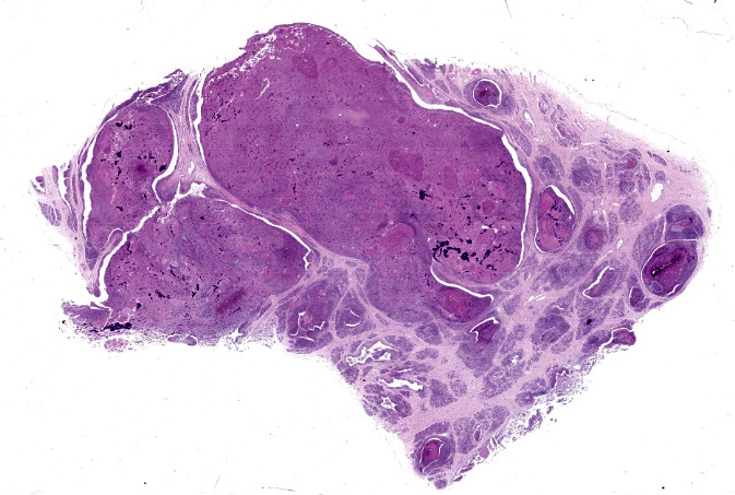
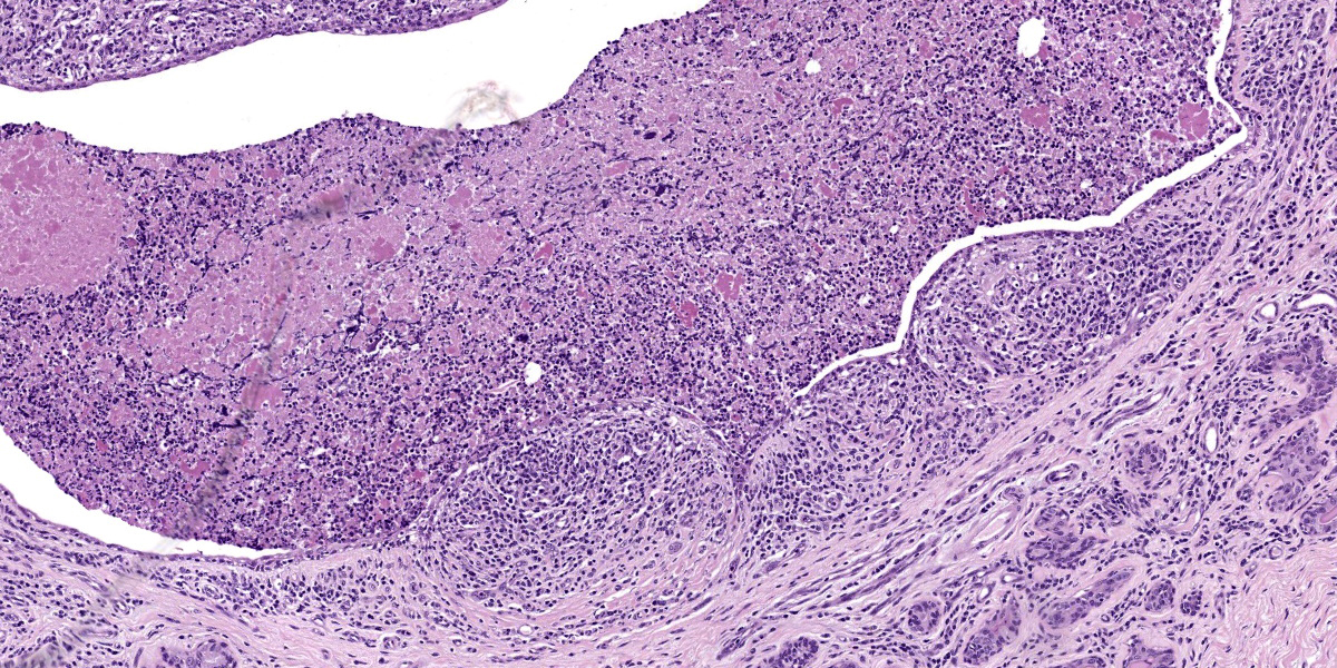
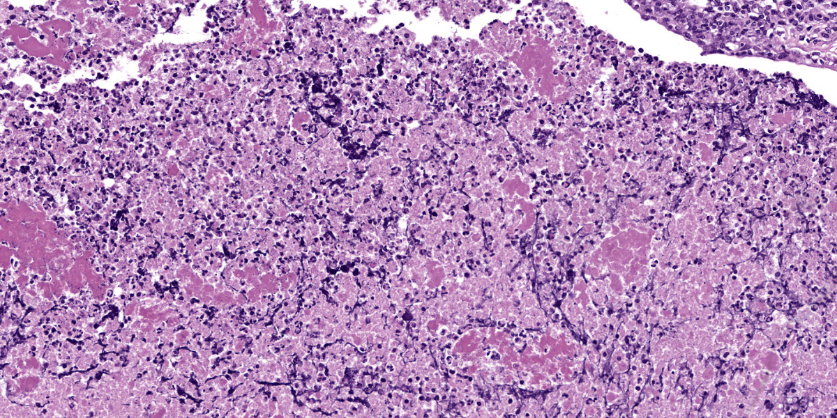
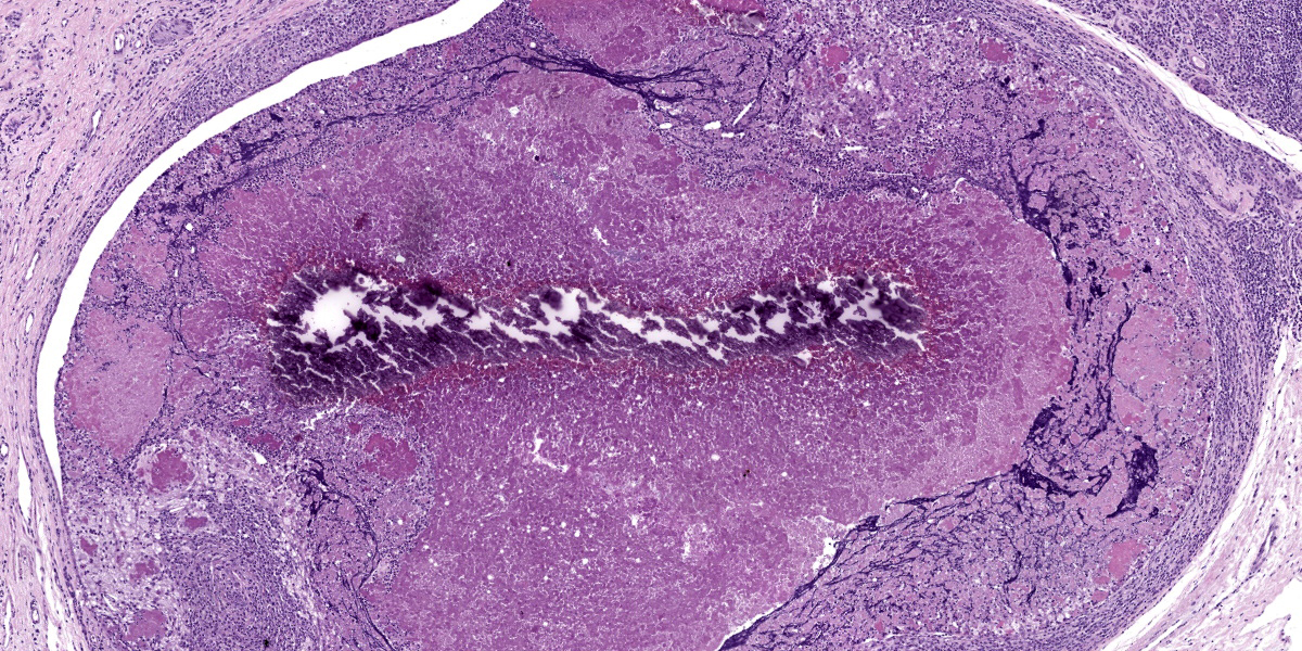
Mammary gland: A severe, chronic active, multifocal inflammatory process comprising 70% of section appears to infiltrate and efface mammary gland lobules and acini. At higher magnification, inflammation is composed of abundant lymphocytes, macrophages, and plasma cells, admixed with fewer neutrophils and scattered eosinophils. Occasionally associated with mammary ducts and acini, lymphocytes and macrophages form round aggregates (tertiary lymphoid follicles). Epithelial ductal cells show one or more of the following changes: intracytoplasmic vacuoles and attenuation (degeneration), shrunken and hypereosinophilic cytoplasm with pyknotic to no nuclei (necrosis) or appear sloughing towards the ductal lumen. Ductal lumens are distended and partially or totally occluded by an abundant amorphous eosinophilic material (proteinaceous secretion), admixed with viable and degenerate neutrophils, macrophages, lymphocytes, and karyorrhectic and cellular debris. Within this secretion, there are occasionally amorphous basophilic granules (mineralization). Diffusely expanding the interlobular interstitium and mammary lobules, there is an increased number of mature collagen fibers (fibrosis).
Contributor’s Morphologic Diagnosis:
Mammary gland: Severe, chronic, multifocal to coalescing, lymphoplasmacytic and histiocytic mastitis with fibrosis and necrosis.
Contributor’s Comment:
Contagious agalactia (CA) is a notifiable disease distributed worldwide that affects goats and sheep.8 The etiological agent in sheep is Mycoplasma agalactiae (Ma), whereas in goats M. mycoides subsp. capri (Mmc), M. capricolum subsp. capricolum (Mcc), and M. putrefaciens (Mp) are also involved.12 Oral, respiratory and mammary (milking parlor) are the main routes of transmission within the herd.1,7,15 Clinical signs and lesions are highly variable and affect mainly postpartum and lactating adult females.14 CA syndrome is characterized by mastitis, polyarthritis, keratoconjunctivitis, pneumonia, and septicemia,6 all of which were observed in our case, with the exception of pneumonia.
Depending on the infection route, the primary site of colonization is the respiratory tract, small intestine, and/or alveoli of mammary glands.1,7,15 Subsequently, Ma is disseminated through the circulation to different organs.13 Microscopically, acute lesions are characterized by a neutrophilic infiltrate with foci of necrosis. Chronic stages develop into an inflammatory infiltrate composed mainly of macrophages, lymphocytes, and plasma cells, together with extensive fibrosis.4
In this case, chronic mastitis, polyarthritis, and keratoconjunctivitis, all characterized by a lymphoplasmacytic and histiocytic infiltrate and severe fibrosis, were observed. Other necropsied goats from the same farm showed acute lesions composed of a severe neutrophilic infiltrate and foci of necrosis in previously mentioned organs. This difference in lesion chronicity among different goats suggests that Ma was chronically present in the herd; however, the outbreak continued actively producing severe and acute cases.
In the typical acute forms, clinical signs and gross and microscopic lesions can facilitate the diagnosis of mycoplasmosis.12 However, to confirm the etiological agent and differentiate among Mycoplasma spp., it is necessary to perform molecular techniques such as PCR.2,5,9,11 Relevant samples are joint fluid, eye swabs, or other affected organs seen at necropsy.10 Bulk tank milk samples can increase the detection sensitivity on a farm.12 Once the outbreak was detected, serology by indirect ELISA and PCRs of Mycoplasma agalactiae of all goats were performed and the positive ones were sacrificed.
Contributing Institution:
Department of Animal Pathology
Universidad de Zaragoza, Spain
http://patologiaanimal.unizar.es/
JPC Diagnosis:
Mammary gland: Mastitis, necrotizing and lymphohistiocytic, chronic, diffuse, marked, with acinar atrophy.
JPC Comment:
Contagious agalactia (CA) is among the most important diseases of small ruminants and is characterized primarily by mammary, joint, and ocular signs, with a drop in milk production followed by increased general morbidity and mortality.3,8 The disease was first reported in Italy in 1816, where it was named “mal di sito,” or “disease of the place,” due to its ability to contaminate and persist in environments and then infect newly-introduced animals.8
Grossly, CA mammary disease may be unilateral or bilateral and is heralded by hot, swollen, and painful mammary glands and enlarged mammary lymph nodes. Infection spreads to the joints, with arthritis characterized by the accumulation of synovial fluid in the carpal or tarsal joints, frequently with concurrent keratoconjunctivitis.3 Other clinical manifestations of CA exist, most notably pneumonia and occasionally abortion, and clinical syndromes may appear in isolation or in various combinations, making it difficult to differentiate CA from other infectious scourges of sheep and goats.
The differential list for CA signs in sheep and goats is long. For mastitis, Staphylococcus, Streptococcus, and Mannheimia spp. should be ruled out, while caprine arthritis encephalitis virus is a prime differential for caprine arthritis.3,8 The Pasteurellacae, along with visna-maedi and peste de petits ruminants viruses, as well as other Mycoplasma spp. such as M. capricolum subsp. capripneumoniae and M. ovipneumoniae should be excluded as causes for respiratory diseases.8 Differentiating among these agents often requires extensive testing, and, as the contributor notes, milk, joint fluid, and eye swabs from several different animals in an infected herd are the recommended diagnostic samples.8
Histologically, CA mastitis is initially characterized by a mononuclear interstitial infiltrate composed primarily of macrophages, though with time, the prominent inflammatory cell becomes CD8+ T lymphocytes.3 Lymphoid follicles are often present just beneath the epithelium, as nicely illustrated in this case.
All participants agreed that this mammary gland, even from subgross, looks “baaad”. Participants appreciated the remarkable ductal ectasia that is a notable histologic feature of this slide. Participants also questioned that nature of the deep purple material present within the ducts, leading to a tangential but informative discussion of oat cells. Oat cells have a similar histologic appearance to the streaming purple material evident in section; however, oat cells are associated with agents that produce a leukotoxin that debilitates the affected cells. These cells remain intact, but their nuclear and cytoplasmic material stream out, particularly when sectioned, producing the characteristic histiologic oat cell appearance. The intraductal material observed here occurs with massive cell death and the resultant commingling of nuclear and cytoplasmic material that adheres together in the necrotic milieu.
Back on task, participants noted the lymphoid nodes which originally made them consider a viral etiology such as arthritis encephalitis virus as a possible cause. While these lymphoid nodules are a reported histologic feature of AC, the moderator noted that mixed mammary gland infections aren’t particularly uncommon, and the presence of one agent doesn’t necessarily preclude the presence of another. Dr. Bender concluded discussion of this case with a review of the various histologic changes associated with mammary development and lactation, noting that evaluating reproductive tissue is inherently challenging as the tissues dynamically respond to the ebb and flow of hormonal tides.
Conference participants felt that necrosis was a significant histologic feature of the examined slide and wanted to include it in the morphologic diagnosis. There was a brief discussion about including fibrosis, however most participants felt that chronicity implies fibrosis and it was omitted in the interest of brevity.
References:
- Agnello S, Chetta DM, Vicari DD, et al. Severe outbreaks of polyarthritis in kids caused by Mycoplasma mycoides subspecies capri in Sicily. Vet Rec. 2012;170 (16):416.
- Amores J, Corrales JC, Martín AG, Sánchez A, Contreras A, de la Fe C. Comparison of culture and PCR to detect Mycoplasma agalactiae and Mycoplasma mycoides subsp. capri in ear swabs taken from goats. Vet Microbiol. 2010;140(1-2):105-108.
- Arteche-Villasol N, Fernandez M, Gutierrez-Exposito D, Perez Valentin. Pathology of the mammary gland in sheep and goats. J Comp Path. 2022;193: 37-49.
- de Azevedo EO, de Alcântara MDB, do Nascimento ER, et al. Contagious agalactia by Mycoplasma agalactiae in small ruminants in Brazil: first report. Braz J Microbiol. 2006;37(4):576-581.
- Becker CAM, Ramos F, Sellal E, Moine S, Poumarat F, Tardy F. Development of a multiplex real-time PCR for contagious agalactia diagnosis in small ruminants. J Microbiol Methods. 2012;90(2):73-79.
- Bergonier D, De Simone F, Russo P, Solsona M, Lambert M, Poumarat F. Variable expression and geographic distribution of Mycoplasma agalactiae surface epitopes demonstrated with monoclonal antibodies. FEMS Microbiol Lett. 1996; 143(2-3):159-165.
- Bergonier D, Berthelot X, Poumarat F. Contagious agalactia of small ruminants: current knowledge concerning epidemiology, diagnosis and control. Rev Sci Tech. 1997;16(3):848-873.
- Jay M, Tardy F. Contagious agalactia in sheep and goats: current perspectives. Vet Med (Auckl). 2019;10:229-247.
- Manso-Silván L, Perrier X, Thiaucourt F. Phylogeny of the Mycoplasma mycoides cluster based on analysis of five conserved protein-coding sequences and possible implications for the taxonomy of the group. Int J Syst Evol Microbiol. 2007; 57(10):2247-2258.
- Marogna G, Rolesu S, Lollai S, Tola S, Leori G. Clinical findings in sheep farms affected by recurrent bacterial mastitis. Small Rum Res. 2010;88:119-125.
- McAuliffe L, Ellis RJ, Lawes JR, Ayling RD, Nicholas RAJ. 16S rDNA PCR and denaturing gradient gel electrophoresis; a single generic test for detecting and differentiating Mycoplasma species. J Med Microbiol. 2005;54(8):731-739.
- Nicholas R, Ayling R, McAuliffe L. Mycoplasma diseases of ruminants. CABI; 2008;76:92-98.
- Razin S, Yogev D, Naot Y. Molecular ciology and pathogenicity of Mycoplasmas. Microbiol Mol Biol Rev. 1998;62 (4):1094-1156.
- Szeredi L, Tenk M, Dán A. Infection of two goatherds with Mycoplasma mycoides subsp. capri in Hungary, evidence of a possible faecal excretion. J Vet Med B Infect Dis Vet Public Health. 2003;50 (4):172-177.
- Todaro M, Puleio R, Sabelli C, Scatassa ML, Console A, Loria GR. Determination of milk production losses in Valle del Belice sheep following experimental infection of Mycoplasma agalactiae. Small Rum Res. 2015;123(1):167-172.


