Results
AFIP Wednesday Slide Conference - No. 9
- 28 October 1998
-
- Conference Moderator:
Dr. Keith Harris, Diplomate, ACVP
Product Safety Assessment, Searle
4901 Searle Parkway
Skokie, IL 60077
NOTE: Click on images for larger views. Use
browser's "Back" button to return to this page.
Return to WSC Case Menu
- Case I - B98-2010 (AFIP 2639018)
-
- Signalment: Adult female B6C3F1 mouse.
History: This mouse was given an intraperitoneal injection
with an experimental material and then necropsied 24 hours later.
-
- Gross Pathology: Gross postmortem examination revealed
wet, edematous lungs.
- Laboratory Results: None.
-
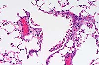
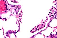
- Contributor's Diagnosis and Comments: Severe acute
diffuse bronchiolar necrosis. Etiology: Naphthalene toxicity.
-
- Several toxic agents are known to induce the selective necrosis
of Clara cells in rodents. These include 4-ipomeanol, 3-methylindole,
and naphthalene (as in this case). Since Clara cells are known
to have a high content of cytochrome P-450 enzymes, they are
more susceptible to toxicant-induced injury than adjacent ciliated
bronchiolar epithelial cells. Mice have a high density of Clara
cells in the lower airways and are more sensitive to Clara cell
toxicants than rats or hamsters. Note that the lesion in this
instance tends to be confined to the lower airways. The dose
of naphthalene used in this experiment was 200 mg/kg.
-
- Recently, coumarin (a natural product utilized in the perfume
industry as a fragrance enhancer) has been shown to produce selective
Clara cell toxicity in the mouse lung. This material has long
been recognized as a potent hepatoxicant in rats, but the pulmonary
toxicity is just beginning to be understood. The pulmonary toxicity
of coumarin may relate to the induction of pulmonary neoplasms
as found in the mouse two year bioassay. However, the mechanism
for this remains to be determined.
 20x
obj
 40x
obj
- Case 9-1 . Lung. There are abundant detached and pyknotic
epithelial cells and neutrophils within small and medium caliber
bronchioles.
 40x
obj
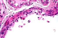
- Case 9-1 . Lung. Larger bronchioles retain intact
ciliated bronchiolar epithelial cells but scattered foci have
bronchiolar epithelial cells with vacuolation, hypereosinophilia,
pyknosis, loss of cilia, and detachment (degeneration & necrosis).
-
- AFIP Diagnosis: Lung, bronchiolar epithelium: Degeneration
and necrosis, acute, diffuse, B6C3F1 mouse, rodent.
-
- Conference Note: The respiratory system is a major
primary target for several classes of toxic compounds including
furans, chlorinated hydrocarbons, aromatic hydrocarbons, pyrolizidine
alkaloids, paraquat, 3-methylindole, and a host of other chemicals.
The lung is exposed to potentially toxic substances both aerogenously
and hematogenously (as the lung receives the entire cardiac output
from the right ventricle). The primary pulmonary lesion resulting
from intoxication with many of these substances, including naphthalene,
is bronchiolar epithelial necrosis due to injury of the nonciliated
Clara cell population. As noted by the contributor, the Clara
cell is a primary target for many toxic substances due to its
high content of P-450 isoenzymes; this system metabolically activates
many organic compounds resulting in the formation of toxic metabolites.
-
- There are definite species specific and dose-dependent differences
in the sensitivity of Clara cells to naphthalene toxicity in
the bronchiolar epithelium of the lung and in the olfactory epithelium
of the nasal cavity among mice, rats, and hamsters. The species
least sensitive to naphthalene-induced Clara cell injury in the
lung, the rat, is most sensitive to injury in the nasal cavity.
The mouse is most sensitive to pulmonary injury with very low
doses of naphthalene, but nasal injury occurs at twice the dose
which produces injury in the rat. The hamster is more sensitive
than the rat to pulmonary injury, but is less sensitive to nasal
injury.
-
- The high degree of anatomical, species, and dose variability
in naphthalene metabolism and injury is probably due to the distribution
of the various P-450 isoenzymes. The initial and obligate step
in naphthalene metabolism is the formation of epoxides and quinones.
Studies in mice indicate that one of these metabolites, 1,2-epoxide,
is the most toxic metabolite and plays an important role in the
cytotoxic actions of naphthalene. The presence of a specific
P-450 isoenzyme within Clara cells, Cyp 2F2, is a key factor
in determining the rates and stereoselectivity of naphthalene
epoxidation. This enzyme is present in mouse but not hamster
or rat Clara cells, and may explain the sensitivity of the mouse
to naphthalene. This differential susceptibility in Clara cell
injury does not occur with most of the organic xenobiotic cytotoxicants.
While dose-dependent and species specific differences occur with
toxic organic agents, a wide variety of compounds injure bronchiolar
Clara cells over a wide range of species.
-
- Contributor: The Procter & Gamble Company, Miami
Valley Laboratories, P.O. Box 398707, Cincinnati, OH 45239-8707.
-
- References:
- 1. Born SL, et al.: Selective Clara cell injury in mouse
lung following acute administration of coumarin. Tox Appl Pharmacol,
1998 (in press).
- 2. Cho M, et al.: Biochemical factors important in Clara
cell selective toxicity in the lung. Drug Metabol Rev 27:369-386,
1995.
- 3. Widdecombe JG, Pack RJ: The Clara cell. Eur J Respir Dis
63:202-220, 1982.
- 4. Plopper CG, et al.: Relationship of cytochrome P-450 activity
to Clara cell cytotoxicity. Histopathologic comparison of the
respiratory tract of mice, rats, and hamsters after parenteral
administration of naphthalene. J Pharmacol Exp Ther 261:353-363,
1992.
- 5. Van Winkle LS, et al.: Cellular response in naphthalene-induced
Clara cell injury and bronchiolar epithelial repair in mice.
Am J Physiol 269:800-818, 1995.
-
- Case II - TAMU1998-1 (AFIP 2641896)
Signalment: Six-year-old, quarter horse stallion.
-
- History: This horse was colicky seven days prior to
presentation in the fall. The colic resolved; however, the horse
became progressively more lethargic. The owner reported the horse
was not urinating. The horse went down in the trailer during
transport to the hospital and had to be anesthetized for removal
from the trailer and admission to the hospital. The bladder was
catheterized and clear urine with dark clots of material presumed
to be blood was observed. The animal died under anesthesia.
- Gross Pathology: The tongue had bilateral, nearly
symmetric ulceration of the ventral tip. The glandular stomach
mucosa had numerous, 2-7 mm ulcerations, and two liters of a
"coffee ground-like" gastric content were observed
in the stomach. Segments of the jejunal mucosa were eroded and
reddened. The kidneys protruded prominently into the abdomen,
were enlarged 1½ times normal size, and the surfaces bulged
when incised. There was mild perirenal edema. Urine was golden
brown and did not contain blood clots, in contrast to the catheterized
urine sample.
Laboratory Results:
1. Blood Values: PCV: 51.5%; WBC: 39,600 (89% neutrophils); BUN:
220; Creatinine: 31.5; Calcium: 6; Phosphorous: 14.9; Potassium:
7.1; Sodium: 126; CPK: ++++; SGOT: ++++.
2. Catheterized urine: Specific gravity: 1.015; Protein: ++++;
Blood: +++.
- Contributor's Diagnosis and Comments: Acute tubular nephrosis
with tubular epithelial necrosis, interstitial edema and casts
of protein and blood.
Etiology: Oak toxicity.
-
- The diagnosis of nephrosis was obvious at necropsy. The horse
had been maintained in a box stall with a paddock without access
to pasture. The severe tubular changes include both tubular cell
necrosis and tubulorrhexis, so that toxins and ischemic acute
tubular nephrosis had to be considered. There was no hemolytic
anemia, and this horse had no access to pigweed or silver maple
trees (unlike in other parts of the country where red maples
cause disease, maple toxicity in Texas is caused by the silver
maple). There was no history of aminoglycoside administration,
and mercury levels were normal. Last year was a banner year for
acorns, and many acorns fell into this horse's paddock from oaks
overhead. Pieces of acorns were recovered from the stallion's
feces.
-
- Although hemoglobinuria is described in some cases of equine
oak nephrosis, the mechanism is not understood, and its presence
was perplexing in this case. It was hypothesized that the severe
necrosis and disruption of the basement membrane and the interstitial
inflammation present caused some hemorrhage. Enterocolitis was
present in this horse histologically, and has been described
in cases of oak toxicity of horses and cattle.
 20x
obj
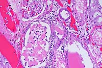
- Case 9-2 . Kidney. Proximal tubule epithelium (right
center) is swollen and vacuolated with nuclear pyknosis (degeneration
& necrosis). Some distal tubules contain homogenous light
pink material (hyaline cast, lower right) and others are filled
with hemoglobin casts (left). Some tubular lumens contain an
admixture of pink protein, sloughed epithelial cells, and neutrophils.
Occassional tubular epithelium has increased basophilia (hyperplasia).
The interstitium is expanded by edema and contains low to moderate
numbers of lymphocytes and macrophages.
-
- AFIP Diagnosis: Kidney: Tubular degeneration and necrosis
(nephrosis), diffuse, with hyaline, granular, and cellular casts,
tubular ectasia, tubular regeneration, and diffuse congestion,
quarter horse, equine.
-
- Conference Note: Prominent histopathologic findings
include degeneration and necrosis of tubular epithelium, sloughing
of necrotic tubular epithelial cells, tubular ectasia, and infiltrates
of neutrophils. In some tubules, erythrocytes and moderate amounts
of a red, granular to globular material, interpreted as protein,
are admixed with sloughed tubular epithelial cells. Within the
interstitium, there are multifocal areas of hemorrhage and edema.
Some tubules are lined by flattened to low cuboidal epithelial
cells that have amphophilic to basophilic cytoplasm and large,
reactive nuclei, interpreted as tubular regeneration by conference
participants.
-
- Oak (Quercus sp.) poisoning has been reported in many regions
of the world, and occurs sporadically in individual animals or
in minor herd epizootics. More than 60 species of oak have been
identified in North America, and all are considered potentially
toxic. Tannins and their metabolites are believed to be the toxic
components. Their levels are highest in young leaves and the
shells of green acorns. Poisoning occurs more commonly in cattle
than horses; this may be related to the gastrointestinal anatomy
in cattle, which allows ingestion of greater quantities of toxic
material at one feeding. Disease outbreaks are often seasonal
with bud and leaf poisoning noted in the spring and acorn poisoning
prominent in autumn. Young buds are much more palatable than
mature leaves, and the levels of tannins become reduced as leaves
mature. Poisoning may also occur during periods of drought when
normal herbage is not available and animals resort to other sources
of food.
-
- While the principal toxins in oak are believed to be the
tannins and their metabolites, the exact mechanism of toxicity
is incompletely understood. Numerous polyphenols are produced
from the metabolism of tannins, the most important of which is
probably digallic acid. Digallic acid is converted to gallic
acid and pyrogallol, both of which are reducing agents and contribute
to toxicosis. Pyrogallol is much more toxic and causes hemorrhagic
gastroenteritis, hematuria, subcutaneous hemorrhage, and hemolysis.
Tannic acid, pyrogallol, and gallic acid administered to rabbits
via stomach tube produce disease similar to that seen in cattle
experimentally fed Q. havardii .
-
- Oak toxicity causes signs of alimentary and urinary disease
in both cattle and horses. Gastrointestinal signs usually occur
early in the course of toxicosis, and animals may be depressed
and lethargic and present with anorexia, tenesmus, constipation,
and colic. After a few days, constipation is often followed by
diarrhea, and fragments of acorns may be present within stools.
Urinary dysfunction usually follows intestinal signs, and may
include polyuria/polydipsia, dyspnea due to hydrothorax, hemoglobinuria,
oliguria, and dependent edema. Mortality rates are often high,
with animals surviving acute disease eventually succumbing to
progressive renal failure. Gastrointestinal lesions may occur
subsequently to renal disease, and uremia and renal failure may
have caused the necropsy findings in the stomach, jejunum, and
tongue.
-
- The most consistent clinicopathologic findings in oak toxicosis
are related to renal disease. Grossly, the kidneys are enlarged,
pale, have petechial hemorrhages, and the medulla is congested.
There may be perirenal, mesenteric and dependent edema, and fluid
accumulation in various body cavities. The most consistent histopathologic
finding is renal tubular necrosis, and proximal convoluted tubules
often contain proteinaceous casts. Adjacent tubules may be unaffected.
The glomeruli are unaffected, and except for congestion, the
medulla remains nearly normal. Common serum clinical chemistry
findings reflect renal failure and include elevated blood urea
nitrogen and creatinine, hypoproteinemia, hypoalbuminemia, hyponatremia,
hypochloremia, hyperkalemia, hypocalcemia, and hyperphosphatemia.
Contributor: Department of Veterinary Pathobiology, College
of Veterinary Medicine, Texas A&M University, College Station,
TX 77843-4467.
-
- References:
- 1. Anderson GA, et al.: Fatal acorn poisoning in a horse:
Pathologic findings and diagnostic considerations. J Am Vet Med
Assoc 182:1105-1110, 1983.
- 2. Duncan CS: Oak leaf poisoning in two horses. Cornell Vet
51:159-162, 1961.
- 3. Panciera RS: Oak poisoning in cattle. In: Effects of Poisonous
Plants on Livestock, Keller RF ed., pp. 499-506, Academic Press,
New York, 1978.
- 4. Schmitz DG: Toxic nephropathy in horses. Compend Cont
Ed Pract Vet 10:104-111,1988.
- 5. Schuh JC, Ross C, Meschter C: Concurrent mercuric blister
and dimethyl sulfoxide (DMSO) application as a cause of mercury
toxicity in two horses. Eq Vet J 20:68-71, 1988.
- 6. Tennant B, Dill SG, Glickman LT, et al.: Acute hemolytic
anemia, methemoglobinemia and Heinz body formation associated
with ingestion of red maple leaves by horses. J Am Vet Med Assoc
179:143-150, 1981.
- 7. Jones TC, Hunt RD, King NW: Diseases due to extraneous
poisons. In: Veterinary Pathology, 6th ed., pp. 704-705, Williams
and Wilkins, 1997.
-
- Case III - AP#1946 (AFIP 2420062)
-
- Signalment: Two-year-old, male, New Zealand white
rabbit.
-
- History: This rabbit was used as a sperm donor for
a reproductive study. Sperm was collected non-invasively with
an artificial vagina. The rabbit had hematuria of one day's duration.
-
- Gross Pathology: The right kidney measured 10 x 10
cm with three 1 cm nodules on surface. The bladder was filled
with bloody urine. The kidney was hollow on cut section and filled
with dark brown fluid. The inner surface of the kidney was necrotic,
with approximately 1 cm of viable cortex. The left kidney was
normal. No other gross lesions were present.
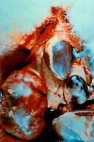
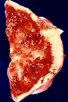
- Case 9-3. Note marked enlargement of right kidney
with locally extensive subcapsular hemorrhage and 3 one centimeter
tan nodules near the capsular surface. In the cut section, there
is diffuse effacement of the cortex by pale white tissue (tumor)
and the medulla is hemorrhagic.
-
- Laboratory Results: The results of special stains,
lectin histochemistry, and immunohistochemistry are outlined
below.
-
- 1. Special stains: An un-decalcified section was stained
by Alizarin red and von Kossa methods to detect mineralization.
With both staining methods, dystrophic calcification was only
present within the necrotic debris. All other parts of the tumor
were negative.
-
- 2. Lectin histochemistry: Ulex europeas agglutinin
was heterogeneously positive with a random distribution of staining.
Peanut agglutinin staining was spotty, but limited only to tumor
cells. Dolichos biflorus and Ricinis communis agglutinins were
negative in all areas.
-
- 3. Immunohistochemistry: Stains included neuron specific
enolase, epithelial membrane antigen, keratins AE1 and AE3, pancytokeratin,
vimentin, S-100 and desmin. Desmin, epithelial membrane antigen,
and S-100 stains were negative within the tumor. Neuron specific
enolase staining was moderately positive, occurring primarily
within the center of tumor lobules. All three keratin antibodies
demonstrated positive staining in neoplastic cells, with stronger
staining in those cells adjacent to necrotic centers. The degree
of staining was moderate with pancytokeratin, and minimal to
moderate with AE1 and AE3. AE3 staining was slightly darker and
more extensive than AE1. Vimentin distribution and degree of
vimentin positivity were similar to that of pancytokeratin, except
that the surrounding stroma was also moderately stained.
-
- Contributor's Diagnosis and Comments: Carcinoma, renal,
NZW rabbit.
-
- Submitted is a section of a neoplastic mass from the right
renal area of the affected rabbit. Within the tumor are large
areas of coagulative necrosis, cellular debris (some of which
is mineralized), and acicular spaces suggestive of cholesterol
crystals. Tumor cells are arranged in lobules, sheets, nests
and acini, and the cells are often associated with an eosinophilic
hyaline matrix resembling osteoid. Round globules of richly eosinophilic
material consistent with a secreted protein are present within
some acini. The hyaline matrix, which is not anisotropic under
polarized light, forms trabeculae next to and between tumor cells
and is also present in larger, acellular areas. Islands of neoplastic
cells are occasionally surrounded by an eosinophilic fibrillar
stroma, which is anisotropic under polarized light. Cell shape
varies from columnar, to low cuboidal, to spindled and stellate.
The columnar and cuboidal cells appear to align along a basement
membrane, while the spindled and stellate cells are more pleomorphic
and less differentiated in appearance. Neoplastic cells are moderate
in size, with a nuclear to cytoplasmic ratio of 1:3. All tumor
nuclei are open-faced with an abundance of euchromatin. Nuclear
shapes are round to oval for the columnar and cuboidal populations.
In the spindled and stellate cells, nuclei are elongate or irregularly-shaped.
The mitotic rate is low at less than 1 per high power field,
and nucleoli are variably present, and generally single and small.
-
- The incidence of spontaneous renal neoplasia in animals is
quite low. Of pri-mary neoplasms, the percentages of those affecting
the kidney are 60, 9.4, 2.3, 1.7 and 0.03, in the pig, horse,
cow, dog and rat respectively1, 2. Carcinomas are the most common
primary renal tumors of dogs, cattle, and sheep3. In mice, spontaneous
renal neoplasms are quite rare except in the case of one inbred
mouse strain. BALB/cf/CD strain mice have a 60-70 percent incidence
of renal carcinoma4. In rats, nephroblastoma is the most common
kidney tumor ob-served. Although spontaneous renal cell carcinomas
are rare in rats, there is a high incidence of renal cell carcinomas
in rats with the Eker mutation. Heterozygotes for the mutation
develop multiple renal cell carcinomas by one year of age.
-
- Renal neoplasms account for 3% of human adult malignancies.
The incidence of human renal neoplasia is higher in males, with
a ratio of 1.6:16. Human renal cell carcinomas commonly metastasize.
The most com-mon metastatic sites are the lungs, bones, lymph
nodes, liver, adrenals, and brain7. Renal cell carcinomas commonly
affect one pole of the kidney. The lesions usually occur as a
single mass. Histologically, the pattern of growth varies from
papillary to solid, trabecular, or tubular. The most common cell
type is the clear cell, hav-ing a rounded or polygonal shape
and abundant clear cytoplasm. Some carcinomas contain granular
cells, which have moderately eosinophilic cytoplasm. Other carcinomas
grow as spindle-shaped cells resembling mes-enchymal tumors.
-
- In rabbits with renal neoplasia, embryonal nephroma occurs
occasionally. An evaluation of rabbit tumors reveals that only
uterine adenocarcinomas occur more frequently than embryonal
nephromas. Weis-broth reviewed the incidence of neoplasia in
rabbits and re-ported 22 cases of embryonal nephroma from 1900-1985,
while over the same period 230 uterine adenocarcinomas occurred.
Secondary polycythemia associated with nephroblastoma has been
re-ported in rabbits. In human Wilm's tumor cases, elevated erythropoietin
is commonly demonstrated. However, this has not been shown in
the rabbit9. A reproducible model for the human Wilm's tumor
was developed by Hard and Fox. A single dose of ethyl-nitrosourea
was given intra-peritoneally to rabbits on the 18th day of gestation.
When rabbits of the strain IIIVO/J were used, there was a greater
than 90% incidence of nephroblastomas in the offspring of treated
dams10. In a report by Carlton and Dietz, renal tumors were described
in two wild rabbits (Sylvilagus floridanus). One tumor was diagnosed
as a hamartoma of urogenital origin. The second tu-mor was diagnosed
as a renal adenocarcinoma11.
-
- Renal cell carcinomas have been reported in the laboratory
rabbit only once12. The tumor was found in a 2½- year-old
female New Zealand white rabbit. The tumor was large and smooth
surfaced, and appeared to arise from the cortex of the right
kidney. The tumor was firm with soft, red, necrotic areas. A
10 cm cyst containing brownish fluid was pre-sent on one side.
No metastases were observed. Cellular morphology varied from
low cuboidal to spindle-shaped. Cells were arranged in irregular
tubular formations, solid sheets, and nests.
-
- Lectins are carbohydrate binding proteins. Different lectins
have a tendency to bind with different sugar moi-eties. In the
kidney, the binding sites of various lectins are sometimes specific
to certain cells in specific neph-ron segments. However, there
are age, developmental, and interspecies differences in the binding
patterns of lectins. Therefore, with few exceptions, the results
obtained from one species will not be applicable to other species.
Holthofer did a comparative study of the lectin binding sites
in the kidney of 14 animal species. In normal rabbit kidney,
he showed that Ricinus communis (castor bean) and Triticum vulgaris
(wheat germ) lectins bound exclusively in proximal renal tubules,
whereas soybean and Arachis hypogaea (peanut) lectins bound dis-tal
tubules and Dolichos biflorus (horse gram) and Ulex europeus
(gorse) lectins bound collecting ducts primarily13. Therefore,
the lectin binding pattern in the kidney tumor from this case
seems to indicate that the site of origin may have been the distal
tubules and collecting duct. However this cannot be confirmed.
Immunohistochemistry is also commonly used to characterize tumors.
The keratin positivity in this case serves to confirm that it
is an epithelial tumor. However, vimentin, the cytoskeletal protein
of mesenchymal cells, was also positive. A similar finding was
previously reported in a study by Holthofer et. al. where simultaneous
expres-sion of vimentin and cytokeratin occurred in 53% of human
renal carcinomas examined14. Although neuron-specific enolase
and S-100 protein positivity has been reported in human renal
cell carcinomas, both markers were negative in this case15.
-
- The gross and microscopic appearance of the tumor in this
report is quite similar to the appearance of the renal cell tumor
described by Kaufmann and Quist. Both tumors were large, smooth
surfaced and firm, and contained a large fluid-filled cyst. In
both cases, cellular morphology varied from low cuboidal to spindled
and stellate, and cells were arranged in irregular tubular formations,
solid sheets, and nests. Tumor metastases were absent in both
animals. The presence of undifferentiated blastema was minimal
to absent in both tumors. Finally, the most significant microscopic
feature was the conspicuous absence of primitive avascular glomerular
formations. The last two points ar-gue against a diagnosis of
embryonal nephroma.
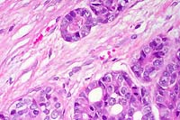 20x
obj
- Case 9-3 . Kidney. The tumor capsule is infiltrated
by nests, cords, and tubule forming pleomorphic polygonal cells.
There are scattered foci of necrosis and occasional mitoses.
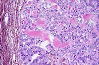 PAMS
20x obj
- Case 9-3. Kidney. A bright pink amorphous to hyaline
material separates tumor cells in many areas. Generally these
areas lack silver staining (black), indicating that this material
is not collagen or basement membrane.
-
- AFIP Diagnosis: Kidney: Renal cell carcinoma, New
Zealand white rabbit, lagomorph.
-
- Conference Note: In humans, renal cell carcinoma occurs
most often in older individuals, usually in the sixth or seventh
decades of life. The use of tobacco is the most prominent risk
factor. Cigarette smokers have twice the incidence of renal cell
carcinoma, and pipe and cigar smokers are also more susceptible16.
Additional predisposing factors may include obesity (especially
in women), hypertension, and exposure to asbestos, heavy metals,
and petroleum products.
-
- While most renal carcinomas are sporadic and occur in older
people, unusual forms of autosomal dominant cancers may occur,
usually in younger individuals. Von Hippel-Lindua (VHL) syndrome
is an autosomal dominant disease in which affected individuals
develop capillary hemangioblastomas at multiple sites within
the central nervous system, including the cerebellum, the retina,
and less commonly the brain stem and spinal cord. Patients also
have cysts involving the pancreas, liver, and kidneys, and a
strong propensity to develop renal cell carcinoma. The VHL gene
is implicated in the development of both familial and sporadic
clear cell tumors.
-
- Renal cell carcinoma in people tends to produce diverse clinical
signs not related to the kidney and is known as one of the great
imitators in human medicine. The tumor causes a number of paraneoplastic
syndromes attributed to hormone production, including polycythemia,
hypercalcemia, hepatic dysfunction, feminization and masculinization,
Cushing syndrome, eosinophilia, leukemoid reactions, and amyloidosis.
Another characteristic of this tumor in people is its tendency
to metastasize widely before the appearance of clinical signs;
there is frequently radiologic evidence of metastasis, primarily
to the lungs and/or bones, at the time of initial presentation.
-
- Contributor: Center for Comparative Medicine, Baylor
College of Medicine, One Baylor Plaza, Houston, TX 77030.
-
- References:
- 1. Baskin GB, De Paoli A: Primary renal neoplasms of the
dog. Vet Path 14:591-605, 1977.
- 2. Hard GC: Tumors of the kidney, renal pelvis and ureter.
In: Pathology of Tumors in Laboratory Ani-mals, Tumors of the
Rat, vol. 1, pp. 301-344, 2nd ed., Turusov and Mohr ed., IARC
Scientific Publica-tions, 1990.
- 3. Maxie MG: The urinary system. In: Pathology of Domestic
Animals, Jubb KVF, Kennedy PC, Palmer N, eds., vol. 2, pp. 518-522,
Acad-emic Press, San Diego, CA, 1993.
- 4. Rabstein LS, Peters RL: Tumors of the kidneys, synovia,
exocrine pancreas, and nasal cavity in BALB/cf/Cd mice. J Nat
Cancer Inst 51:999-1006, 1973.
- 5. Eker R, Mossige J, Johannessen JV, Aars H: Hereditary
renal adenomas and adenocarcinomas in rats. Diagnostic Histopathology
4:99-110, 1981.
- 6. Murphy WM, Beckwith JB, Farrow GM: Tumors of the Kidney,
Bladder, and Related Urinary Struc-tures. In: Atlas of Tumor
Pathology, pp. 92-131, Fascicle 11, 3rd series, Armed Forces
Institute of Pathology, Washington DC, 1994.
- 7. Weisbroth SH: Neoplastic Diseases. In: The Biology of
the Laboratory Rabbit, Manning, Ringler, Newcomer, eds., 2nd
ed., pp. 259-292, Academic Press, San Diego, CA, 1994.
- 8. Wardrop KJ, Nakamura J, Giddens WE Jr.: Nephroblastoma
with secondary polycythemia in a New Zealand white rabbit. Lab
Anim Sci 32:280-282, 1982.
- 9. Hard GC, Fox, RR: Histologic characterization of renal
tumors (nephroblastomas) induced transplacentally in IIIVO/J
and WH/J rabbits by N-ethylnitrosourea. Am J Path 113:8-18, 1983.
- 10. Carlton WW, Dietz JM: Two renal tumors in cottontail
rabbits (Sylvilagus floridanus). Vet Pa-th 14:29-35, 1977.
- 11. Kaufmann AF, Quist KD: Spontaneous renal carcinoma in
a New Zealand white rabbit. Lab Anim Care 20:530-531, 1970.
- 12. Holthöfer H: Lectin binding sites in kidney: A comparative
study of 14 animal species. J Histochemistry Cytochemistry 31:531-537,
1983.
- 13. Holthöfer H, et al.: Cellular origin and differentiation
of renal carcinomas: A fluorescence microscopic study with kidney-specific
antibodies, antiintermediate filament antibodies, and lectins.
Lab Inves 49:317-326, 1983.
- 14. Kusama K, et al.: Tumor markers in human renal cell carcinoma.
Tumor Biology 12:189-197, 1991.
- 15. Cotran RS, Kumar V, Collins, Robbins SL: The kidney.
In: Robbins Pathologic Basis of Disease, 6th ed., pp. 991-994,
WB Saunders, Philadelphia, 1999.
-
- Case IV - P98-4500/N (AFIP 2641836)
-
- Signalment: Six-year-old, cross-bred, gelding, equine,
Equus caballus.
-
- History: The horse suffered from dysphagia, sweating,
intermittent colic, and weakness for a few weeks. Because the
onset of clinical signs was insidious, the duration of clinical
disease could not be determined with certainty. Physical examination
revealed weakness, sweating, dehydration, emaciation, reflux
and colonic impaction. The horse stood with an arched back and
tucked-up abdomen. No treatment was provided, and the horse was
euthanized due to very poor general condition.
-
- Gross Pathology: At necropsy, the horse was emaciated
and in very poor condition. The coat in the area of neck and
shoulders was bilaterally moist. Internal organs were normal
except for the gastrointestinal tract. The stomach contained
only minimal amounts of water, and no ingesta was present. The
entire large bowel was severely dilated and filled with dry feces.
Feces and mucosa were covered by thick, sticky mucus.
-
- Laboratory Results: None.
-
- Contributor's Diagnosis and Comments:
- 1. Coeliaco-mesenteric ganglion, neurons: Chromatolysis and
eosinophilia, diffuse, marked; margination, pyknosis and loss
of nuclei, diffuse, marked; vacuolization, multifocal, mild;
lipopigment accumulation, diffuse, mild, cross-bred, equine.
2. Coeliaco-mesenteric ganglion: Lymphocytic infiltration, multifocal,
minimal.
- Lesions are consistent with equine dysautonomia (grass sickness).
-
- Equine dysautonomia (ED) is a profoundly debilitating, almost
invariably fatal disease of undetermined etiology that affects
equidae in northwestern Europe and South America. The European
variant is also known as grass sickness, grass disease and autonomic
polyganglionopathy. The South American variant is also known
as "mal seco". Similar dysautonomic diseases occur
in other species including cats, dogs and hares.
-
- The clinical course of the disease in horses can be acute
or chronic. Horses with the acute form of ED usually die within
48 hours after the onset of clinical signs, which are predominated
by abdominal pain, gastric reflux, tachycardia, intestinal atony
and dysphagia. The chronic form may be insidious in onset, and
horses may survive for several weeks. Anorexia leading to progressive
weight loss and emaciation, intermittent colic due to colonic
impaction, sweating, tremor, weakness and mild dysphagia may
occur. In some cases diarrhea is observed.
-
- Gross lesions are variable and non-specific, but may be indicative
of gastrointestinal atony. Aspiration pneumonia due to dysphagia
may occur. Histologically, the myenteric and submucosal alimentary
plexuses and peripheral ganglia are affected by neuronal degeneration.
In chronic cases, there may be a numerical decrease of neurons,
while the number of non-neuronal cells is increased. Lymphocytic
infiltration, as in the submitted case, is a common finding in
horses and seems to be unrelated to the development of ED. Neuronal
degeneration is not limited to the autonomic plexuses and ganglia,
and may also occur in the dorsal root ganglia, the intermediolateral
nucleus, and the ventral horns of the spinal cord. Specific brain
stem nuclei may be affected including the nucleus (N.) motoricus
nervi hypoglossi, N. dorsalis nervi vagi, N. ambiguus, N. vestibularis
lateralis, N. occulomotorius, and formatio reticularis.
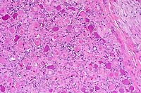 10x
obj
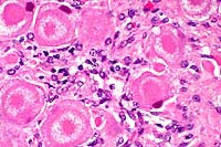 40x
obj
- Case 9-4 . Ganglion. Multifocally neurons have loss
of Nissle substance, with coalescence of the cell cytoplasm as
a central homogenous eosinophilic aggregate surrounded by a clear
to slightly fibrillar halo zone. Nuclei are karyolytic with poorly
defined nuclear outlines. Low to moderate numbers of lymphocytes
and macrophages infiltrate these zones of neuronal degeneration.
-
- AFIP Diagnosis: Ganglion: Neuronal degeneration and
necrosis, diffuse, with satellite cell proliferation and mild
multifocal lymphocytic ganglioneuritis, mixed breed horse, equine.
-
- Conference Note: Conference participants noted several
histologic changes affecting neurons including loss of Nissl
substance (chromatolysis), swollen nuclei, karyolysis, and hypereosinophilia.
Some neurons are pale and contain indistinct or faded nuclei.
Occasional neurons contain multiple, peripheral, clear, discreet
intracytoplasmic vacuoles that surround a central zone of hypereosinophilia.
Axonal swelling with occasional spheroids is present multifocally.
There is also a diffuse increase in satellite cells. Nuclear
placement and chromatin pattern may be varied depending upon
anatomical location of neurons, and participants were cautious
in placing emphasis on location and morphology of nuclei.
-
- The nature and distribution of microscopic lesions are very
similar in the species which suffer from dysautonomia, and the
findings in this case are fairly representative of the condition.
The cytoplasm of affected neurons loses the basophilic granularity
of Nissl substance and takes on a homogenous, "ground glass",
appearance. Initially, neurons are rounded and swollen in the
early stages of disease, but eventually become shrunken and irregular
with pyknotic and faded nuclei. Progression of the disease leads
to neuronal loss and satellite cell proliferation, especially
prominent in the cat in which the disease is also known as Key-Gaskell
syndrome (KGS).
-
- While the lesions are similar in several species, significant
differences in clinical presentation are observed. Horses with
ED have increased heart rate and suffer from patchy sweating,
while 50% of cats with KGS have bradycardia. Cats and dogs develop
dry mucous membranes, while horses drool thick saliva, probably
due to the inability to swallow. Cats also have fixed, dilated
pupils and greatly reduced lacrimation, while these abnormalities
are not present in horses.
-
- The etiology of ED is unknown, although an ingested neurotoxin
is suspected. Characteristic autonomic neuronal degeneration
was produced in peripheral autonomic ganglia, and spinal cord
and brain stem nuclei in several healthy horses following intraperitoneal
injection of serum from acute cases of ED, although the experimental
recipients did not develop clinical signs of ED. Research thus
far supports the hypothesis that ED is caused by a neurotoxin
that may gain access to the circulation, and may also injure
autonomic neurons through retrograde axonal transport.
-
- The suspected neurotoxin may be ingested, produced during
metabolic transformation of an ingested agent, or synthesized
by the bacterial flora in the gut. Damage to the neurons in the
intestinal myenteric and submucosal plexuses occurs more extensively
than at other sites (such as the coeliaco-mesenteric ganglion),
and probably reflects the higher concentrations of neurotoxin
that are related to the site of absorption. The degeneration
and depletion of neurons is most severe in the ileum, and this
may be the main site of entry for the toxin. Lesions in autonomic
neurons distant to the intestinal tract are less severe, and
hypothetically are due to neurotoxin transferred by retrograde
axonal flow from the site of absorption. Neurotoxin absorbed
into the systemic circulation may also potentially damage neurons
which are not protected by the blood-brain barrier. Regardless
of the etiopathogenesis, the histologic lesions lead to disordered
autonomic innervation of the alimentary tract from the pharynx
to the rectum.
-
- Contributor: Department of Veterinary Pathology, Faculty
of Veterinary Medicine, Utrecht University, Postbox 80158, 3508
TD Utrecht, The Netherlands.
-
- References:
- 1. Johnson PJ: Equine dysautonomia. Equine Pract 17:25-32,
1995.
- 2. Fatzer R, et al.: Sind equine motorische Nervenzell-Degeneration
(EMND) und Graskrankheit des Pferdes unterschiedliche Manifestationen
der gleichen Grundkrankheit? Pferdeheilk 11:17-29, 1995.
- 3. Pinsent PJN: Grass sickness of horses (grass disease:
equine dysautonomia). Vet Annual 29:169-174, 1989.
- 4. Pollin MM, Griffiths IR: A review of the primary dysautonomias
of domestic animals. J Comp Pathol 106:99-119, 1992.
- 5. Schulze C, Venner M, Pohlenz J: Chronische Graskrankheit
(Equine Dysautonomie) bei einer zweieinhalb-jährigen Isländer-Stute
auf einer nordfrisischen Insel. Pferdeheilk 12:345-350, 1997.
- 6. Jubb KVF, Huxtable CR: The nervous system. In: Pathology
of Domestic Animals, Jubb, Kennedy, Palmer eds., 4th ed., vol.
1, pp. 365-366, Academic Press, San Diego, 1993.
-
- Ed Stevens, DVM
Captain, United States Army
Registry of Veterinary Pathology*
Department of Veterinary Pathology
Armed Forces Institute of Pathology
(202)782-2615; DSN: 662-2615
Internet: STEVENSE@afip.osd.mil
-
- * The American Veterinary Medical Association and the American
College of Veterinary Pathologists are co-sponsors of the Registry
of Veterinary Pathology. The C.L. Davis Foundation also provides
substantial support for the Registry.
Return to WSC Case Menu
 20x
obj
 40x
obj
 40x
obj
 20x
obj


 20x
obj
 PAMS
20x obj
 10x
obj
 40x
obj