Signalment: Adult, female, mixed breed dog.
History: This dog was housed in a standard indoor run. The dog was to be quarantined and conditioned for experimental use. Over a two month period, the general condition of the dog declined. The dog subsequently developed a large, oval, firm mass protruding from the vulva along with a serosanguineous vaginal discharge. The dog was euthanized and necropsied for diagnostic purposes.
Gross Pathology: Necropsy revealed a pale-white, firm but friable, nodular, multi-lobulated mass that measured 10 cm x 10 cm.
Laboratory Results: Complete blood count and serum chemistry profile were within normal limits.
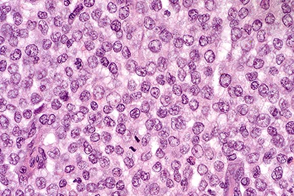
Contributor's Diagnosis and Comments: Transmissible venereal tumor.
Canine transmissible venereal tumor (CTVT) is a naturally occurring tumor that is most often seen in young, roaming, sexually active dogs. CTVT is coitally transmitted and affects the external genitalia of either sex. Common locations for the tumor in the male dog are the caudal part of the penis and the glans penis. Common locations for the tumor in the female dog are the posterior part of the vagina and at the junction of the vestibule and vagina. Extragenital occurrence of CTVT has been reported in lymph nodes, skin, lips, buccal and nasal mucosa and less frequently in the tonsils, liver, pancreas, spleen, lung, and kidney.
CTVT is currently classified as a round cell tumor of reticuloendothelial origin. The tumor continues to grow until immune mechanisms cause regression of the tumor or the animal succumbs to secondary complications. Regression is an IgG-mediated process.
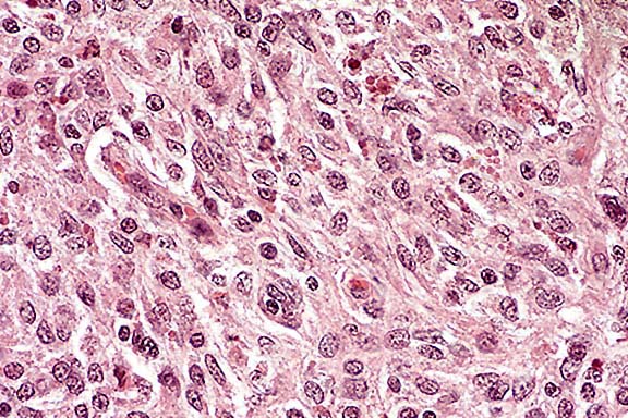
Histologically, the cells are round, ovoid or polyhedral and may be arranged in compact sheets. The cells contain moderate eosinophilic cytoplasm; nuclei have marginal chromatin and few mitotic figures. Differential diagnosis for this neoplastic mass includes lipoma, mast cell tumor, epidermoid carcinoma, squamous cell carcinoma, fibroma, and histiocytoma.
Conference Note: Transmission of this tumor occurs only by transplantation of viable tumor cells. Oncogenic viral particles have never been demonstrated in the neoplastic cells, and cell-free filtrates are non-transmissible.1 The karyotype of the tumor cells is constantly and significantly different from that of normal canine cells. The normal chromosome count of the dog is 78, with 76 of the chromosomes acrocentric; in CTVT cells there are usually 58 to 59 chromosomes, with 13 to 17 metacentric and 42 acrocentric. This is a constant karyotypic variation and is found worldwide.1,4 The tumor is also transmissible to the fox, coyote, and jackal.5
The precise mechanisms by which CTVT cells can be transplanted
across major histocompatibility (MHC) barriers and survive for
a substantial time before spontaneously regressing are not known.5,6
Infected dogs develop anti-tumor antibodies. However, CTVT cells
produce a tumor-associated antigen which may block some of those
antibodies in dogs with growing tumors. Also, cells from growing
tumors lack expression of Class I or Class II MHC antigens, whereas
30 to 40% of those from early regressor tumors express both Class
I and II MHC antigens.6 This may provoke additional immune reactions
of the host to speed up the rejection process and cause the tumor
mass to regress in 2 to 3 weeks.6
The cellular origin of CTVT is uncertain. Immunohistochemical
studies have shown that many of the neoplastic cells express lysozyme
and/or a1-antitrypsin, supporting the hypothesis that some of
these neoplasms are of histiocytic origin.2 Although these markers
are not reliable for all CTVT's, they can be helpful in excluding
lymphomas, amelanotic melanomas, poorly differentiated carcinomas
and mast cell tumors from the differential diagnosis.2 Differentiating
CTVT from canine cutaneous histiocytoma should be based on clinical
and histopathologic criteria.
Contributor: Emory University, G 70 Rollins Research Center, Atlanta, GA 30322.
References:
Signalment: Fetus (gestational age @ 7.5 months), female, Holstein, bovine.
History: This fetus was born moribund and euthanized in extremis. The cow was serologically positive for bovine immunodeficiency virus (BIV) and bovine leukosis virus (BLV) at least one year before the abortion. She experienced a dislocated hip and a rib fracture 3 months previously. She was thin (450kg), had an abscess on the medial side of the left stifle and laminitis affecting all feet one month after abortion.
Gross Pathology: Necrotic areas and hemorrhages were
seen mainly on the cotyledonary areas of the placenta. Intercotyledonary
areas surrounding the placentomes had a thickened leathery aspect.
The fetus had an enlarged liver with rounded edges, excessive
fluid in the peritoneum and pericardium. Petechial hemorrhages
were in the lung. Hematocysts were on the right auriculo-ventricular
valve.
Laboratory Results:
Bacteriology: No bacteria were isolated from the liver, lung and stomach content. Contaminants were cultured from the placenta.
Virology/rickettsiology: Fluorescent antibody testing of frozen lung sections was negative for infectious bovine rhinotracheitis (IBR) herpesvirus and bovine virus diarrhea (BVD) pestivirus.
Chlamydia psittaci was isolated after inoculation of placental extracts into Vero cells.
Polymerase chain reaction targeted at the BIV gag gene was positive from samples of blood from the dam and from an homogenate of placenta, whereas fetal lung tissue was negative.
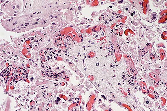
Contributor's Diagnosis and Comments: Placenta, placentitis, necrotizing, multifocal, acute to subacute, severe, with vasculitis and intratrophoblastic intracytoplasmic inclusions.
Cause: Chlamydia psittaci.
Histopathologic Description: The cotyledonary villi are
multifocally necrotic, with hemorrhage. Mineralization is in a
few necrotic areas. The small cotyledonary arterioles and the
large chorionic arteries are segmentally thrombosed, have medial
fibrinoid necrosis, moderate to marked transmural infiltrates
of neutrophils, and/or are surrounded by sheaths of inflammatory
cells, mainly neutrophils. Clumps of necrotic material are often
seen at the base of cotyledonary villi. Capillaries of the cotyledonary
villi have hyaline microthrombi. The chorion is multifocally infiltrated
by neutrophils and is edematous. Trophoblasts and syncytiotrophoblasts
uncommonly have intracytoplasmic vacuoles that push the pyknotic
nuclei to the periphery and are composed of foamy to granular
~ 1-2 mm pale basophilic organisms. All these elementary changes,
due to the multifocal nature of the lesion, may not be present
in every section. The Machiavello stain performed from fixed placental
tissues was negative.
This is an additional, fully documented case of chlamydial abortion in cattle. Unlike sheep and goats, chlamydial urogenital disease is believed to be rare in bovine species.1 Although chlamydial abortions have been experimentally produced in cows, the importance of Chlamydia as a natural abortigenic pathogen in cows is presently not clear.1,2 The condition might be under-diagnosed in this species. Several outbreaks have been recently described 3,4 and scattered cases are reported from several countries over the world, including the United States5, Japan, Australia, India3, Slovakia, Italy, Germany and the United Kingdom.4
This particular case occurred in an emaciated, debilitated cow persistently infected with bovine immunodeficiency virus (BIV) and bovine leukosis virus (BLV), which was euthanized and subsequently necropsied by one of the contributors. This cow is from a herd where retroviral infections are endemic and are associated with several problems.6,7 The serotype of the chlamydial isolate was determined to be serovar B, mainly isolated in pigeons.
Conference Note: Conference participants agreed that this lesion is compatible with chlamydiosis. Although special stains viewed in conference were negative, the contributor's culture results are considered to be significant.
Numerous pathogens can cause placentitis and abortion in cattle. Bacterial and fungal infections are more common than viral causes. The differential etiologic diagnosis includes Chlamydia psittaci, Brucella abortus, Campylobacter spp., Listeria monocytogenes, Leptospira spp., Ureaplasma diversum, Actinomyces pyogenes, Salmonella spp., Yersinia pseudotuberculosis, Hemophilus somnus, Coxiella burnetti, Borrelia coriaceae, and Aspergillus spp.
Significant viral and protozoal causes of bovine abortion include bovine pestivirus (BVD virus), bovine herpesvirus 1, bovine orbivirus (bluetongue virus), and Neospora caninum.
Contributor: Department of Veterinary Pathology, School of Veterinary Medicine, Louisiana State University, Baton Rouge, Louisiana 70803
References:
Signalment: 8-year-old, male, canine.
History: The dog had a progressive left forelimb lameness of 6 months duration which was non-responsive to anti-inflammatory therapy. The dog was euthanized at the owner's request.
Gross Pathology: A circumscribed reddish-grey firm intramedullary
mass,
8 mm in diameter, was located at the level of the seventh cervical
vertebra and was centered to the left of the midline.
Contributor's Diagnosis and Comments: Canine, spinal cord (C7), intramedullary, hemangioma.
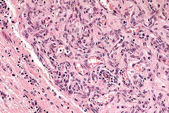
Conference Note: This case was also studied by the Department of Neuropathology of the AFIP. While it is agreed that this neoplasm resembles that reported by Cordy as hemangioma, the morphologic and immunohistochemical staining characteristics more closely resemble those of hemangioblastoma. Immunohistochemical stains performed at the AFIP demonstrated that many of the stromal cells are positive for neuron-specific enolase (NSE) and negative for factor VIII-related antigen (FVIII-rag) while the spindle cells that line the vascular spaces are positive for FVIII-rag. The NSE-positive stromal cells are not a component of "hemangioma" as the term is normally applied.
To the best of our knowledge, hemangioblastoma has not been reported in dogs. In humans, hemangioblastoma is a capillary-rich neoplasm containing variably lipid-rich interstitial or stromal cells.5 It occurs predominantly in middle-aged males and is most often located in the cerebellum. However, these tumors also arise in the medulla oblongata and spinal cord, particularly when they occur in association with Von Hippel-Lindau syndrome. This human disease syndrome includes intracranial or intraspinal hemangioblastoma; retinal hemangioblastoma; cystic lesions of the liver, kidney, pancreas, and epididymis; and renal cell tumors.
Spinal hemangioblastomas are discrete intramedullary tumors that abut the leptomeninges. Microscopically, they vary considerably in their cellular density, but generally consist of large polygonal interstitial cells, the exact nature of which is unknown, that are packed between abundant, often criss-crossing capillary channels. There are at least 3 variants of this tumor described.5
At least two morphologic differences were noted between the canine tumor and human hemangioblastoma. First, in humans, mast cells are commonly distributed rather uniformly throughout the tumor. Second, the stromal cells in the canine tumor do not contain an appreciable amount of lipid.
Contributor: Department of Veterinary Pathology, Faculty of Veterinary Medicine, University College Dublin, Ballsbridge, Dublin 4, Ireland
References:
Signalment: 10-week-old pig.
History: In a 450 sow, farrow-to-finish swine production facility, poor growth rate, dyspnea, and increased mortality among nursery pigs was investigated. Necropsy of three affected pigs was performed.
Gross Pathology: Lymph nodes were enlarged. Lungs were diffusely swollen and rubbery; in cranioventral portions, they were consolidated and gray.
Laboratory Results: Circovirus was isolated (on porcine fetal kidney cells) from lymph node and spleen. Porcine circovirus was demonstrated by immunohistochemistry in the lung, spleen, and kidney (performed by Dr. E.G. Clark, Western College of Veterinary Medicine, University of Saskatchewan, Saskatoon, SK). Fluorescent antibody tests of the lung were negative for Mycoplasma hyopneumoniae, swine influenza virus, pseudorabies virus, and coronavirus.
Contributor's Diagnosis and Comments: Inguinal lymph node: Lymphadenitis, granulomatous, multifocal-coalescing, with polymorphous amphophilic to basophilic inclusion bodies in the cytoplasm of macrophages, consistent with porcine circovirus infection.
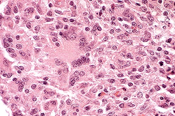
Conference Note: Conference participants considered a differential diagnosis for granulomatous lymphadenitis in swine, which included tuberculosis, brucellosis, and fungal infections. A variety of special stains did not demonstrate bacteria or fungi in the lymph node.
The exact role that porcine circovirus (PCV) plays in pig disease remains to be determined. Early epidemiologic studies showed that antibodies to PCV were widespread in slaughter hogs and wild boars in Germany, and was apparently not associated with disease.4,5 A later study found evidence suggesting that transplacental infection occurs; however, it was concluded that PCV is probably not a significant cause of reproductive disorders in pigs in Northern Ireland.1 In contrast, a recent report suggests that, in Western Canada, PCV may be responsible for numerous lesions seen in several herds in a "post-weaning multisystemic wasting syndrome".3
Contributor: Animal Disease Diagnostic Laboratory - 1175, Purdue University, West Lafayette, IN 47907-1175
References:
Terrell W. Blanchard
Major, VC, USA
Registry of Veterinary Pathology*
Department of Veterinary Pathology
Armed Forces Institute of Pathology
(202)782-2615; DSN: 662-2615
Internet: blanchard@email.afip.osd.mil
* The American Veterinary Medical Association and the American College of Veterinary Pathologists are co-sponsors of the Registry of Veterinary Pathology. The C.L. Davis Foundation also provides substantial support for the Registry.